 Germán G. Sgro1†
 Gabriel U. Oka1†
 Diorge P. Souza1‡
 William Cenens1
 Ethel Bayer-Santos1‡
 Bruno Y. Matsuyama1Natalia F. Bueno1Thiago Rodrigo dos Santos1
 Cristina E. Alvarez-Martinez2
 Roberto K. Salinas1
 Chuck S. Farah1*
- 1Departamento de Bioquímica, Instituto de Química, Universidade de São Paulo, São Paulo, Brazil
- 2Departamento de Genética, Evolução, Microbiologia e Imunologia, Instituto de Biologia, University of Campinas (UNICAMP), Campinas, Brazil
Bacteria have been constantly competing for nutrients and space for billions of years. During this time, they have evolved many different molecular mechanisms by which to secrete proteinaceous effectors in order to manipulate and often kill rival bacterial and eukaryotic cells. These processes often employ large multimeric transmembrane nanomachines that have been classified as types I–IX secretion systems. One of the most evolutionarily versatile are the Type IV secretion systems (T4SSs), which have been shown to be able to secrete macromolecules directly into both eukaryotic and prokaryotic cells. Until recently, examples of T4SS-mediated macromolecule transfer from one bacterium to another was restricted to protein-DNA complexes during bacterial conjugation. This view changed when it was shown by our group that many Xanthomonas species carry a T4SS that is specialized to transfer toxic bacterial effectors into rival bacterial cells, resulting in cell death. This review will focus on this special subtype of T4SS by describing its distinguishing features, similar systems in other proteobacterial genomes, and the nature of the effectors secreted by these systems and their cognate inhibitors.
Introduction
Type IV secretion systems (T4SSs) have been studied since the birth of modern molecular biology, starting with the description of bacterial conjugation over 70 years ago (Lederberg and Tatum, 1946). It quickly became evident that the T4SS-mediated horizontal transfer of genetic material is a major contributor to bacterial evolution, making it necessary to consider lateral connections between lineages for a complete description of the bacterial tree of life (de la Cruz and Davies, 2000). Horizontal gene transfer is also one of the principal mechanisms for the spread of genes conferring resistance to antibiotics (Cabezon et al., 2015). Moreover, many pathogenic bacteria use T4SSs to facilitate their proliferation and survival inside eukaryotic hosts, typically by the secretion of protein effectors or protein-DNA complexes (Gonzalez-Rivera et al., 2016). T4SSs are thus important virulence factors in a variety of human diseases, including whooping cough (Bordetella pertussis; Locht et al., 2011; Carbonetti, 2015), cat-scratch fever (Bartonella henselae; Siamer and Dehio, 2015), brucellosis (Brucella spp.; Ke et al., 2015), Legionnaire’s pneumonia (Legionella pneumophila; Finsel and Hilbi, 2015), Q fever (Coxiella burnetii; Moffatt et al., 2015) and peptic ulcer and gastric cancer (Helicobacter pylori; Naumann et al., 2017). One of the most well-characterized T4SSs is that of Agrobacterium tumefaciens which injects nucleoprotein complexes and protein factors into plant cells (Alvarez-Martinez and Christie, 2009; Li and Christie, 2018). Furthermore, specialized T4SSs from Neisseria gonorrhoeae or H. pylori secrete DNA to the extracellular milieu or uptake DNA from the environment to the bacterial cytoplasm, respectively (Hofreuter et al., 2001; Hamilton et al., 2005; Callaghan et al., 2017). Finally, the plant pathogen Xanthomonas citri (Oliveira et al., 2016; Sgro et al., 2018; Souza et al., 2015) and, more recently, the opportunistic human pathogen Stenotrophomonas maltophilia (preprint: Bayer-Santos et al., 2019), have been shown to use a T4SS to inject toxic effectors into target bacteria, thus inducing the death of rival cells (Figure 1).
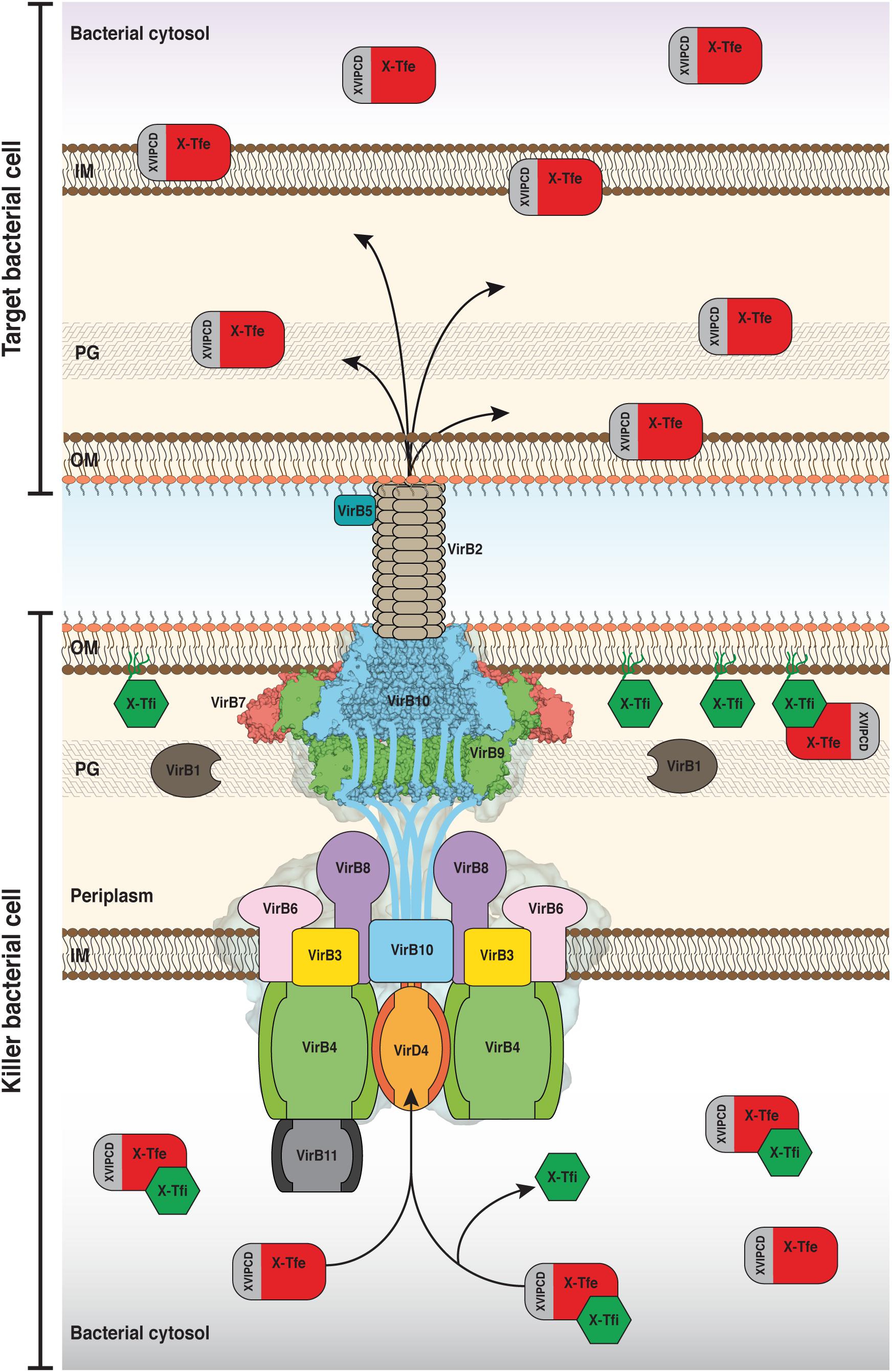
Figure 1. Schematic model of the structure and function of the bacteria-killing Xanthomonadales-like Type IV secretion systems (X-T4SSs). The model shows the interface between two bacterial cells. The killer cell (below) is armed with an X-T4SS whose general architecture is based on the negative-stained electron microscope map of the R388 T4SS shown in the background (Low et al., 2014; Redzej et al., 2017) and the cryo-EM structure of the X. citri core complex (VirB7, VirB9, and VirB10; Sgro et al., 2018) associated with the outer membrane (OM). The disordered N-terminal domains of the VirB10 subunits extend down from the core complex and pass through the inner membrane. The inner membrane (IM) complex is made up of VirB3, VirB6, VirB8, the three ATPases VirB4, VirB11, and VirD4 as well as the aforementioned N-terminal segments of VirB10. Pili, made up of VirB2 and VirB5, mediate intercellular contacts. X-T4SS effectors (X-Tfes) interact, via their XVIPCD domains, with VirD4 and are subsequently transferred to the T4SS for translocation into the target cell where they will degrade target structures such as membrane phospholipids or carbohydrate and peptide linkages in the peptidoglycan (PG) layer. Prior to secretion, X-Tfes whose activities could target cytosolic substrates can be inhibited by cytosolic variants of their cognate immunity proteins (X-Tfis). If X-Tfes make their way into the periplasm, either by leakage from the secretion channel or by injection by neighboring cells of the same species, they will be inhibited by the periplasmic lipoprotein forms of the cognate X-Tfi. Portions of the Figure were adapted from Low et al. (2014) and Sgro et al. (2018) with permission from the publishers.
T4SSs are structurally very diverse. For example, the related pKM101 and R388 plasmid-encoded conjugation systems (Chandran et al., 2009; Fronzes et al., 2009; Rivera-Calzada et al., 2013) and the pathogenic Legionella Dot/Icm (Ghosal et al., 2017; Chetrit et al., 2018) and H. pylori Cag (Frick-Cheng et al., 2016; Chang et al., 2018) effector-secreting systems, while all exhibiting an outer membrane-associated core complex with 14-fold or 13-fold symmetry, present significantly different features in terms of their overall size. These systems also display a varied set of both functional and structural subunits, and even the homologous subunits have very low sequence similarity and frequently present modified domain architectures (Alvarez-Martinez and Christie, 2009; Christie et al., 2014; Guglielmini et al., 2014; Christie, 2016; Grohmann et al., 2017). For these reasons, the T4SSs from Gram-negative bacteria have been divided into two major classes, denoted A and B (Christie and Vogel, 2000), and classification systems based on detailed phylogenetic analysis have divided Gram-negative and Gram-positive T4SSs into up to 8 classes (Guglielmini et al., 2014).
The canonical class A, best represented by the A. tumefaciens vir system and those coded by conjugative plasmids pKM101, R388, and RP4, have the basic set of 12 conserved subunits, named VirB1 to VirB11 plus VirD4 (Tzfira and Citovsky, 2006). The overall organization of the canonical class A T4SSs has been revealed in electron microscopy studies (Low et al., 2014; Redzej et al., 2017) and can be divided into two general (sub)complexes (Figure 1). The inner membrane complex is made up of subunits embedded in, or associated with, the inner membrane: VirB3, VirB4, VirB6, VirB8, VirB11, and VirD4. The outer membrane or “core” complex is comprised of the subunits VirB7, VirB9, and VirB10. These two complexes are connected by a flexible “stalk” of unknown composition, though it has been proposed to be made up, at least in part, by the disordered N-terminal domain of VirB10 (which also has an N-terminal transmembrane helix embedded in the inner membrane) and/or the C-terminal domain of VirB8 (Christie, 2016; Waksman, 2019). In addition to this transmembrane structure, there are extracellular pili made of subunits VirB2 and VirB5, that are presumably involved in making contact with the membrane of the target cell or organelle (Alvarez-Martinez and Christie, 2009). Even within the class A T4SSs, a large degree of sequence and size diversity has been observed for many of the subunits in different species. This is perhaps most starkly exemplified when considering the H. pylori Cag T4SS which, in addition to orthologs of the basic set of canonical class A subunits, possesses another five subunits that are required for proper function (Backert et al., 2015; Frick-Cheng et al., 2016).
The even more distantly related class B includes T4SSs found in the pathogens L. pneumophila, C. burnetii, and Rickettsiella grylli as well as in the IncI conjugative plasmids R64 and ColIb-P9 (Sexton and Vogel, 2002). L. pneumophila causes Legionnaire’s disease in humans by infecting alveolar macrophages where it replicates within a specialized vacuole (Backert and Meyer, 2006; Ensminger and Isberg, 2009). Its Dot/Icm T4SS is made up of 27 components and secretes more than 300 effector proteins that manipulate signal transduction pathways in the host cell, primarily affecting organelle trafficking (Hilbi et al., 2017; Qiu and Luo, 2017). The bacteria-killing T4SSs, which is the topic of this review, belong to the canonical class A T4SSs, although they do have some structurally distinguishing features as described below.
The Xanthomonadales order of Gammaproteobacteria (Saddler and Bradbury, 2007), recently divided into two orders, Xanthomonadales (families Xanthomonadaceae and Rhodanobacteraceae) and Nevskiales (Naushad et al., 2015), include several hundred phytopathogenic species of the genera Xanthomonas and Xylella as well as important and ubiquitous soil, water and plant-associated bacteria of the genera Stenotrophomonas, Lysobacter, Luteimonas, Pseudoxanthomonas, Rhodanobacter, Luteibacter, Dyella, Frateuria, Aquimonas, and others (Van Sluys et al., 2002; Saddler and Bradbury, 2007; Looney et al., 2009; Mansfield et al., 2012). Some Stenotrophomonas strains are opportunistic pathogens of immunosuppressed human patients (Chang et al., 2015) and some Stenotrophomonas and Lysobacter strains have been recognized as potential biological control agents in combating plant diseases caused by fungi or other bacteria (Hayward et al., 2010; Mukherjee and Roy, 2016; Panthee et al., 2016). Other species from the genera Lysobacter and Luteimonas have been isolated from extreme environments (Brito et al., 2013; Zhang et al., 2015). Although the role of types II and III secretion systems in the virulence of species of the genus Xanthomonas is already well established (Buttner and Bonas, 2010), until a few years ago there was little information available on the functions of other secretion systems in these bacterial species. An accompanying article in this series deals with the recently discovered role of the X. citri Type VI secretion system (T6SS) in protection against predation by phagocytic amoebas (Bayer-Santos et al., 2018). In this review, we will focus on the special characteristics of the Xanthomonadales Type IV secretion systems, first described in X. citri, and their role in the contact-dependent killing of rival Gram-negative species. The review will focus on describing the distinguishing structural features of the T4SS components encoded by the chromosomal virB locus of X. citri, their conservation in homologous systems in the order Xanthomonadales and other proteobacterial genomes, the nature of the effectors secreted by these systems and the cognate inhibitors of these effectors.
The Chromosomally Coded T4SS of Xanthomonas citri
The T4SS encoded by the chromosomal vir locus of X. citri contains the canonical set of 12 structural components found in other class A T4SSs (Figure 2; Alegria et al., 2005; Souza et al., 2011). The presence of chromosomally encoded homologs in several other Xanthomonas species (see below) suggested an important function in Xanthomonas biology. A role in bacterial conjugation or nucleic acid transfer was deemed unlikely since the chromosomal virB locus does not contain genes coding for homologs of the DNA processing components of the relaxosome or characteristic palindromic oriT sites (Alegria et al., 2005). Furthermore, a knockout of the virB7 gene in X. citri did not affect the development of canker symptoms in citrus plants (Souza et al., 2011) and the deletion of a large part of the homologous operon in Xanthomonas campestris pv. campestris 8004 did not modify the phenotype of infection in several plants of the Brassicaceae family (He et al., 2007), ruling out a direct involvement of the T4SS in Xanthomonas virulence (at least in these two species). Our group subsequently demonstrated that this secretion system confers to X. citri the capacity to kill other Gram-negative cells in a contact-dependent manner (Souza et al., 2015). The first bacterial killing experiments were performed by confronting X. citri with common laboratory strains of Escherichia coli as well as the Betaproteobacterium Chromobacterium violaceum (Souza et al., 2015) and subsequent experiments demonstrated similar T4SS-dependent killing of several other Gram-negative bacteria but not Gram-positive bacteria (DPS, GUO, WC, and CSF; unpublished). Recently, a CPRG-based colorimetric assay has been employed to monitor the real time kinetics of T4SS-dependent bacterial killing by both X. citri (Sgro et al., 2018) and S. maltophilia (Preprint: Bayer-Santos et al., 2019). Time-lapse microscopy clearly showed that bacterial killing by X. citri and S. maltophilia requires cell-cell contact and that the death of target cells is evidenced by the loss of cell turgor and contents over a very short period of time (Souza et al., 2015; Preprint: Bayer-Santos et al., 2019). These T4SSs share with some T6SSs the ability to transfer their toxic effectors directly into rival bacterial species of different orders and phyla and so are important factors for interspecies competition. In this sense, they differ from contact-dependent growth inhibition (CDI) systems (Hayes et al., 2014) and the Staphylococcus aureus Type VII secretion system (T7SS; Cao et al., 2016) that seem to be important for competition between cells of the same or closely related species (intraspecies competition).
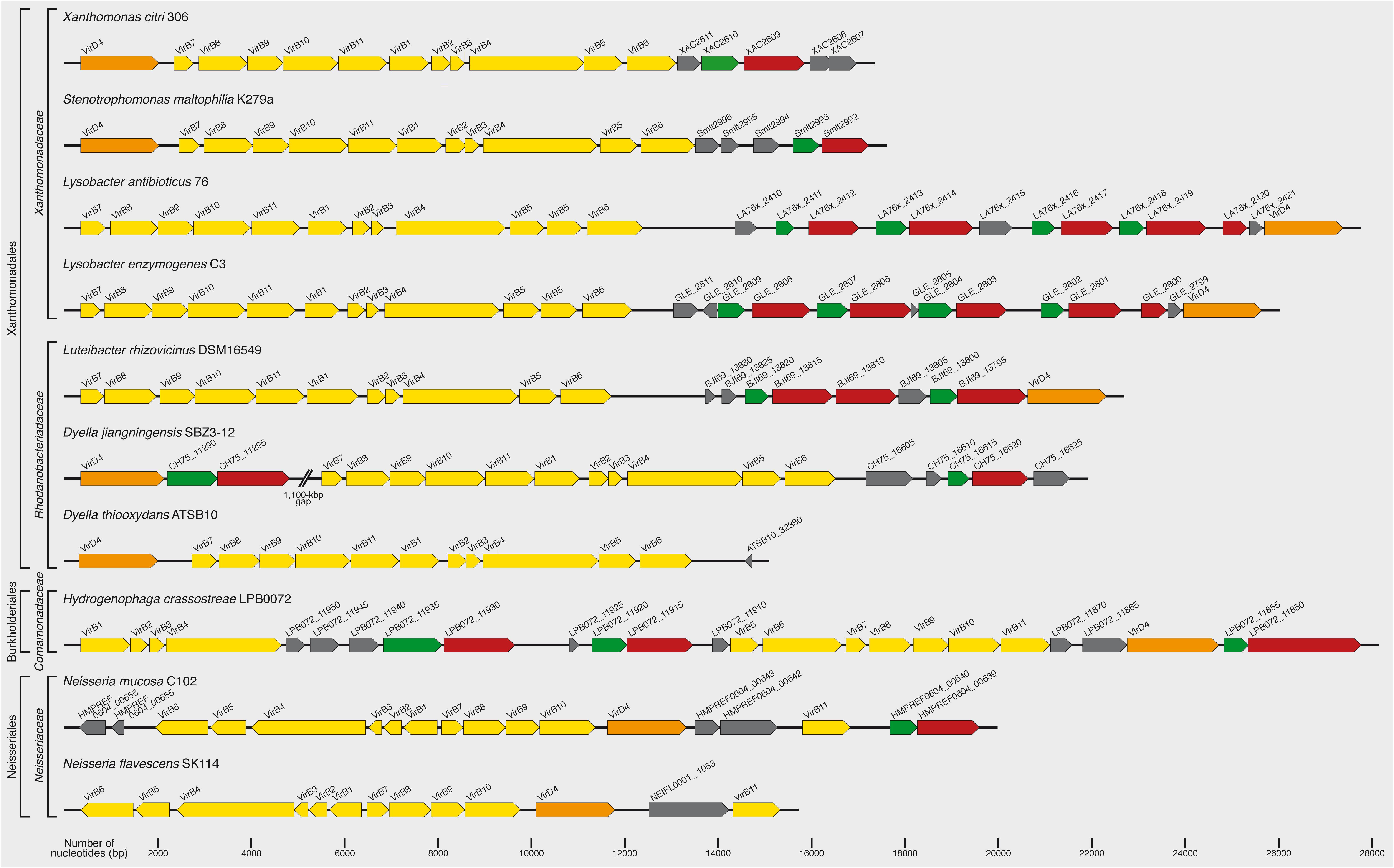
Figure 2. Xanthomonas citri chromosomal vir locus and its homologs in other species. The top line presents the T4SS encoded by the chromosomal vir locus of X. citri 306 (Da Silva et al., 2002; Alegria et al., 2005). It contains the canonical set of 12 components found in other class A T4SSs. Genes coding for T4SSs with similar characteristics to that of X. citri can be identified in the chromosomes of many other species (see Table 1 for an extensive list). Shown here are representative examples from Stenotrophomonas maltophilia K279a (Crossman et al., 2008), Lysobacter antibioticus 76 (de Bruijn et al., 2015), Lysobacter enzymogenes C3 (unpublished; GenBank accession CP013140), Luteibacter rhizovicinus DSM16549 (unpublished; GenBank accession CP017480), Dyella jiangningensis SBZ3-12 (Bao et al., 2014), Dyella thiooxydans ATSB10 (unpublished; GenBank accession CP014841), Hydrogenophaga crassostreae LPB0072 (unpublished; GenBank accession LVWD01000013), Neisseria mucosa C102 (unpublished, GenBank accession GCA_000186165) and Neisseria flavescens SK114 (unpublished; GenBank accession ACQV01000009). VirB and VirD4 genes are shown in yellow and orange, respectively. Xanthomonadales-like T4SS effectors (X-Tfes) and immunity proteins (X-Tfis) are colored red and green, respectively. Other open reading frames coding for proteins of unknown function are shown in gray.
Identification of Homologous Systems in the Order Xanthomonadales and Other Proteobacterial Genomes
Several of the X. citri T4SS components have some interesting features that distinguish them from their homologs in other more distantly related class A T4SSs involved in horizontal transfer of genetic material. For example, the VirB7 and VirB8 subunits have C-terminal extensions absent in most of their more distantly related homologs (see below). Genes coding for T4SSs with similar characteristics to that of X. citri can be identified in the chromosomes of many other Xanthomonas species (Figure 2 and Table 1), for example Xanthomonas campestris pv. campestris B100 (Vorholter et al., 2008), Xanthomonas albilineans GPEPC73 (Pieretti et al., 2009), X. campestris pv. vasculorum NCPPB702 (Studholme et al., 2010) and X. campestris pv. musacearum NCPPB2005 (Wasukira et al., 2012). The corresponding locus is fragmented in X. campestris pv. campestris strains ATCC33913 (Da Silva et al., 2002) and 8004 (Qian et al., 2005), with the virB5 and virB6 genes found in other regions of the genomes. X. campestris pv. vesicatoria 85-10 (Thieme et al., 2005) lacks a significant part of the vir locus (all that remains is the 5′ region coding for VirD4, VirB7, VirB8, and VirB9). The system is also absent in Xanthomonas oryzae strains KACC10331 (Lee et al., 2005), MAFF311018 (Ochiai et al., 2005), PXO99A (Salzberg et al., 2008), and BLS256 (Salzberg et al., 2007), in Xanthomonas fuscans subsp. aurantifolii strains 10535 and 11122 (Moreira et al., 2010) and in all Xylella species sequenced to date. Homologous loci can be found in some Xanthomonadales species of the genera Stenotrophomonas, Pseudoxanthomonas, Luteimonas, Lysobacter, Thermomonas, Rhodanobacter, Dyella, Frateuria, and Luteibacter (Table 1). Interestingly, homologous systems are also found in some species of the Betaproteobacteria orders Burkholderiales (genera Hydrogenophaga, Variovorax) and Neisseriales (genera Neisseria and Morococcus) (Table 1). This is consistent with the observation that in some phylogenetic analyses, Xanthomonadales species are observed to branch anomalously with Betaproteobacteria, most probably due to horizontal gene transfer events (Martins-Pinheiro et al., 2004; Comas et al., 2006; Naushad and Gupta, 2013). We will therefore employ the term X-T4SS to designate all Xanthomonadales-like Type IV secretion systems.
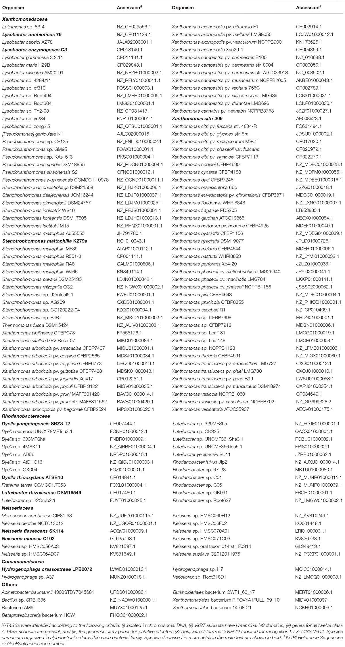
Table 1. Bacterial strains that code for a putative X-T4SS and X-Tfes substrates.
Figure 2 presents the organization of genetic loci, homologous to the X. citri chromosomal vir locus, that are found in the genomes of a few bacterial species selected from genera of the Xanthomonadaceae family (Xanthomonas, Stenotrophomonas, and Lysobacter), and Rhodanobacteraceae family (Dyella and Luteibacter) within the order Xanthomonadales. Also shown are examples of genetic loci that code for X-T4SSs found in the more distant Betaproteobacteria genera of the Comamonadaceae family (Hydrogenophaga) and Neisseriaceae family (Neisseria). One interesting observation is that the loci in species from the Xanthomonadales order seem to have one operon coding for all 11 VirB components, beginning with the virB7 gene. On the other hand, in Hydrogenophaga crassostreae, the operon has been divided into two segments (virB7-11 and virB1-6) and in Neisseria mucosa and N. flavescens it has been divided into three or more segments (Figure 2). The positions of the virD4 genes also vary: in most Xanthomonas and Stenotrophomonas species it is found immediately upstream of the virB7 gene while in the more distantly related species it appears upstream, downstream or inserted between segments coding for the virB genes (Figure 2).
Distinguishing Structural Features of the Bacteria-Killing Xanthomonadales-Like T4SSs (X-T4SSs)
Supplementary Figures S1–S12 present multiple amino acid sequence alignments of VirB1–VirB11 and VirD4 (respectively) X-T4SS components coded by the homologous loci presented in Figure 2. What follows in this section is a brief description of some interesting structural features that can be identified from these alignments and, in some cases, their correlations with known structures and site-directed mutagenesis studies. The observations gleaned from these comparisons are likely to apply to most of the X-T4SSs listed in Table 1.
VirB7, VirB9, and VirB10: Components of the Core Complex
The 2.9 Å resolution crystal structure of the outer membrane layer of the pKM101 core complex (Chandran et al., 2009) and the recently published 3.3 Å resolution cryo-electron microscopy (cryo-EM) structure of the complete X. citri core complex (Sgro et al., 2018), along with the lower resolution EM maps of the complete pKM101 (Fronzes et al., 2009; Rivera-Calzada et al., 2013), R388 (Low et al., 2014) and A. tumefaciens (Gordon et al., 2017) core complexes have provided us with the greatest detail as yet of the periplasmic channel that connects the inner and outer membranes of class A T4SSs. These structures, are all made of 14 copies of VirB7–VirB9–VirB10 heterotrimers (named TraN-TraO-TraF in pKM101 and TrwH-TrwF-TrwE in R388) and can be divided into two layers: the O-layer associated with the outer membrane, consisting of VirB7 and the C-terminal domains of VirB9 and VirB10, and the I-layer made up of the N-terminal domains of VirB9 and VirB10 (Figures 3, 4; Fronzes et al., 2009).
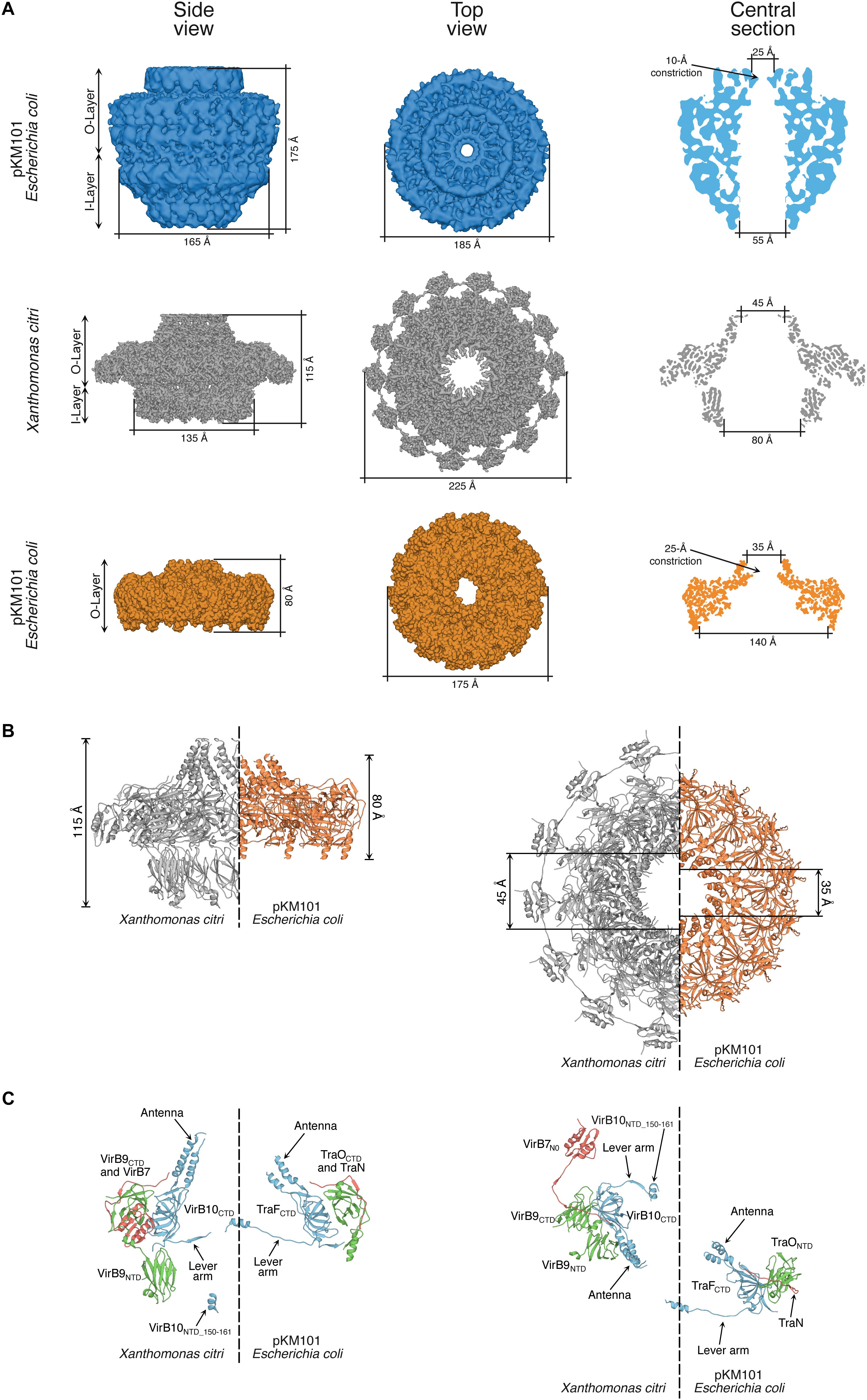
Figure 3. Comparison of core complex structures. (A) Comparison of the electron microscopy maps of the full-length core complexes from pKM101 (12.4 Å resolution; top row; Rivera-Calzada et al., 2013) and X. citri T4SSs (3.3 Å resolution; middle row; Sgro et al., 2018). Also shown is the electron density map of the O-layer of the pKM101 core complex obtained by X-ray crystallography (2.9 Å resolution; lower row; Chandran et al., 2009). General features and dimensions are shown for side and top views, and for a central section. (B) Side-by-side comparison of the atomic models of the X. citri core complex (gray) and pKM101 (orange) O-layers. General features and dimensions are shown for side and top views. (C) Side-by-side comparison of the atomic models of the VirB7–VirB9–VirB10 trimer and TraN-TraO-TraF trimer in the X. citri core complex and pKM101 O-layer, respectively. Colors: VirB10 and TraF (blue), VirB9 and TraO (green), VirB7 and TraN (red). Side (left) and top (right) views are shown of diametrically opposed trimers taken from the side-by-side comparisons shown in B. NTD, N-terminal domain; CTD, C-terminal domain. Portions of the Figure were adapted from Rivera-Calzada et al. (2013) and Sgro et al. (2018) with permission from the publishers.
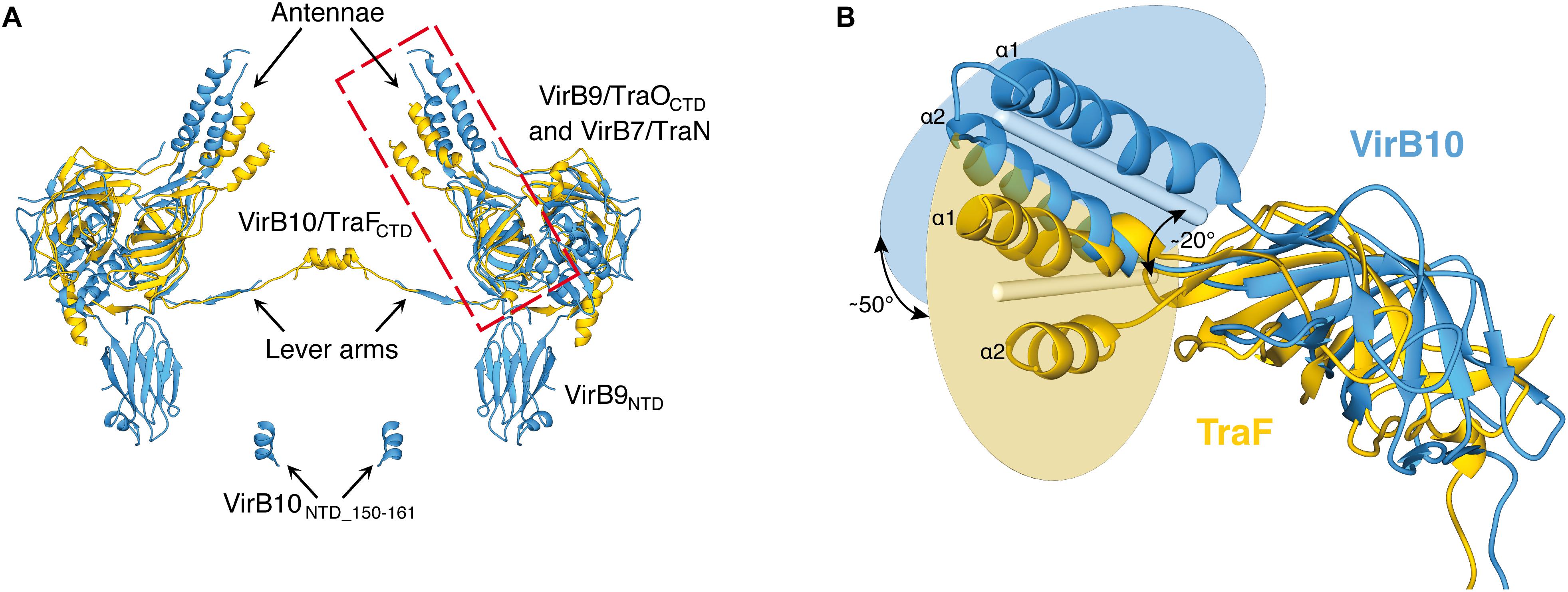
Figure 4. Relative orientations of the antennae that form the outer membrane pore in the X. citri and pKM101 core complexes. (A) Superposition of the atomic models of diametrically opposed VirB7–VirB9–VirB10 trimers of the X. citri core complex (blue) and TraN-TraO-TraF trimers of the pKM101 O-layer (yellow). (B) Details of the relative orientations of the VirB10 and TraF C-terminal domains. The structures shown correspond to the red rectangle in (A). The blue and yellow circles represent the planes that contain the central axes of the two antenna helices (α1 and α2). The blue and yellow rods represent the average vector between the two helices in each protein. The angles between the planes (∼50°) and between the rods (∼20°) are shown. Figure derived from Sgro et al. (2018) with modifications. NTD, N-terminal domain; CTD, C-terminal domain. This Figure was adapted from and Sgro et al. (2018) with permission from the publishers.
The VirB7 lipoprotein component of X-T4SSs is much larger (ranging from 130 to 185 amino acids; Supplementary Figure S7) than that found in other class A T4SSs (normally ∼40 amino acids). This large size is due to an extra globular C-terminal domain called N0 (Souza et al., 2011). Interestingly, similar N0 domains are also found in a myriad of transport systems located in Gram-negative bacterial outer membranes, ranging from secretins of T2SSs, Type IV pilus biogenesis machineries (Korotkov et al., 2009), T3SSs (Spreter et al., 2009), filamentous phages (Spagnuolo et al., 2010), long-tailed bacteriophages (Kanamaru et al., 2002; Kondou et al., 2005), signal-transduction domains in TonB-dependent receptors (Garcia-Herrero and Vogel, 2005; Ferguson et al., 2007) and membrane-penetrating devices in T6SSs (Leiman et al., 2009). The N0 domain is also the C-terminal domain of the outer membrane lipoprotein DotD of the class B T4SSs found in the human pathogens L. pneumophila and C. burnetii (Nakano et al., 2010). The presence of this domain in many outer membrane transport systems could reflect an unexplored evolutionary relationship between them (Souza et al., 2011). The function of the VirB7 N0 domain is possibly related to the observation that it mediates VirB7 oligomerization and, as the VirB7 subunits are highly concentrated in the context of the core complex, it was predicted that the VirB7 domain could assemble an extra peripheral ring in the O-layer of the X-T4SS (Souza et al., 2011), subsequently confirmed by the resolution of the X. citri core complex structure (Sgro et al., 2018). This external ring of N0 domains give the X. citri core complex its characteristic profile that resembles a flying saucer (Figure 3; Sgro et al., 2018). The motifs that mediate VirB7 oligomerization (specific residues in the N0 domain and a [T/S]EIPL) motif that immediately precedes it, contribute to T4SS assembly in the X. citri periplasm and are essential for its antibacterial activity (Souza et al., 2011; Oliveira et al., 2016; Sgro et al., 2018). The multiple sequence alignment in Supplementary Figure S7 shows that these motifs, as well as the region involved in interaction with VirB9, are conserved among X-T4SS VirB7 proteins.
Like their homologs in other class A T4SSs, the X-T4SS VirB9 proteins have two domains connected by a central linker. They all possess an N-terminal signal peptide with a cleavage site immediately after a highly conserved alanine residue (Supplementary Figure S9). The cryo-EM structure of the X. citri core complex (Sgro et al., 2018) and the NMR solution structure of the X. citri VirB7–VirB9 binary complex (Oliveira et al., 2016) showed that the X. citri VirB9 C-terminal domain interacts with VirB7 and with the VirB10 C-terminal domain in the O-layer of the core complex in a manner similar to that observed for pKM101 (Figure 4). The X. citri core complex structure provided us with the first high resolution structure of the I-layer, composed of 14 VirB9 N-terminal β-sandwich domains that pack against each other side-by-side, forming a ring with an internal diameter of 80 Å (Figure 3; Sgro et al., 2018). At the base of the I-layer, small 12-residue helices from the N-terminal domains of VirB10 fit into grooves at the interfaces between VirB9 subunits (Sgro et al., 2018). The multiple sequence alignment of X-T4SS VirB9 proteins shows that the N- and C-terminal domains are quite well conserved but the central linker is variable in both sequence and length (Supplementary Figure S9). This is consistent with the observation that deletion of six residues in the linker region did not affect the stability or function of the T4SS in X. citri (Sgro et al., 2018).
VirB10 subunits in class A T4SSs can be divided into three substructures: the N-terminal cytosolic portion with its contiguous transmembrane helix that spans the bacterial inner membrane, the periplasmic portion of the N-terminal domain that is largely unstructured in the NMR analysis of the isolated domain and the cryo-EM structure of the X. citri core complex (Sgro et al., 2018) and the C-terminal domain localized in the O-layer of the core complex. The alignments shown in Supplementary Figure S10 reveal that the C-terminal domains of X-T4SS VirB10 subunits are very well conserved. The sequences are also very similar to their counterparts in pKM101, R388 and A. tumefaciens (Sgro et al., 2018). One region in this domain that presents a relatively high degree of variability is the “antenna” (Chandran et al., 2009), made up of two alpha helices (α1 and α2) that form the actual pore through the outer membrane. Figure 4 presents a comparison of the relative orientations of the VirB10 antennae when the X. citri core complex and pKM101 O-layer structures are superposed. In the X. citri core complex structure, the antenna helices are twisted clockwise (∼50°) and tilted vertically (∼20°) with respect to the corresponding helices in pKM101, producing a more open outer membrane pore in the X. citri structure (45 Å versus 35 Å; Figure 4; Sgro et al., 2018). We also observe variability in sequence and length of the linker between the alpha helices that is expected to form a loop on the extracellular side of the outer membrane. This loop is particularly rich in threonine, serine and glycine residues and is longer in the Xanthomonadales VirB10 proteins than in their counterparts in A. tumefaciens and pKM101 (Supplementary Figure S10; Sgro et al., 2018). In both X. citri core complex and pKM101 O-layer structures, these loops are disordered with no significant density in the corresponding maps. Since 14 such loops are expected to be in close proximity at the extracellular face of the outer membrane, they could interact to facilitate closing and opening of the pore. However, an X. citri VirB10-msfGFP chimera with an 11 residue deletion in this loop is still partially functional (Sgro et al., 2018).
In contrast to the VirB10 C-terminal domain, its N-terminal domain is known to be highly variable in size and sequence. Xanthomonadales VirB10 proteins have relatively long N-terminal cytosolic segments that precede the transmembrane helix that passes through the inner membrane. While this cytosolic peptide is only 38 or 22 residues long and lacks proline residues in pKM101 and A. tumefaciens, it is 57–68 residues long and is rich in proline residues (around 15%) in most X-T4SS VirB10 subunits (Supplementary Figure S10). The N-terminal region of VirB10 has been observed to interact with VirD4 of the T4SSs from A. tumefaciens (Garza and Christie, 2013) and plasmids R388 (Llosa et al., 2003; Segura et al., 2013) and R27 (Gilmour et al., 2003). Whether similar interactions occur in the X-T4SS remains to be investigated.
The periplasmic portions of the N-terminal domains (NTDs) of VirB10 subunits in many T4SSs are particularly rich in prolines (Jakubowski et al., 2009) and this is also the case for the X-T4SSs (Supplementary Figure S10; Sgro et al., 2018). NMR studies of the periplasmic VirB10 NTD revealed it to be intrinsically disordered but with a 12 residue segment that samples a helical conformation and suffers conformational perturbations when mixed with a near 2:1 excess of isolated N-terminal domain of VirB9 at sub-millimolar concentrations (Sgro et al., 2018). This VirB10 N-terminal helix (residues 151–162 in X. citri; Supplementary Figure S10) fits into the groove between two VirB9 N-terminal domains in the X. citri core complex structure (Sgro et al., 2018). The VirB9NTD-VirB10NTD interaction, in addition to the VirB7–VirB7 interactions mentioned above (Souza et al., 2011) are two examples of interactions that are relatively weak when measured in isolation but reveal themselves to be physiologically relevant in the context of a large multi-subunit complex whose assembly is expected to be highly cooperative. The helical region is in fact the only well-conserved sequence in the N-terminal domains of the Xanthomonadales and H. crassostreae X-T4SS VirB10 subunits and can be described as a P[S/T]Lh[E/D/Q]RRh motif where h is a hydrophobic residue (Supplementary Figure S10). The VirB10 subunits from the two Neisseria species shown in Supplementary Figure S10 do not seem to carry this motif.
One interesting observation is the very small number of stereospecific contacts between the I- and O-layers in the X. citri core complex. This immediately brings up the question as to what maintains the relative orientations between the two layers. The answer may be that individually weak interactions, multiplied fourteen times in the mature complex, could together be strong enough to favor specific conformational states between the two layers. Another interesting observation is that the long flexible N-terminal linkers that emerge from the VirB9 and VirB10 C-terminal domains in the direction of the I-layer point in opposite directions and pass by each other with their main chain atoms coming within 7 Å of each other (Figure 3C; Sgro et al., 2018). Therefore, the covalent linkages between the VirB9 and VirB10 N- and C-domains can be looked upon as forming an intricate cross-weave pattern at the interface of the I- and O-layers. This detail could have a role in maintaining the two layers in a preferential orientation by restricting the excessive relative rotations in both clockwise and counter-clockwise directions. Enigmatically, however, small (6 or 8 residue) deletions in these linkers had only moderate effects on T4SS-dependent bacterial killing by X. citri (Sgro et al., 2018).
VirB1
In A. tumefaciens, VirB1 undergoes cleavage of its N-terminal signal peptide upon transport to the periplasm and a second cleavage reaction that produces two fragments: (i) an N-terminal SLT (soluble lytic transglycosylase) domain predicted to be involved in peptidoglycan remodeling during T4SS biogenesis and (ii) a 76 residue C-terminal fragment (named VirB1∗) that is subsequently transported to the extracellular milieu (Baron et al., 1997; Llosa et al., 2000). The VirB1 proteins in the X-T4SSs all have a well-conserved 150 residue N-terminal domain with predicted SLT activity (Supplementary Figure S1). These proteins lack an N-terminal signal sequence, however, and so their mechanism of transport into the periplasm is not yet known. The X-T4SSs VirB1 C-terminal domains vary in length from 130 to over 200 residues and are highly variable in sequence (Supplementary Figure S1). Whether X-T4SS VirB1 proteins undergo C-terminal processing in a manner analogous to that observed in A. tumefaciens remains to be investigated.
VirB2 and VirB5: The Components of the T4SS Pilus
The VirB2 pilin subunits in the Xanthomonadales species and more distant H. crassostreae, N. flavescens, and N. mucosa all have similar sizes and a very well conserved central hydrophobic region as well as a predicted cleavable 31–39 residue long N-terminal signal peptide (Supplementary Figure S2). After removal of the signal peptide, these pilins are predicted to have lengths between 80 and 104 residues. This is significantly greater than the 70 and 64 residue long mature F-pilin and its close homolog from plasmid pED208 (respectively) whose cryo-EM structures have been determined in the context of the assembled sex pilus (Costa et al., 2016). Sequence-based secondary structure predictions for the X-T4SS VirB2 subunits correspond well with the two pilin structures and this allowed us to predict the positions of the two major helices (α2 and α3) in X-T4SS VirB2 as well as the intervening positively charged loop which interacts with the head groups of bound phospholipids in the pilus lumen (Supplementary Figure S2; Costa et al., 2016). The larger size of the X-T4SS VirB2 subunits is due to highly variable C-terminal extensions (Supplementary Figure S2). It is not clear whether these C-terminal extensions decorate the external pilus surface in X-T4SSs, perhaps providing binding sites for species-specific targets, or whether they are processed as has been observed for some P-pili (Eisenbrandt et al., 1999).
VirB5 is thought to be associated with the T4SS pilus, perhaps as a minor pilin at the pilus tip (Schmidt-Eisenlohr et al., 1999; Aly and Baron, 2007; Alvarez-Martinez and Christie, 2009). Due to the very high sequence variability in VirB5 proteins, its annotation as a bona-fide T4SS component in deposited genomic sequences is often ambiguous. In the bacteria-killing T4SSs under consideration here, these proteins have between 200 and 280 residues and are highly variable in sequence. They all have a predicted N-terminal cleavable signal peptide as well as a pair of cysteine residues found in the central portion of the amino acid sequence that are separated by 11 to 33 residues (Supplementary Figure S5). Interestingly, the genetic loci coding for X-T4SSs in the two Lysobacter species shown in Figure 2 each carry two virB5 genes in tandem. These protein pairs are 73% and 43% identical in Lysobacter antibioticus and Lysobacter enzymogenes, respectively. The predicted involvement of VirB5 in mediating the binding of the pilus to specific structures on the target cell (Alvarez-Martinez and Christie, 2009), could be a causative factor in this subunit’s accelerated evolution.
VirB8, VirB6, and VirB3: Integral Membrane Proteins of the Inner Membrane Complex
VirB8 is an integral membrane protein with an N-terminal cytosolic peptide, a transmembrane helix and a globular C-terminal domain localized in the periplasm, the latter of which has been shown to interact with several other T4SS components, including VirB6, VirB9, and VirB10 (Alvarez-Martinez and Christie, 2009; Sivanesan et al., 2010; Villamil Giraldo et al., 2012). High resolution structures of the soluble C-terminal region of VirB8 proteins and homologs from diverse T4SSs have been determined: A. tumefaciens (Bailey et al., 2006), pKM101 (Casu et al., 2016), H. pylori (ComB10) and Brucella suis (Terradot et al., 2005), Clostridium perfringens, Rickettsia typhi and several Bartonella species (Gillespie et al., 2015), L. pneumophila and plasmid R64 (Kuroda et al., 2015). All these structures present the same fold, a β-sheet juxtaposed against a group of α-helices, and in most cases have been shown to oligomerize to different degrees (see references above). Since there are an estimated 12 copies of VirB8 in each class A T4SS (Low et al., 2014), these domains could associate to form an as-yet unknown structure in the bacterial periplasm. Interestingly, the bacteria-killing X-T4SSs have highly distinctive VirB8 components that are significantly longer (between 290 and 370 residues in length) than the canonical VirB8 components observed in the species listed above (all less than 250 residues). This greater size is, in large part, due to a C-terminal extension enriched in Ala, Gln, Gly, and Pro (AQGP) residues (Table 2 and Supplementary Figure S8). The length of this extension and its enrichment in these residues are particularly evident in the Xanthomonadales order (53–73%) and H. crassostreae (45%) and less so in the X-T4SS VirB8 proteins of Neisseria species (Table 2). The role, if any, of the AQGP-rich extensions in these proteins is not yet known.
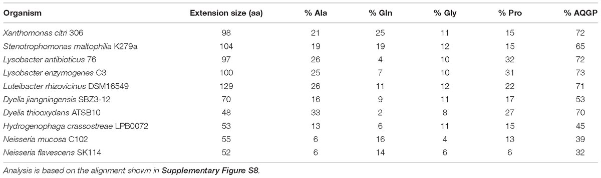
Table 2. Characteristics of C-terminal extensions in VirB8 proteins from X-T4SSs.
VirB6 is predicted to be a polytopic integral protein. In both A. tumefaciens and B. suis, the VirB6 N-terminus is located in the periplasm and the C-terminus is located in the cytosol, implying an odd number of transmembrane helices, estimated to be five in A. tumefaciens (Jakubowski et al., 2004) and seven in B. suis (Villamil Giraldo et al., 2015). Supplementary Figure S6 presents the multiple sequence alignment of X-T4SS VirB6 proteins that also align well with their homologs in A. tumefaciens and B. suis (data not shown). The precise number and positions of the transmembrane helices is again ambiguous since transmembrane helix pairs 3/4 and 5/6 could alternatively be longer single helices (Supplementary Figure S6). Another common feature of these proteins is a large loop between transmembrane helices 2 and 3, predicted to be localized in the periplasm for both A. tumefaciens and B. suis. The periplasmic loops of the estimated 24 copies of VirB6 per T4SS (Low et al., 2014) are expected to interact with other periplasmic components of the inner membrane complex (Figure 1; Ding et al., 2003; Alvarez-Martinez and Christie, 2009; Villamil Giraldo et al., 2012, 2015; Christie, 2016).
X-T4SS VirB3 proteins are predicted to be bitopic or polytopic membrane proteins with one or two transmembrane helices located within the region encompassed by residues 15 and 57 (see Supplementary Figure S3 for details). This is consistent with the difficulty in defining VirB3 topology in other T4SSs (Alvarez-Martinez and Christie, 2009). In some organisms, gene fusions have been observed between VirB3 and VirB4 (Batchelor et al., 2004) but this does not seem to be the case in the X-T4SSs. These gene fusions and the highly conserved synteny of virB3 and virB4 genes in most organisms suggests that these two proteins could interact with each other at or within the inner membrane (Peña et al., 2012). This hypothesis is supported by the observation that A. tumefaciens VirB4 is required to maintain normal levels of VirB3 (Jones et al., 1994) and that recombinant R388 VirB4 and VirB3 can be co-purified as a 1:1 complex (Low et al., 2014).
VirB4, VirB11, and VirD4: The ATPases of the Inner Membrane Complex
VirB4, VirB11, and VirD4 are P-loop ATPases with conserved Walker A and Walker B motifs and, in most T4SSs characterized to date, all three proteins are required for biogenesis and/or function (Alvarez-Martinez and Christie, 2009; Christie et al., 2014; Christie, 2016). These proteins use the hydrolysis of ATP to carry out mechanical work, expected to manifest itself in substrate unfolding, transfer and/or extrusion through the T4SS channel (Atmakuri et al., 2004).
VirB4 is the most well conserved T4SS subunit and has been used in phylogenetic analyses to trace evolutionary relationships and propose models for the emergence of T4SS subclasses (Guglielmini et al., 2014). Studies on different T4SSs have reported evidence that VirB4 interacts, at least transiently, with all of the other inner membrane complex components (Jones et al., 1994; Atmakuri et al., 2004; Cascales and Christie, 2004; Jakubowski et al., 2004; Paschos et al., 2006; Ripoll-Rozada et al., 2013; Low et al., 2014; Christie, 2016).
VirB4 has been localized to two 3-tiered barrel-like pedestals at the base of the inner membrane complex in EM reconstructions of the complete R388 T4SS, with each barrel corresponding to a VirB4 hexamer (Low et al., 2014; Redzej et al., 2017). VirB4 proteins can be divided into N- and C-terminal domains. The C-terminal domain carries all the conserved motifs required for nucleotide binding and hydrolysis (Walker A and Walker B boxes and motifs C, D, and E) and these motifs are present in X-T4SS VirB4 proteins (Supplementary Figure S4). The N-terminal domain, known to mediate interactions with the inner membrane, is expected to correspond to the upper tier in the R388 T4SS structure (Low et al., 2014; Redzej et al., 2017). One variable in the family of VirB4 proteins is the presence or absence of one or more predicted N-terminal transmembrane helices and the question as to their requirement for VirB4 function has proven to be controversial (Rabel et al., 2003), with the additional caveat that VirB4/TraB from pKM101 can be purified in soluble and membrane-bound forms (Durand et al., 2010). Therefore, it is not clear whether VirB4 should be considered an integral or peripheral membrane protein, or both (Arechaga et al., 2008; Christie, 2016; Waksman, 2019). The X-T4SS VirB4 proteins do not have predicted transmembrane helices using the TMHMM v2.0 (Krogh et al., 2001) and PSort (Nakai and Horton, 1999) prediction algorithms.
VirB11 is a soluble membrane-associated AAA+ ATPase that has been shown to interact with both VirD4 and VirB4 (Ripoll-Rozada et al., 2013). The crystal structures of VirB11 homologs from B. suis (Hare et al., 2006) and H. pylori (Yeo et al., 2000) are both two-tiered hexameric rings in which each ring layer consists of six N- or C-terminal domains from the constituent monomers. Within VirB11 monomers, the domains are connected by a central linker of varying length, short in H. pylori and 17 residues longer (linker B and α2C) in B. suis, resulting in a domain-swapped architecture in the latter (Hare et al., 2006). As a consequence, the nucleotide binding site in H. pylori VirB11 is located at the interface between the two domains of the same monomer while in B. suis the nucleotide binds at the interface between the N-terminal domain of one monomer and the C-terminal domain of the neighboring monomer (Hare et al., 2006). VirB11 proteins from X-T4SSs show a high degree of sequence similarity and all have the long version of the linker, which aligns well with the B. suis linker B and α2C sequences (Supplementary Figure S11). We can therefore expect that the X-T4SS VirB11 proteins exhibit a domain-swapped structure similar to that of B. suis (Hare et al., 2006).
The VirD4 ATPase and its homologs are often called coupling proteins due to their role in selecting substrates for export by the T4SS. Analysis of the X-T4SS VirD4 proteins suggests that they have a canonical VirD4-like architecture (Llosa and Alkorta, 2017) with two N-terminal transmembrane helices with a predicted intervening ∼30 amino acid periplasmic loop and a cytosolic C-terminal domain. The cytosolic domain can be separated into a nucleotide binding domain (NBD), with conserved Walker A and Walker B motifs, and a so-called all alpha domain (AAD) (Supplementary Figure S12; Gomis-Ruth et al., 2002). The VirD4 N-terminal transmembrane domain helices have been implicated in interacting with the VirB10 N-terminal region that includes its transmembrane helix (Segura et al., 2013) and the VirD4 all alpha domain is involved in substrate recognition (Gomis-Ruth et al., 2002; Whitaker et al., 2015). TrwB, the VirD4 homolog of the conjugative plasmid R388, has been crystallized as a hexameric ring (Gomis-Ruth et al., 2002). It has been proposed that VirB4 and VirD4 could form transient heterohexameric complexes during substrate transport (Peña et al., 2012; Waksman, 2019) and low resolution electron microscopy studies of the intact VirB3-B10/D4 T4SS from R388 observed VirD4 dimers sandwiched between the two hexameric VirB4 barrels (Redzej et al., 2017). Therefore, the oligomeric structure of VirD4 in a fully assembled and functioning T4SS is still not clear (Redzej et al., 2017; Chetrit et al., 2018; Waksman, 2019).
Xanthomonadales-Like T4SS Effectors (X-Tfes) and Their Cognate Inhibitors (X-Tfis)
X-Tfes
The first clues regarding the physiological role of the X. citri T4SS came from the identification of T4SS substrates using the VirD4 coupling protein as a bait in yeast two-hybrid assays against a Xanthomonas genomic library (Alegria et al., 2005). The strategy was based on the reasoning that in other well-characterized T4SSs, the VirD4 component is known to interact with the macromolecular substrates prior to transport (Llosa and Alkorta, 2017). This screening originally identified 12 so-called “Xanthomonas VirD4 interacting proteins,” or XVIPs (Alegria et al., 2005), later called Xanthomonadaceae T4SS effectors (Souza et al., 2015) and from here on Xanthomonadales-like T4SS effectors (X-Tfes, Figure 5). In X. citri, the gene for one X-Tfe (XAC2609) is found in the vir locus while the remaining X-Tfe genes are dispersed throughout the genome. Interestingly, all of these proteins have a common C-terminal domain entitled “XVIP conserved domain” or XVIPCD (Figures 1, 5), typically around 120 residues long, required for interaction with VirD4 (Alegria et al., 2005) and for secretion in a T4SS-dependent manner (Souza et al., 2015). The XVIPCD is characterized by a few conserved motifs in its N-terminal region and a glutamine-rich C-terminal region (Alegria et al., 2005).
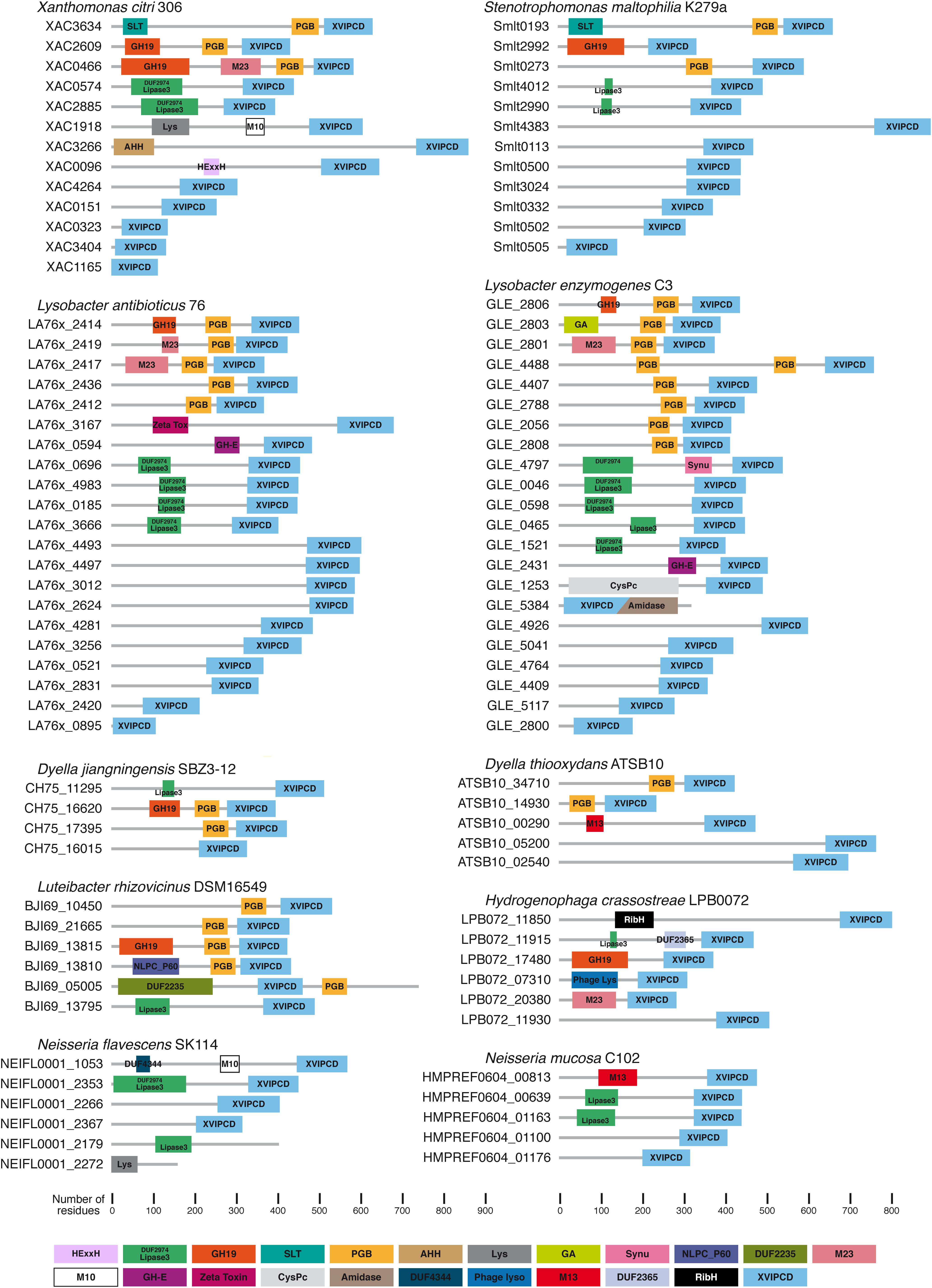
Figure 5. Putative Xanthomonadales-like T4SS effectors (X-Tfes) found in selected species that carry an X-T4SS. Shown here are representative examples from Xanthomonas citri 306 (Da Silva et al., 2002), Stenotrophomonas maltophilia K279a (Crossman et al., 2008), Lysobacter antibioticus 76 (de Bruijn et al., 2015), Lysobacter enzymogenes C3 (unpublished; GenBank accession CP013140), Luteibacter rhizovicinus DSM16549 (unpublished; GenBank accession CP017480), Dyella jiangningensis SBZ3-12 (Bao et al., 2014), Dyella thiooxydans strain ATSB10 (unpublished; GenBank accession CP014841), Hydrogenophaga crassostreae LPB0072 (unpublished; GenBank accession LVWD01000013), Neisseria mucosa C102 (unpublished, GenBank accession GCA_000186165) and Neisseria flavescens SK114 (unpublished; GenBank accession ACQV01000009). Protein domains were identified by sequence comparison with the Pfam (El-Gebali et al., 2019) and/or CDD databases (Marchler-Bauer et al., 2015) and are colored according to the scheme presented at the bottom of the Figure. Domain abbreviations: M10 (Pfam accession PF08548), M13 (Pfam accession PF01431), M23 (Pfam accession PF01551), Lipase3 (Pfam accession PF01764), DUF2974 (Pfam accession PF11187), GH-E (Pfam accession PF14410), GH19 (Pfam accession PF00182), Zeta Toxin (Pfam accession PF06414), SLT (CDD accession cd00254), CysPc (CDD accession cd00044), PGB (Pfam accession PF01471), Amidase (Pfam accession PF01510), AHH (Pfam accession PF14412), DUF4344 (Pfam accession PF14247), Lys (CDD accession cl00222), Phage lyso (Pfam accession PF00959), GA (Pfam accession PF01832), Synu (CDD accession cl03193), DUF2365 (Pfam accession PF10157), NLPC_P60 (Pfam accession PF00877), RibH (Pfam accession PF02267), DUF2235 (Pfam accession PF09994), HExxH (HExxH motif in putative metalloprotease domain; Firczuk and Bochtler, 2007).
The discovery of the XVIPCD as the secretion signal for the X. citri X-Tfes allowed for a large-scale bioinformatics identification of X-Tfe genes present in other bacterial genomes (Souza et al., 2015). Figure 5 shows the domain architectures of the X-Tfes identified by bioinformatics analysis of bacterial genomes whose X-T4SSs are described in Figure 2. The N-terminal portions of the X. citri X-Tfes are highly variable in size and architecture and most are predicted to function within the periplasm as peptidoglycan (PG) glycohydrolases, lytic transglycosylases, PG peptidases or lipases (Figure 5). Therefore, these bacterial species probably use their X-T4SSs to inject not one, but a diverse cocktail of X-Tfes that will simultaneously attack multiple structures in the target cell (Figure 1). Two purified X. citri X-Tfes (XAC2609 and XAC0466) with predicted PG hydrolase activities have been shown to lyse PG and induce the lysis of Gram-positive cells, which have exposed bacterial cell walls (Souza et al., 2015). It is interesting that a considerable fraction of X-Tfes have N-terminal sequences with no identifiable domains, opening the possibility that new domain families with antibacterial activities could be characterized in the future. One such X-Tfe, Smlt3024 from S. maltophilia K279a (Figure 5), has been shown to inhibit E. coli growth when heterologously expressed and directed to the periplasm (preprint: Bayer-Santos et al., 2019).
It is worth noting that we often encounter several open reading frames that code for small proteins, sometimes possessing little more than an intact XVIPCD; for example XAC0323, XAC1165, and XAC3404 in X. citri (respectively 136, 127, and 132 residues in length; Figure 5; Souza et al., 2015). In some cases, these open reading frames appear to be fragments of ancestral X-Tfes genes that suffered frameshift mutations. One example of this phenomenon is provided by the XAC1165 gene whose first 37 nucleotides overlap with the 3′ end of the upstream XAC1164 gene which codes for a 437 protein of unknown function. The amino acid sequences of XAC1164 and XAC1165 align very well with the N-terminal and XVIPCD regions, respectively, of the Smlt0113 X-Tfe protein from S. maltophilia (Figure 5). Thus X. citri XAC1164 and XAC1165 proteins are homologous, and probably the non-functional, fragments of a functional X-Tfe (Smlt0113) in S. maltophilia.
X-Tfis
To protect against the toxicity of endogenous or exogenous X-Tfes, X. citri and S. maltophilia produce specific immunity proteins that bind to their cognate toxins (Alegria et al., 2005; Souza et al., 2015; preprint: Bayer-Santos et al., 2019). These inhibitors have been termed Xanthomonadaceae T4SS immunity proteins (Souza et al., 2015) and from here on Xanthomonadales-like T4SS immunity proteins (X-Tfis). The genes coding for X-Tfis are usually found upstream and are probably co-transcribed with their cognate X-Tfe (Souza et al., 2015; preprint: Bayer-Santos et al., 2019). All bacterial species identified so far that carry an X-T4SS also code for multiple X-Tfe/X-Tfi pairs and, in most cases, the genes for at least one pair is found within, or in close proximity to, the locus that codes for the structural components of the X-T4SS (see Figure 2). Furthermore, in almost all cases where the X-Tfe is predicted to act upon periplasmic structures (glycosidic and peptide bonds in peptidoglycan or ester linkages in phospholipids), the cognate X-Tfi carries an N-terminal signal peptide and lipobox for periplasmic localization and anchoring in the outer membrane (Souza et al., 2015; preprint: Bayer-Santos et al., 2019). On the other hand, some X-Tfes with N-terminal domains predicted to act in the cytosol of the target cell (for example the X. citri X-Tfe XAC3266 with an N-terminal AHH domain with predicted nuclease activity; Figure 5) have a cognate X-Tfi (for example XAC3267) lacking a lipoprotein signal (Souza et al., 2015). Finally, some X-Tfes are expected to be active in both the cytosol and periplasm, as are the cases of the X-Tfes with predicted lipase domains with phospholipase activities (NFB, DPS, BM, and CSF, manuscript in preparation; Figure 5). This brings up the question regarding the cellular localization of the X-Tfis. An analysis of the X. citri X-Tfis with putative N-terminal signal peptide and Lipobox sites indicates that their coding genes have potential alternative downstream start (ATG) codons with associated ribosome binding sites (Figure 6). This raises the possibility that many X-Tfes can be produced in two versions: (i) a membrane-associated periplasmic lipoprotein and (ii) a soluble cytosolic protein (Figure 1). Thus, if X-Tfes make their way into the periplasm, either by leakage from the secretion channel or by injection by neighboring cells of the same species, they will be inhibited by the periplasmic lipoprotein forms of the cognate X-Tfi. On the other hand, X-Tfes whose activities could target cytosolic substrates can be inhibited by cytosolic variants of their cognate X-Tfis. If this is in fact the case, for at least this latter subset of X-Tfes, transport will necessarily involve previous dissociation of the X-Tfe/X-Tfi cytosolic pair.
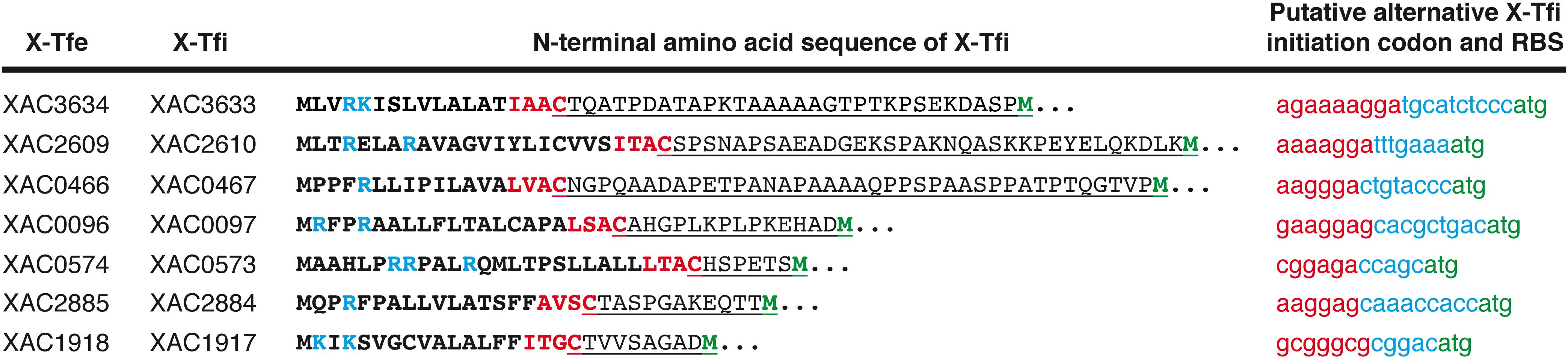
Figure 6. Possible alternative translation start codons that could lead to the production of soluble cytosolic X-Tfis in Xanthomonas citri. The first two columns list the names of X. citri X-Tfe/X-Tfi pairs in which the X-Tfi is predicted to be a lipoprotein (Souza et al., 2015). The third column presents the N-terminal amino acid sequence of the X-Tfi in which the signal sequence and Lipobox are shown in bold. The basic nucleotides at the N-terminus of the signal sequence are shown in blue. The four Lipobox residues are shown in red. Underlined residues are those from the absolutely conserved Cys residue at the site of cleavage in the Lipobox to the next Met residue (green) in the protein sequence. The last column presents the nucleotide sequence (lowercase letters) immediately upstream of the putative alternative start codon (green). The putative Shine–Dalgarno sequence (ribosome binding site) for this alternative start codon is shown in red.
Parallels Between X-T4SS X-Tfe/X-Tfi, T6SS Effector/Immunity Protein and Plasmid-Encoded Toxin/Antitoxin (TA) Pairs
X-Tfe/X-Tfi pairs share many of the characteristics observed for T6SS effectors and their inhibitors (Russell et al., 2011, 2013). For example, one immunity protein from X. citri (X-TfiXAC2610) inhibits the GH19 family PG hydrolase X-TfeXAC2609 and has a very similar topology, though very little sequence similarity, to the PG hydrolase inhibitors PliI and Tsi1 (Van Herreweghe et al., 2010; Russell et al., 2011) the latter of which is an inhibitor of the T6SS effector Tse1 from Pseudomonas aeruginosa (Souza et al., 2015). Another example is provided by the X-Tfe Smlt0332 from S. maltophilia K279a, which has many X-Tfe homologs in other Xanthomonadales species (data not shown) but whose N-terminal region has no similarity with annotated domains in the Pfam or CCD databases. Interestingly, Blast searches against the curated KEGG database (Kanehisa et al., 2017) using this domain identified a large number of homologous sequences fused to VgrG domains in effectors predicted to be secreted by T6SSs of Metakosakonia, Pantoea, Kosakonia, Enterobacter, Burkholderia, Methylocaldum, Ralstonia, Cronobacter, and Xanthomonas species (Table 3). Searches against the non-redundant protein sequence database (Altschul et al., 1990) identified many more such homologs in other bacterial species (data not shown). These findings raise interesting questions regarding the evolution, distribution and exchange of T4SS and T6SS effectors in the biosphere. In fact, we can make a general observation that anti-bacterial Type IV and Type VI secretion systems share many enzymatic effector and cognate inhibitory modules and differ only in the specific sequences recognized for transport. It also raises the possibility that the acquisition of immunity proteins could be advantageous even in the absence of the cognate effector by offering a defense against the toxic activity of substrates launched by both T4SS and T6SSs during encounters with rival bacteria. We have in fact recently observed the reciprocal T4SS-dependent dueling between S. maltophilia and X. citri cells which could mimic similar encounters between soil- and/or plant-associated bacteria in the environment (preprint: Bayer-Santos et al., 2019). The differences in X-Tfe and X-Tfi repertoires between rival species will contribute to the outcome of these encounters.
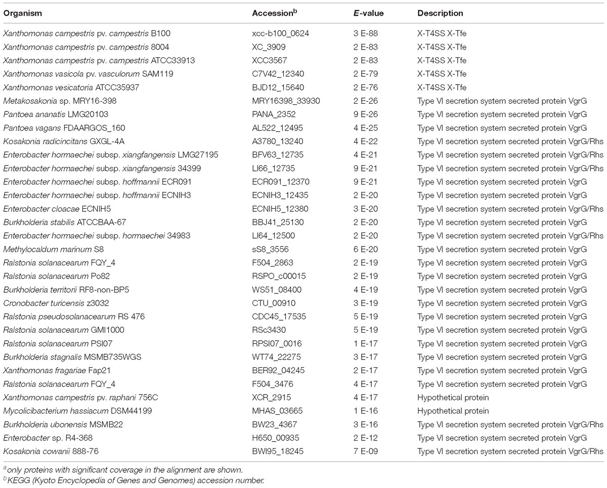
Table 3. List of proteins in the KEGG database with greatest similaritya to the N-terminal domain (residues 1–240) of Smlt0332 from S. maltophilia K279a.
Effector/immunity protein pairs associated with T4SS and T6SSs show intriguing parallels with toxin/anti-toxin modules that function to guarantee vertical transmission of mobile elements (Jensen and Gerdes, 1995). For example, Harms et al. (2017) have shown that the pVbh plasmid of Bartonella schoenbuchensis, codes for a toxin/antitoxin module in which the toxin component, VbhT, acquired a C-terminal BID (Bep Intracellular Delivery) domain that confers its transfer to recipient cells during conjugation. They propose that its function may be to support intercellular DNA transfer by pre-emptively addicting the recipient cell to the plasmid, which also carries the gene for the antitoxin antidote. It is not difficult to imagine a scenario in which the T4SS coded by such a plasmid could lose its capacity to transfer DNA but retain its capacity to transfer the toxin, and in this way evolve into a single-purpose bacteria-killing T4SS. Therefore, we can expect bactericidal T4SSs, perhaps with very different recognition signals, to have arisen on multiple occasions in distantly related bacterial species.
Closing Remarks
These are early days in the characterization of antibacterial T4SSs. The structure of the X. citri core complex has provided a good reference for comparison with other T4SSs, and has illustrated the structural variability that we can expect to encounter even within the class A T4SSs. While X-T4SS activities have only been experimentally verified for X. citri and S. maltophilia, bioinformatics analysis allowed us to confidently expand the list of bacterial families both within the Gammaproteobacteria class as well as to other families within the Betaproteobacteria (Table 1) that carry proteins with many of the characteristic X-T4SSs features, including VirB7 proteins with N0 domains, VirB8 proteins with AQGP-rich extensions as well as recognizable X-Tfe/X-Tfi pairs in which the effector carries a C-terminal XVIPCD. The list of X-T4SSs will most surely expand significantly in the future. However, it is unlikely that bactericidal T4SSs are restricted to the X-T4SSs described here. It is more probable that many other bacterial species carry as yet uncharacterized and perhaps unrecognized T4SSs that recruit effectors with recognition signals significantly different from the XVIPCDs associated with X-T4SSs as predicted (Souza et al., 2015) and as illustrated by the results obtained for the T4SS encoded by the B. schoenbuchensis pVbh plasmid (Harms et al., 2017).
Author Contributions
GS, GO, EB-S, and CF produced the figures and tables. CF wrote the manuscript. All authors contributed with critical discussions and revisions that led to the final version of the manuscript.
Funding
This work was supported by grants from the Fundação de Amparo à Pesquisa do Estado de São Paulo (FAPESP) to CF (Grant # 2017/17303-7), CA-M (Grant # 2018/01852-4), and EB-S (Grant # 2017/02178-2).
Conflict of Interest Statement
The authors declare that the research was conducted in the absence of any commercial or financial relationships that could be construed as a potential conflict of interest.
Acknowledgments
GS, GO, DS, WC, BM, and EB-S acknowledge scholarships from FAPESP. DS, TdS, and NB acknowledge fellowships from Coordenação de Aperfeiçoamento de Pessoal de Nível Superior (CAPES).
Supplementary Material
The Supplementary Material for this article can be found online at: https://www.frontiersin.org/articles/10.3389/fmicb.2019.01078/full#supplementary-material
References
Alegria, M. C., Souza, D. P., Andrade, M. O., Docena, C., Khater, L., Ramos, C. H. I., et al. (2005). Identification of new protein-protein interactions involving the products of the chromosome- and plasmid-encoded type IV secretion loci of the phytopathogen Xanthomonas axonopodis pv. citri. J. Bacteriol. 187, 2315–2325. doi: 10.1128/JB.187.7.2315-2325.2005
Altschul, S. F., Gish, W., Miller, W., Myers, E. W., and Lipman, D. J. (1990). Basic local alignment search tool. J. Mol. Biol. 215, 403–410. doi: 10.1016/S0022-2836(05)80360-2
Alvarez-Martinez, C. E., and Christie, P. J. (2009). Biological diversity of prokaryotic type IV secretion systems. Microbiol. Mol. Biol. Rev. 73, 775–808. doi: 10.1128/MMBR.00023-09
Aly, K. A., and Baron, C. (2007). The VirB5 protein localizes to the T-pilus tips in Agrobacterium tumefaciens. Microbiology 153, 3766–3775. doi: 10.1099/mic.0.2007/010462-0
Arechaga, I., Pena, A., Zunzunegui, S., del Carmen Fernandez-Alonso, M., Rivas, G., and de la Cruz, F. (2008). ATPase activity and oligomeric state of TrwK, the VirB4 homologue of the plasmid R388 type IV secretion system. J. Bacteriol. 190, 5472–5479. doi: 10.1128/JB.00321-08
Atmakuri, K., Cascales, E., and Christie, P. J. (2004). Energetic components VirD4, VirB11 and VirB4 mediate early DNA transfer reactions required for bacterial type IV secretion. Mol. Microbiol. 54, 1199–1211. doi: 10.1111/j.1365-2958.2004.04345.x
Backert, S., and Meyer, T. F. (2006). Type IV secretion systems and their effectors in bacterial pathogenesis. Curr. Opin. Microbiol. 9, 207–217. doi: 10.1016/j.mib.2006.02.008
Backert, S., Tegtmeyer, N., and Fischer, W. (2015). Composition, structure and function of the Helicobacter pylori cag pathogenicity island encoded type IV secretion system. Future Microbiol. 10, 955–965. doi: 10.2217/fmb.15.32
Bailey, S., Ward, D., Middleton, R., Grossmann, J. G., and Zambryski, P. C. (2006). Agrobacterium tumefaciens VirB8 structure reveals potential protein-protein interaction sites. Proc. Natl. Acad. Sci. U.S.A. 103, 2582–2587. doi: 10.1073/pnas.0511216103
Bao, Y., Kwok, A. H. Y., He, L., Jiang, J., Huang, Z., Leung, F. C. C., et al. (2014). Complete genome sequence of Dyella jiangningensis strain SBZ3-12, isolated from the surfaces of weathered rock. Genome Announc. 2:e00416-14. doi: 10.1128/genomeA.00416-14
Baron, C., Llosa, M., Zhou, S., and Zambryski, P. C. (1997). VirB1, a component of the T-complex transfer machinery of Agrobacterium tumefaciens, is processed to a C-terminal secreted product, VirB1. J. Bacteriol. 179, 1203–1210. doi: 10.1128/jb.179.4.1203-1210.1997
Batchelor, R. A., Pearson, B. M., Friis, L. M., Guerry, P., and Wells, J. M. (2004). Nucleotide sequences and comparison of two large conjugative plasmids from different Campylobacter species. Microbiology 150(Pt 10), 3507–3517. doi: 10.1099/mic.0.27112-0
Bayer-Santos, W., Matsuyama, B. Y., Di Sessa, G., Mininel, I. D. V., and Farah, C. S. (2019). The opportunistic pathogen Stenotrophomonas maltophilia utilizes a type IV secretion system for interbacterial killing. bioRxiv [Preprint]. doi: 10.1101/557322
Bayer-Santos, E., Lima, L., dos, P., Ceseti, L., de, M., Ratagami, C. Y., et al. (2018). Xanthomonas citri T6SS mediates resistance to Dictyostelium predation and is regulated by an ECF σ factor and cognate Ser/Thr kinase. Environ. Microbiol. 20, 1562–1575. doi: 10.1111/1462-2920.14085
Brito, E. M., Pinon-Castillo, H. A., Guyoneaud, R., Caretta, C. A., Gutierrez-Corona, J. F., Duran, R., et al. (2013). Bacterial biodiversity from anthropogenic extreme environments: a hyper-alkaline and hyper-saline industrial residue contaminated by chromium and iron. Appl. Microbiol. Biotechnol. 97, 369–378. doi: 10.1007/s00253-012-3923-5
Buttner, D., and Bonas, U. (2010). Regulation and secretion of Xanthomonas virulence factors. FEMS Microbiol. Rev. 34, 107–133. doi: 10.1111/j.1574-6976.2009.00192.x
Cabezon, E., Ripoll-Rozada, J., Pena, A., de la Cruz, F., and Arechaga, I. (2015). Towards an integrated model of bacterial conjugation. FEMS Microbiol. Rev. 39, 81–95. doi: 10.1111/1574-6976.12085
Callaghan, M. M., Heilers, J. H., van der Does, C., and Dillard, J. P. (2017). Secretion of chromosomal DNA by the Neisseria gonorrhoeae type IV secretion system. Curr. Top. Microbiol. Immunol. 413, 323–345. doi: 10.1007/978-3-319-75241-9_13
Cao, Z., Casabona, M. G., Kneuper, H., Chalmers, J. D., and Palmer, T. (2016). The type VII secretion system of Staphylococcus aureus secretes a nuclease toxin that targets competitor bacteria. Nat. Microbiol. 2:16183. doi: 10.1038/nmicrobiol.2016.183
Carbonetti, N. H. (2015). Contribution of pertussis toxin to the pathogenesis of pertussis disease. Pathog. Dis. 73:ftv073. doi: 10.1093/femspd/ftv073
Cascales, E., and Christie, P. J. (2004). Definition of a bacterial type IV secretion pathway for a DNA substrate. Science 304, 1170–1173. doi: 10.1126/science.1095211
Casu, B., Smart, J., Hancock, M. A., Smith, M., Sygusch, J., and Baron, C. (2016). Structural analysis and inhibition of TraE from the pKM101 type IV secretion system. J. Biol. Chem. 291, 23817–23829. doi: 10.1074/jbc.M116.753327
Chandran, V., Fronzes, R., Duquerroy, S., Cronin, N., Navaza, J., and Waksman, G. (2009). Structure of the outer membrane complex of a type IV secretion system. Nature 462, 1011–1015. doi: 10.1038/nature08588
Chang, Y. T., Lin, C. Y., Chen, Y. H., and Hsueh, P. R. (2015). Update on infections caused by Stenotrophomonas maltophilia with particular attention to resistance mechanisms and therapeutic options. Front. Microbiol. 6:893. doi: 10.3389/fmicb.2015.00893
Chang, Y. W., Shaffer, C. L., Rettberg, L. A., Ghosal, D., and Jensen, G. J. (2018). In vivo structures of the Helicobacter pylori cag type IV secretion system. Cell Rep. 23, 673–681. doi: 10.1016/j.celrep.2018.03.085
Chetrit, D., Hu, B., Christie, P. J., Roy, C. R., and Liu, J. (2018). A unique cytoplasmic ATPase complex defines the Legionella pneumophila type IV secretion channel. Nat. Microbiol. 3, 678–686. doi: 10.1038/s41564-018-0165-z
Christie, P. J. (2016). The mosaic type IV secretion systems. EcoSal Plus 7:ecosalplus.ESP-0020-2015. doi: 10.1128/ecosalplus.ESP-0020-2015
Christie, P. J., and Vogel, J. P. (2000). Bacterial type IV secretion: conjugation systems adapted to deliver effector molecules to host cells. Trends Microbiol. 8, 354–360. doi: 10.1016/s0966-842x(00)01792-3
Christie, P. J., Whitaker, N., and González-Rivera, C. (2014). Mechanism and structure of the bacterial type IV secretion systems. Biochim. Biophys. Acta 1843, 1578–1591. doi: 10.1016/j.bbamcr.2013.12.019
Comas, I., Moya, A., Azad, R. K., Lawrence, J. G., and Gonzalez-Candelas, F. (2006). The evolutionary origin of Xanthomonadales genomes and the nature of the horizontal gene transfer process. Mol. Biol. Evol. 23, 2049–2057. doi: 10.1093/molbev/msl075
Costa, T. R. D., Ilangovan, A., Ukleja, M., Redzej, A., Santini, J. M., Smith, T. K., et al. (2016). Structure of the bacterial sex F pilus reveals an assembly of a stoichiometric protein-phospholipid complex. Cell 166, 1436–1444.e10. doi: 10.1016/j.cell.2016.08.025
Crossman, L. C., Gould, V. C., Dow, J. M., Vernikos, G. S., Okazaki, A., Sebaihia, M., et al. (2008). The complete genome, comparative and functional analysis of Stenotrophomonas maltophilia reveals an organism heavily shielded by drug resistance determinants. Genome Biol. 9:R74. doi: 10.1186/gb-2008-9-4-r74
Da Silva, A. C. R., Ferro, J. A., Reinach, F. C., Farah, C. S., Furlan, L. R., Quaggio, R. B., et al. (2002). Comparison of the genomes of two Xanthomonas pathogens with differing host specificities. Nature 417, 459–463. doi: 10.1038/417459a
de Bruijn, I., Cheng, X., de Jager, V., Expósito, R. G., Watrous, J., Patel, N., et al. (2015). Comparative genomics and metabolic profiling of the genus Lysobacter. BMC Genomics 16:991. doi: 10.1186/s12864-015-2191-z
de la Cruz, F., and Davies, J. (2000). Horizontal gene transfer and the origin of species: lessons from bacteria. Trends Microbiol. 8, 128–133. doi: 10.1016/s0966-842x(00)01703-0
Ding, Z., Atmakuri, K., and Christie, P. J. (2003). The outs and ins of bacterial type IV secretion substrates. Trends Microbiol. 11, 527–535. doi: 10.1016/j.tim.2003.09.004
Durand, E., Oomen, C., and Waksman, G. (2010). Biochemical dissection of the ATPase TraB, the VirB4 homologue of the Escherichia coli pKM101 conjugation machinery. J. Bacteriol. 192, 2315–2323. doi: 10.1128/JB.01384-09
Eisenbrandt, R., Kalkum, M., Lai, E. M., Lurz, R., Kado, C. I., and Lanka, E. (1999). Conjugative pili of IncP plasmids, and the Ti plasmid T pilus are composed of cyclic subunits. J. Biol. Chem. 274, 22548–22555. doi: 10.1074/jbc.274.32.22548
El-Gebali, S., Mistry, J., Bateman, A., Eddy, S. R., Luciani, A., Potter, S. C., et al. (2019). The Pfam protein families database in 2019. Nucleic Acids Res. 47, D427–D432. doi: 10.1093/nar/gky995
Ensminger, A. W., and Isberg, R. R. (2009). Legionella pneumophila Dot/Icm translocated substrates: a sum of parts. Curr. Opin. Microbiol. 12, 67–73. doi: 10.1016/j.mib.2008.12.004
Ferguson, A. D., Amezcua, C. A., Halabi, N. M., Chelliah, Y., Rosen, M. K., Ranganathan, R., et al. (2007). Signal transduction pathway of TonB-dependent transporters. Proc. Natl. Acad. Sci. U.S.A. 104, 513–518. doi: 10.1073/pnas.0609887104
Finsel, I., and Hilbi, H. (2015). Formation of a pathogen vacuole according to Legionella pneumophila: how to kill one bird with many stones. Cell. Microbiol. 17, 935–950. doi: 10.1111/cmi.12450
Firczuk, M., and Bochtler, M. (2007). Folds and activities of peptidoglycan amidases. FEMS Microbiol. Rev. 31, 676–691. doi: 10.1111/j.1574-6976.2007.00084.x
Frick-Cheng, A. E., Pyburn, T. M., Voss, B. J., McDonald, W. H., Ohi, M. D., and Cover, T. L. (2016). Molecular and structural analysis of the Helicobacter pylori cag type IV secretion system core complex. mBio 7:e02001-15. doi: 10.1128/mBio.02001-15
Fronzes, R., Schafer, E., Wang, L., Saibil, H. R., Orlova, E. V., and Waksman, G. (2009). Structure of a type IV secretion system core complex. Science 323, 266–268. doi: 10.1126/science.1166101
Garcia-Herrero, A., and Vogel, H. J. (2005). Nuclear magnetic resonance solution structure of the periplasmic signalling domain of the TonB-dependent outer membrane transporter FecA from Escherichia coli. Mol. Microbiol. 58, 1226–1237. doi: 10.1111/j.1365-2958.2005.04889.x
Garza, I., and Christie, P. J. (2013). A putative transmembrane leucine zipper of Agrobacterium VirB10 is essential for T-Pilus biogenesis but not type IV secretion. J. Bacteriol. 195, 3022–3034. doi: 10.1128/JB.00287-13
Ghosal, D., Chang, Y. W., Jeong, K. C., Vogel, J. P., and Jensen, G. J. (2017). In situ structure of the Legionella Dot/Icm type IV secretion system by electron cryotomography. EMBO Rep. 18, 726–732. doi: 10.15252/embr.201643598
Gillespie, J. J., Phan, I. Q. H., Scheib, H., Subramanian, S., Edwards, T. E., Lehman, S. S., et al. (2015). Structural insight into how bacteria prevent interference between multiple divergent type IV secretion systems. mBio 6:e01867-15. doi: 10.1128/mBio.01867-15
Gilmour, M. W., Gunton, J. E., Lawley, T. D., and Taylor, D. E. (2003). Interaction between the IncHI1 plasmid R27 coupling protein and type IV secretion system: TraG associates with the coiled-coil mating pair formation protein TrhB. Mol. Microbiol. 49, 105–116. doi: 10.1046/j.1365-2958.2003.03551.x
Gomis-Ruth, F. X., Moncalian, G., de la Cruz, F., and Coll, M. (2002). Conjugative plasmid protein TrwB, an integral membrane type IV secretion system coupling protein. Detailed structural features and mapping of the active site cleft. J. Biol. Chem. 277, 7556–7566. doi: 10.1074/jbc.m110462200
Gonzalez-Rivera, C., Bhatty, M., and Christie, P. J. (2016). Mechanism and function of type IV secretion during infection of the human host. Microbiol. Spectr. 4:VMBF-0024-2015. doi: 10.1128/microbiolspec.VMBF-0024-2015
Gordon, J. E., Costa, T. R. D., Patel, R. S., Gonzalez-Rivera, C., Sarkar, M. K., Orlova, E. V., et al. (2017). Use of chimeric type IV secretion systems to define contributions of outer membrane subassemblies for contact-dependent translocation. Mol. Microbiol. 105, 273–293. doi: 10.1111/mmi.13700
Grohmann, E., Christie, P. J., Waksman, G., and Backert, S. (2017). Type IV secretion in Gram-negative and Gram-positive bacteria. Mol. Microbiol. 107, 455–471. doi: 10.1111/mmi.13896
Guglielmini, J., Neron, B., Abby, S. S., Garcillan-Barcia, M. P., de la Cruz, F., and Rocha, E. P. (2014). Key components of the eight classes of type IV secretion systems involved in bacterial conjugation or protein secretion. Nucleic Acids Res. 42, 5715–5727. doi: 10.1093/nar/gku194
Hamilton, H. L., Dominguez, N. M., Schwartz, K. J., Hackett, K. T., and Dillard, J. P. (2005). Neisseria gonorrhoeae secretes chromosomal DNA via a novel type IV secretion system. Mol. Microbiol. 55, 1704–1721. doi: 10.1111/j.1365-2958.2005.04521.x
Hare, S., Bayliss, R., Baron, C., and Waksman, G. (2006). A large domain swap in the VirB11 ATPase of Brucella suis leaves the hexameric assembly intact. J. Mol. Biol. 360, 56–66. doi: 10.1016/j.jmb.2006.04.060
Harms, A., Liesch, M., Körner, J., Québatte, M., Engel, P., and Dehio, C. (2017). A bacterial toxin-antitoxin module is the origin of inter-bacterial and inter-kingdom effectors of Bartonella. PLoS Genet. 13:e1007077. doi: 10.1371/journal.pgen.1007077
Hayes, C. S., Koskiniemi, S., Ruhe, Z. C., Poole, S. J., and Low, D. A. (2014). Mechanisms and biological roles of contact-dependent growth inhibition systems. Cold Spring Harb. Perspect. Med. 4:a010025. doi: 10.1101/cshperspect.a010025
Hayward, A. C., Fegan, N., Fegan, M., and Stirling, G. R. (2010). Stenotrophomonas and Lysobacter: ubiquitous plant-associated gamma-proteobacteria of developing significance in applied microbiology. J. Appl. Microbiol. 108, 756–770. doi: 10.1111/j.1365-2672.2009.04471.x
He, Y. Q., Zhang, L., Jiang, B. L., Zhang, Z. C., Xu, R. Q., Tang, D. J., et al. (2007). Comparative and functional genomics reveals genetic diversity and determinants of host specificity among reference strains and a large collection of Chinese isolates of the phytopathogen Xanthomonas campestris pv. campestris. Genome Biol. 8:R218.
Hilbi, H., Nagai, H., Kubori, T., and Roy, C. R. (2017). Subversion of host membrane dynamics by the legionella Dot/Icm type IV secretion system. Curr. Top. Microbiol. Immunol. 413, 221–242. doi: 10.1007/978-3-319-75241-9_9
Hofreuter, D., Odenbreit, S., and Haas, R. (2001). Natural transformation competence in Helicobacter pylori is mediated by the basic components of a type IV secretion system. Mol. Microbiol. 41, 379–391. doi: 10.1046/j.1365-2958.2001.02502.x
Jakubowski, S. J., Kerr, J. E., Garza, I., Krishnamoorthy, V., Bayliss, R., Waksman, G., et al. (2009). Agrobacterium VirB10 domain requirements for type IV secretion and T pilus biogenesis. Mol. Microbiol. 71, 779–794. doi: 10.1111/j.1365-2958.2008.06565.x
Jakubowski, S. J., Krishnamoorthy, V., Cascales, E., and Christie, P. J. (2004). Agrobacterium tumefaciens VirB6 domains direct the ordered export of a DNA substrate through a type IV secretion system. J. Mol. Biol. 341, 961–977. doi: 10.1016/j.jmb.2004.06.052
Jensen, R. B., and Gerdes, K. (1995). Programmed cell death in bacteria: proteic plasmid stabilization systems. Mol. Microbiol. 17, 205–210. doi: 10.1111/j.1365-2958.1995.mmi_17020205.x
Jones, A. L., Shirasu, K., and Kado, C. I. (1994). The product of the VirB4 gene of Agrobacterium tumefaciens promotes accumulation of VirB3 protein. J. Bacteriol. 176, 5255–5261. doi: 10.1128/jb.176.17.5255-5261.1994
Kanamaru, S., Leiman, P. G., Kostyuchenko, V. A., Chipman, P. R., Mesyanzhinov, V. V., Arisaka, F., et al. (2002). Structure of the cell-puncturing device of bacteriophage T4. Nature 415, 553–557. doi: 10.1038/415553a
Kanehisa, M., Furumichi, M., Tanabe, M., Sato, Y., and Morishima, K. (2017). KEGG: new perspectives on genomes, pathways, diseases and drugs. Nucleic Acids Res. 45, D353–D361. doi: 10.1093/nar/gkw1092
Ke, Y., Wang, Y., Li, W., and Chen, Z. (2015). Type IV secretion system of Brucella spp. and its effectors. Front. Cell. Infect. Microbiol. 5:72. doi: 10.3389/fcimb.2015.00072
Kondou, Y., Kitazawa, D., Takeda, S., Tsuchiya, Y., Yamashita, E., Mizuguchi, M., et al. (2005). Structure of the central hub of bacteriophage Mu baseplate determined by X-ray crystallography of gp44. J. Mol. Biol. 352, 976–985. doi: 10.1016/j.jmb.2005.07.044
Korotkov, K. V., Pardon, E., Steyaert, J., and Hol, W. G. (2009). Crystal structure of the N-terminal domain of the secretin GspD from ETEC determined with the assistance of a nanobody. Structure 17, 255–265. doi: 10.1016/j.str.2008.11.011
Krogh, A., Larsson, B., von Heijne, G., and Sonnhammer, E. L. (2001). Predicting transmembrane protein topology with a hidden Markov model: application to complete genomes. J. Mol. Biol. 305, 567–580. doi: 10.1006/jmbi.2000.4315
Kuroda, T., Kubori, T., Thanh Bui, X., Hyakutake, A., Uchida, Y., Imada, K., et al. (2015). Molecular and structural analysis of Legionella DotI gives insights into an inner membrane complex essential for type IV secretion. Sci. Rep. 5:10912. doi: 10.1038/srep10912
Lederberg, J., and Tatum, E. L. (1946). Gene recombination in Escherichia coli. Nature 158:558. doi: 10.1038/158558a0
Lee, B. M., Park, Y. J., Park, D. S., Kang, H. W., Kim, J. G., Song, E. S., et al. (2005). The genome sequence of Xanthomonas oryzae pathovar oryzae KACC10331, the bacterial blight pathogen of rice. Nucleic Acids Res. 33, 577–586. doi: 10.1093/nar/gki206
Leiman, P. G., Basler, M., Ramagopal, U. A., Bonanno, J. B., Sauder, J. M., Pukatzki, S., et al. (2009). Type VI secretion apparatus and phage tail-associated protein complexes share a common evolutionary origin. Proc. Natl. Acad. Sci. U.S.A. 106, 4154–4159. doi: 10.1073/pnas.0813360106
Li, Y. G., and Christie, P. J. (2018). The Agrobacterium VirB/VirD4 T4SS: mechanism and architecture defined through in vivo mutagenesis and chimeric systems. Curr. Top. Microbiol. Immunol. 418, 233–260. doi: 10.1007/82_2018_94
Llosa, M., and Alkorta, I. (2017). Coupling proteins in type IV secretion. Curr. Top. Microbiol. Immunol. 413, 143–168. doi: 10.1007/978-3-319-75241-9_6
Llosa, M., Zunzunegui, S., and de la Cruz, F. (2003). Conjugative coupling proteins interact with cognate and heterologous VirB10-like proteins while exhibiting specificity for cognate relaxosomes. Proc. Natl. Acad. Sci. U.S.A. 100,10465–10470. doi: 10.1073/pnas.1830264100
Llosa, M., Zupan, J., Baron, C., and Zambryski, P. (2000). The N- and C-terminal portions of the Agrobacterium VirB1 protein independently enhance tumorigenesis. J. Bacteriol. 182, 3437–3445. doi: 10.1128/jb.182.12.3437-3445.2000
Locht, C., Coutte, L., and Mielcarek, N. (2011). The ins and outs of pertussis toxin. FEBS J. 278, 4668–4682. doi: 10.1111/j.1742-4658.2011.08237.x
Looney, W. J., Narita, M., and Mühlemann, K. (2009). Stenotrophomonas maltophilia: an emerging opportunist human pathogen. Lancet Infect. Dis. 9, 312–323. doi: 10.1016/S1473-3099(09)70083-0
Low, H. H., Gubellini, F., Rivera-Calzada, A., Braun, N., Connery, S., Dujeancourt, A., et al. (2014). Structure of a type IV secretion system. Nature 508, 550–553. doi: 10.1038/nature13081
Mansfield, J., Genin, S., Magori, S., Citovsky, V., Sriariyanum, M., Ronald, P., et al. (2012). Top 10 plant pathogenic bacteria in molecular plant pathology. Mol. Plant Pathol. 13, 614–629. doi: 10.1111/j.1364-3703.2012.00804.x
Marchler-Bauer, A., Derbyshire, M. K., Gonzales, N. R., Lu, S., Chitsaz, F., Geer, L. Y., et al. (2015). CDD: NCBI’s conserved domain database. Nucleic Acids Res. 43, D222–D226. doi: 10.1093/nar/gku1221
Martins-Pinheiro, M., Galhardo, R. S., Lage, C., Lima-Bessa, K. M., Aires, K. A., and Menck, C. F. M. (2004). Different patterns of evolution for duplicated DNA repair genes in bacteria of the Xanthomonadales group. BMC Evol. Biol. 4:29. doi: 10.1186/1471-2148-4-29
Moffatt, J. H., Newton, P., and Newton, H. J. (2015). Coxiella burnetii: turning hostility into a home. Cell. Microbiol. 17, 621–631. doi: 10.1111/cmi.12432
Moreira, L. M., Almeida, N. F. Jr., Potnis, N., Digiampietri, L. A., Adi, S. S., Bortolossi, J. C., et al. (2010). Novel insights into the genomic basis of citrus canker based on the genome sequences of two strains of Xanthomonas fuscans subsp. aurantifolii. BMC Genomics 11:238. doi: 10.1186/1471-2164-11-238
Mukherjee, P., and Roy, P. (2016). Genomic potential of Stenotrophomonas maltophilia in bioremediation with an assessment of its multifaceted role in our environment. Front. Microbiol. 7:967. doi: 10.3389/fmicb.2016.00967
Nakai, K., and Horton, P. (1999). PSORT: a program for detecting sorting signals in proteins and predicting their subcellular localization. Trends Biochem. Sci. 24, 34–35. doi: 10.1016/S0968-0004(98)01336-X
Nakano, N., Kubori, T., Kinoshita, M., Imada, K., and Nagai, H. (2010). Crystal structure of Legionella DotD: insights into the relationship between type IVB and type II/III secretion systems. PLoS Pathog. 6:e1001129. doi: 10.1371/journal.ppat.1001129
Naumann, M., Sokolova, O., Tegtmeyer, N., and Backert, S. (2017). Helicobacter pylori: a paradigm pathogen for subverting host cell signal transmission. Trends Microbiol. 25, 316–328. doi: 10.1016/j.tim.2016.12.004
Naushad, H. S., and Gupta, R. S. (2013). Phylogenomics and molecular signatures for species from the plant pathogen-containing order Xanthomonadales. PLoS One 8:e55216. doi: 10.1371/journal.pone.0055216
Naushad, S., Adeolu, M., Wong, S., Sohail, M., Schellhorn, H. E., and Gupta, R. S. (2015). A phylogenomic and molecular marker based taxonomic framework for the order Xanthomonadales: proposal to transfer the families Algiphilaceae and Solimonadaceae to the order Nevskiales ord. nov. and to create a new family within the order Xanthomonadales, the family Rhodanobacteraceae fam. nov., containing the genus Rhodanobacter and its closest relatives. Antonie Van Leeuwenhoek 107, 467–485. doi: 10.1007/s10482-014-0344-8
Ochiai, H., Inoue, V., Takeya, M., Sasaki, A., and Kaku, H. (2005). Genome sequence of Xanthomonas oryzae pv. oryzae suggests contribution of large numbers of effector genes and insertion sequences to its race diversity. Jpn. Agric. Res. Q. 39, 275–287. doi: 10.6090/jarq.39.275
Oliveira, L. C., Souza, D. P., Oka, G. U., Lima, F., da, S., Oliveira, R. J., et al. (2016). VirB7 and VirB9 interactions are required for the assembly and antibacterial activity of a type IV secretion system. Structure 24, 1707–1718. doi: 10.1016/j.str.2016.07.015
Panthee, S., Hamamoto, H., Paudel, A., and Sekimizu, K. (2016). Lysobacter species: a potential source of novel antibiotics. Arch. Microbiol. 198, 839–845. doi: 10.1007/s00203-016-1278-5
Paschos, A., Patey, G., Sivanesan, D., Gao, C., Bayliss, R., Waksman, G., et al. (2006). Dimerization and interactions of Brucella suis VirB8 with VirB4 and VirB10 are required for its biological activity. Proc. Natl. Acad. Sci. U.S.A. 103, 7252–7257. doi: 10.1073/pnas.0600862103
Peña, A., Matilla, I., Martín-Benito, J., Valpuesta, J. M., Carrascosa, J. L., De La Cruz, F., et al. (2012). The hexameric structure of a conjugative VirB4 protein ATPase provides new insights for a functional and phylogenetic relationship with DNA translocases. J. Biol. Chem. 287, 39925–39932. doi: 10.1074/jbc.M112.413849
Pieretti, I., Royer, M., Barbe, V., Carrere, S., Koebnik, R., Cociancich, S., et al. (2009). The complete genome sequence of Xanthomonas albilineans provides new insights into the reductive genome evolution of the xylem-limited Xanthomonadaceae. BMC Genomics 10:616. doi: 10.1186/1471-2164-10-616
Qian, W., Jia, Y., Ren, S. X., He, Y. Q., Feng, J. X., Lu, L. F., et al. (2005). Comparative and functional genomic analyses of the pathogenicity of phytopathogen Xanthomonas campestris pv. campestris. Genome Res. 15, 757–767. doi: 10.1101/gr.3378705
Qiu, J., and Luo, Z. Q. (2017). Legionella and Coxiella effectors: strength in diversity and activity. Nat. Rev. Microbiol. 15, 591–605. doi: 10.1038/nrmicro.2017.67
Rabel, C., Grahn, A. M., Lurz, R., and Lanka, E. (2003). The VirB4 family of proposed traffic nucleoside triphosphatases: common motifs in plasmid RP4 TrbE are essential for conjugation and phage adsorption. J. Bacteriol. 185, 1045–1058. doi: 10.1128/JB.185.3.1045-1058.2003
Redzej, A., Ukleja, M., Connery, S., Trokter, M., Felisberto-Rodrigues, C., Cryar, A., et al. (2017). Structure of a VirD4 coupling protein bound to a VirB type IV secretion machinery. EMBO J. 36, 3080–3095. doi: 10.15252/embj.201796629
Ripoll-Rozada, J., Zunzunegui, S., de la Cruz, F., Arechaga, I., and Cabezón, E. (2013). Functional interactions of VirB11 traffic ATPases with VirB4 and VirD4 molecular motors in type IV secretion systems. J. Bacteriol. 195, 4195–4201. doi: 10.1128/JB.00437-13
Rivera-Calzada, A., Fronzes, R., Savva, C. G., Chandran, V., Lian, P. W., Laeremans, T., et al. (2013). Structure of a bacterial type IV secretion core complex at subnanometre resolution. EMBO J. 32, 1195–1204. doi: 10.1038/emboj.2013.58
Russell, A. B., Hood, R. D., Bui, N. K., LeRoux, M., Vollmer, W., and Mougous, J. D. (2011). Type VI secretion delivers bacteriolytic effectors to target cells. Nature 475, 343–347. doi: 10.1038/nature10244
Russell, A. B., LeRoux, M., Hathazi, K., Agnello, D. M., Ishikawa, T., Wiggins, P. A., et al. (2013). Diverse type VI secretion phospholipases are functionally plastic antibacterial effectors. Nature 496, 508–512. doi: 10.1038/nature12074
Saddler, G. S., and Bradbury, J. F. (2007). “Xanthomonadales ord. nov,” in Bergey’s Manual of Systematic Bacteriology, eds D. J. Brenner, N. R. Krieg, J. T. Staley, G. M. Garrity, D. R. Boone, P. Vos, et al. (Boston, MA: Springer). doi: 10.1007/0-387-28022-7_3
Salzberg, S., Schatz, M., Delcher, A., Roberts, M., Rabinowicz, P., Inman, J., et al. (2007). Xanthomonas oryzae pv. oryzicola BLS256, whole genome shotgun sequencing project. Submitt. to GenBank database (AAQN00000000.1).
Salzberg, S. L., Sommer, D. D., Schatz, M. C., Phillippy, A. M., Rabinowicz, P. D., Tsuge, S., et al. (2008). Genome sequence and rapid evolution of the rice pathogen Xanthomonas oryzae pv. oryzae PXO99A. BMC Genomics 9:204. doi: 10.1186/1471-2164-9-204
Schmidt-Eisenlohr, H., Domke, N., Angerer, C., Wanner, G., Zambryski, P. C., and Baron, C. (1999). Vir proteins stabilize VirB5 and mediate its association with the T pilus of Agrobacterium tumefaciens. J. Bacteriol. 181, 7485–7492.
Segura, R. L., Águila-Arcos, S., Ugarte-Uribe, B., Vecino, A. J., De La Cruz, F., Goñi, F. M., et al. (2013). The transmembrane domain of the T4SS coupling protein TrwB and its role in protein-protein interactions. Biochim. Biophys. Acta 1828, 2015–2025. doi: 10.1016/j.bbamem.2013.05.022
Sexton, J. A., and Vogel, J. P. (2002). Type IVB secretion by intracellular pathogens. Traffic 3, 178–185. doi: 10.1034/j.1600-0854.2002.030303.x
Sgro, G. G., Costa, T. R. D., Cenens, W., Souza, D. P., Cassago, A., Coutinho de Oliveira, L., et al. (2018). Cryo-EM structure of the bacteria-killing type IV secretion system core complex from Xanthomonas citri. Nat. Microbiol. 3, 1429–1440. doi: 10.1038/s41564-018-0262-z
Siamer, S., and Dehio, C. (2015). New insights into the role of Bartonella effector proteins in pathogenesis. Curr. Opin. Microbiol. 23, 80–85. doi: 10.1016/j.mib.2014.11.007
Sivanesan, D., Hancock, M. A., Villamil Giraldo, A. M., and Baron, C. (2010). Quantitative analysis of VirB8-VirB9-VirB10 interactions provides a dynamic model of type IV secretion system core complex assembly. Biochemistry 49, 4483–4493. doi: 10.1021/bi902201y
Souza, D. P., Andrade, M. O., Alvarez-Martinez, C. E., Arantes, G. M., Farah, C. S., and Salinas, R. K. (2011). A component of the Xanthomonadaceae type IV secretion system combines a VirB7 motif with a N0 domain found in outer membrane transport proteins. PLoS Pathog. 7:e1002031. doi: 10.1371/journal.ppat.1002031
Souza, D. P., Oka, G. U., Alvarez-Martinez, C. E., Bisson-Filho, A. W., Dunger, G., Hobeika, L., et al. (2015). Bacterial killing via a type IV secretion system. Nat. Commun. 6:6453. doi: 10.1038/ncomms7453
Spagnuolo, J., Opalka, N., Wen, W. X., Gagic, D., Chabaud, E., Bellini, P., et al. (2010). Identification of the gate regions in the primary structure of the secretin pIV. Mol. Microbiol. 76, 133–150. doi: 10.1111/j.1365-2958.2010.07085.x
Spreter, T., Yip, C. K., Sanowar, S., Andre, I., Kimbrough, T. G., Vuckovic, M., et al. (2009). A conserved structural motif mediates formation of the periplasmic rings in the type III secretion system. Nat. Struct. Mol. Biol. 16, 468–476. doi: 10.1038/nsmb.1603
Studholme, D. J., Kemen, E., Maclean, D., Schornack, S., Aritua, V., Thwaites, R., et al. (2010). Genome-wide sequencing data reveals virulence factors implicated in banana Xanthomonas wilt. FEMS Microbiol. Lett. 310, 182–192. doi: 10.1111/j.1574-6968.2010.02065.x
Terradot, L., Bayliss, R., Oomen, C., Leonard, G. A., Baron, C., and Waksman, G. (2005). Structures of two core subunits of the bacterial type IV secretion system, VirB8 from Brucella suis and ComB10 from Helicobacter pylori. Proc. Natl. Acad. Sci. U.S.A. 102, 4596–4601. doi: 10.1073/pnas.0408927102
Thieme, F., Koebnik, R., Bekel, T., Berger, C., Boch, J., Buttner, D., et al. (2005). Insights into genome plasticity and pathogenicity of the plant pathogenic bacterium Xanthomonas campestris pv. vesicatoria revealed by the complete genome sequence. J. Bacteriol. 187, 7254–7266. doi: 10.1128/JB.187.21.7254-7266.2005
Tzfira, T., and Citovsky, V. (2006). Agrobacterium-mediated genetic transformation of plants: biology and biotechnology. Curr. Opin. Biotechnol. 17, 147–154. doi: 10.1016/j.copbio.2006.01.009
Van Herreweghe, J. M., Vanderkelen, L., Callewaert, L., Aertsen, A., Compernolle, G., Declerck, P. J., et al. (2010). Lysozyme inhibitor conferring bacterial tolerance to invertebrate type lysozyme. Cell. Mol. Life Sci. 67, 1177–1188. doi: 10.1007/s00018-009-0241-x
Van Sluys, M. A., Monteiro-Vitorello, C. B., Camargo, L. E. A., Menck, C. F. M., da Silva, A. C., Ferro, J. A., et al. (2002). Comparative genomic analysis of plant-associated bacteria. Annu. Rev. Phytopathol. 40, 169–189. doi: 10.1146/annurev.phyto.40.030402.090559
Villamil Giraldo, A. M., Mary, C., Sivanesan, D., and Baron, C. (2015). VirB6 and VirB10 from the Brucella type IV secretion system interact via the N-terminal periplasmic domain of VirB6. FEBS Lett. 589, 1883–1889. doi: 10.1016/j.febslet.2015.05.051
Villamil Giraldo, A. M., Sivanesan, D., Carle, A., Paschos, A., Smith, M. A., Plesa, M., et al. (2012). Type IV secretion system core component VirB8 from Brucella binds to the globular domain of VirB5 and to a periplasmic domain of VirB6. Biochemistry 51, 3881–3890. doi: 10.1021/bi300298v
Vorholter, F. J., Schneiker, S., Goesmann, A., Krause, L., Bekel, T., Kaiser, O., et al. (2008). The genome of Xanthomonas campestris pv. campestris B100 and its use for the reconstruction of metabolic pathways involved in xanthan biosynthesis. J. Biotechnol. 134, 33–45. doi: 10.1016/j.jbiotec.2007.12.013
Waksman, G. (2019). From conjugation to T4S systems in Gram-negative bacteria: a mechanistic biology perspective. EMBO Rep. 20:e47012. doi: 10.15252/embr.201847012
Wasukira, A., Tayebwa, J., Thwaites, R., Paszkiewicz, K., Aritua, V., Kubiriba, J., et al. (2012). Genome-wide sequencing reveals two major sub-lineages in the genetically monomorphic pathogen Xanthomonas campestris pathovar musacearum. Genes 3, 361–377. doi: 10.3390/genes3030361
Whitaker, N., Chen, Y., Jakubowski, S. J., Sarkar, M. K., Li, F., and Christie, P. J. (2015). The all-alpha domains of coupling proteins from the Agrobacterium tumefaciens VirB/VirD4 and Enterococcus faecalis pCF10-encoded type IV secretion systems confer specificity to binding of cognate DNA substrates. J. Bacteriol. 197, 2335–2349. doi: 10.1128/JB.00189-15
Yeo, H. J., Savvides, S. N., Herr, A. B., Lanka, E., and Waksman, G. (2000). Crystal structure of the hexameric traffic ATPase of the Helicobacter pylori type IV secretion system. Mol. Cell 6, 1461–1472. doi: 10.1016/s1097-2765(00)00142-8
Keywords: bacterial competition, Xanthomonadales, type IV immunity protein, type IV secretion effector, type IV secretion system, X-Tfe, X-Tfi, X-T4SS
Citation: Sgro GG, Oka GU, Souza DP, Cenens W, Bayer-Santos E, Matsuyama BY, Bueno NF, dos Santos TR, Alvarez-Martinez CE, Salinas RK and Farah CS (2019) Bacteria-Killing Type IV Secretion Systems. Front. Microbiol. 10:1078. doi: 10.3389/fmicb.2019.01078
Received: 15 March 2019; Accepted: 29 April 2019;
Published: 21 May 2019.
Edited by:
Ignacio Arechaga, University of Cantabria, SpainReviewed by:
Elisabeth Grohmann, Beuth Hochschule für Technik Berlin, GermanyXiancai Rao, Army Medical University, China
Copyright © 2019 Sgro, Oka, Souza, Cenens, Bayer-Santos, Matsuyama, Bueno, dos Santos, Alvarez-Martinez, Salinas and Farah. This is an open-access article distributed under the terms of the Creative Commons Attribution License (CC BY). The use, distribution or reproduction in other forums is permitted, provided the original author(s) and the copyright owner(s) are credited and that the original publication in this journal is cited, in accordance with accepted academic practice. No use, distribution or reproduction is permitted which does not comply with these terms.
*Correspondence: Chuck S. Farah, chsfarah@iq.usp.br
†These authors have contributed equally to this work
‡Present address: Diorge P. Souza, MRC Laboratory for Molecular Cell Biology, University College London, London, United Kingdom Ethel Bayer-Santos, Departamento de Microbiologia, Instituto de Ciências Biomédicas, Universidade de São Paulo, São Paulo, Brazil