Advertisement

Main menu

  • Home
  • Articles
    • Newest Articles
    • Current Issue
    • Methods & Resources
    • Archive
    • Subjects
  • Collections
  • Submit
    • Submit a Manuscript
    • Author Guidelines
    • License, Copyright, Fee
    • FAQ
    • Why Submit
  • About
    • About Us
    • Editors & Staff
    • Board Members
    • Licensing and Reuse
    • Reviewer Guidelines
    • Privacy Policy
    • Advertise
    • Contact Us
    • LSA LLC
  • Alerts
  • Other Publications
    • EMBO Press
    • The EMBO Journal
    • EMBO reports
    • EMBO Molecular Medicine
    • Molecular Systems Biology
    • Rockefeller University Press
    • Journal of Cell Biology
    • Journal of Experimental Medicine
    • Journal of General Physiology
    • Cold Spring Harbor Laboratory Press
    • Genes & Development
    • Genome Research

User menu

  • My alerts

Search

  • Advanced search
Life Science Alliance
  • Other Publications
    • EMBO Press
    • The EMBO Journal
    • EMBO reports
    • EMBO Molecular Medicine
    • Molecular Systems Biology
    • Rockefeller University Press
    • Journal of Cell Biology
    • Journal of Experimental Medicine
    • Journal of General Physiology
    • Cold Spring Harbor Laboratory Press
    • Genes & Development
    • Genome Research
  • My alerts
Life Science Alliance

Advanced Search

  • Home
  • Articles
    • Newest Articles
    • Current Issue
    • Methods & Resources
    • Archive
    • Subjects
  • Collections
  • Submit
    • Submit a Manuscript
    • Author Guidelines
    • License, Copyright, Fee
    • FAQ
    • Why Submit
  • About
    • About Us
    • Editors & Staff
    • Board Members
    • Licensing and Reuse
    • Reviewer Guidelines
    • Privacy Policy
    • Advertise
    • Contact Us
    • LSA LLC
  • Alerts
  • Follow lsa Template on Twitter
Research Article
Transparent Process
Open Access

Loss of Nup210 results in muscle repair delays and age-associated alterations in muscle integrity

View ORCID ProfileStephen Sakuma, Ethan YS Zhu, Marcela Raices, Pan Zhang, Rabi Murad, View ORCID ProfileMaximiliano A D’Angelo  Correspondence email
Stephen Sakuma
1Cell and Molecular Biology of Cancer Program, National Cancer Institute (NCI)-designated Cancer Center, Sanford Burnham Prebys Medical Discovery Institute, La Jolla, CA, USA
Roles: Conceptualization, Data curation, Formal analysis, Validation, Investigation, Visualization, Methodology, Writing—original draft, review, and editing
  • Find this author on Google Scholar
  • Find this author on PubMed
  • Search for this author on this site
  • ORCID record for Stephen Sakuma
Ethan YS Zhu
1Cell and Molecular Biology of Cancer Program, National Cancer Institute (NCI)-designated Cancer Center, Sanford Burnham Prebys Medical Discovery Institute, La Jolla, CA, USA
Roles: Data curation, Software, Formal analysis, Investigation
  • Find this author on Google Scholar
  • Find this author on PubMed
  • Search for this author on this site
Marcela Raices
1Cell and Molecular Biology of Cancer Program, National Cancer Institute (NCI)-designated Cancer Center, Sanford Burnham Prebys Medical Discovery Institute, La Jolla, CA, USA
Roles: Investigation, Methodology
  • Find this author on Google Scholar
  • Find this author on PubMed
  • Search for this author on this site
Pan Zhang
2Bioinformatics Core, NCI-Designated Cancer Center, Sanford Burnham Prebys Medical Discovery Institute, La Jolla, CA, USA
Roles: Resources, Data curation
  • Find this author on Google Scholar
  • Find this author on PubMed
  • Search for this author on this site
Rabi Murad
2Bioinformatics Core, NCI-Designated Cancer Center, Sanford Burnham Prebys Medical Discovery Institute, La Jolla, CA, USA
Roles: Resources, Data curation
  • Find this author on Google Scholar
  • Find this author on PubMed
  • Search for this author on this site
Maximiliano A D’Angelo
1Cell and Molecular Biology of Cancer Program, National Cancer Institute (NCI)-designated Cancer Center, Sanford Burnham Prebys Medical Discovery Institute, La Jolla, CA, USA
Roles: Conceptualization, Data curation, Formal analysis, Supervision, Funding acquisition, Methodology, Project administration, Writing—original draft, review, and editing
  • Find this author on Google Scholar
  • Find this author on PubMed
  • Search for this author on this site
  • ORCID record for Maximiliano A D’Angelo
  • For correspondence: mdangelo@sbpdiscovery.org
Published 15 December 2021. DOI: 10.26508/lsa.202101216
  • Article
  • Figures & Data
  • Info
  • Metrics
  • Reviewer Comments
  • PDF
Loading

Article Figures & Data

Figures

  • Supplementary Materials
  • Figure S1.
    • Download figure
    • Open in new tab
    • Download powerpoint
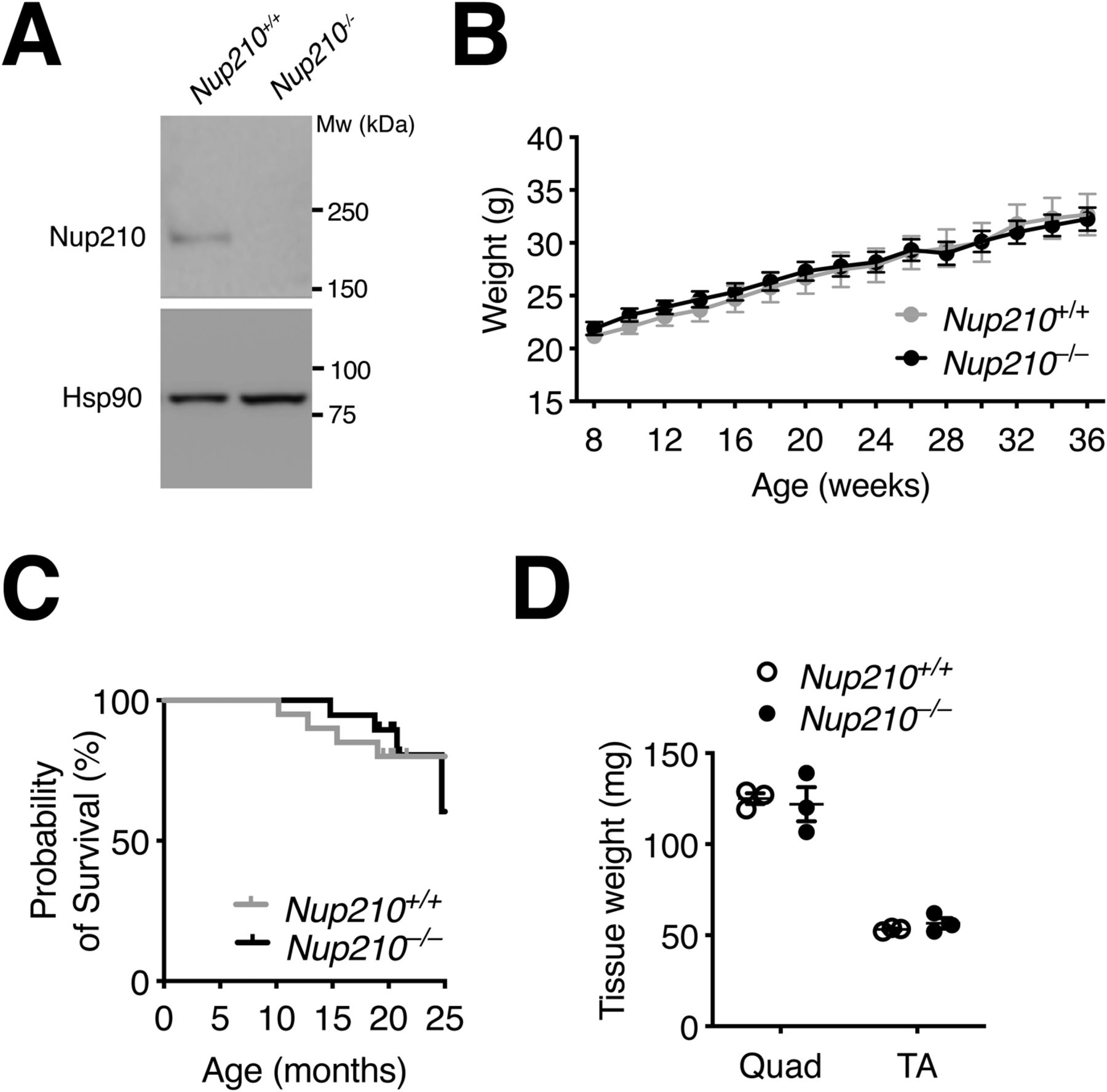
    Figure S1. Nup210 is dispensable for muscle growth and survival.

    (A) Total protein was isolated from control and Nup210 knockout quad muscle and Nup210 levels were analyzed by Western blots. Hsp90 was used as loading control. (B) Weight measurements of Nup210+/+ and Nup210−/− mice (n = 14–15) were taken every 2 wk. (C) Kaplan–Meier plot of Nup210+/+ and Nup210−/− mice (n = 19–20) measuring survival for up to 25 mo. (D) Fresh tissue weights for Quad and TA muscles isolated from Nup210+/+ and Nup210−/− mice (n = 3). Data are plotted as mean ± SEM.

  • Figure 1.
    • Download figure
    • Open in new tab
    • Download powerpoint
    Figure 1. Nup210 is dispensable for mouse development.

    (A) Schematic illustration of the generation of the Nup210 knockout (Nup210−/−) mouse from the HprtCre-mediated recombination of the loxP flanked exon 2 of Nup210 (Nup210f/f). (B) Nup210 was immunoprecipitated from quadricep (Quad), tibialis anterior (TA), and gastrocnemius (GAS) muscles and the amount of protein in the immunoprecipitated was analyzed by Western blot. Hsp90 in the input is used as loading control for the immunoprecipitation and IgG shows the levels of anti-Nup210 used. Asterisk shows oligomeric forms of Nup210 previously described (13). (C) Representative images of Nup210 immunofluorescence staining in TA muscle longitudinal sections obtained from Nup210+/+ and Nup210−/− mice. Scale bar, 25 μm. (D) Representative images of H&E full projections (top) and detailed views (bottom) of TA muscle transverse sections isolated from 6- to 8-wk-old Nup210+/+ and Nup210−/− mice. Scale bar 100 μm. (E) Representative images of immunofluorescence staining for Laminin in quadricep muscles from 6- to 8-wk-old mice. Scale bar, 100 μm. (F) Quantification of myofiber cross-sectional area distribution in quadricep muscles from 6- to 8-wk-old mice (n = 3). Data are binned in 250 μm2 bins and plotted as mean.

  • Figure 2.
    • Download figure
    • Open in new tab
    • Download powerpoint
    Figure 2. Primary muscle satellite cells from Nup210 knockout mice efficiently differentiate into myotubes.

    (A) Schematic illustration (top) and representative images immunofluorescence staining (bottom) of sorted satellite cells from Nup210+/+ and Nup210−/− mice after induction of myogenic differentiation. Arrows indicate undifferentiated muscle progenitor cells which lack Nup210 expression. Scale bar, 50 μm. (B) Representative immunofluorescence images of differentiated satellite cells from Nup210+/+ and Nup210−/− mice stained for myosin heavy chain (MHC) and Hoechst. Scale bar, 300 μm. (C) Nup210+/+ and Nup210−/− satellite cells were sorted, differentiated, and stained for MHC. The percentage of nuclei in MHC-positive cells (differentiation index) was quantified. (D) Nup210+/+/Pax7-CreERT2 and Nup210f/f/Pax7-CreERT2 were treated with tamoxifen and satellite cells were isolated and differentiated in vitro. (C) The differentiation index (percentage of nuclei in MHC-positive cells) was quantified as in (C). (D, E) Representative immunofluorescence images of cells from (D) stained for Nup210 and MHC. Scale bar, 100 μm. Data are plotted as mean ± SD.

  • Figure S2.
    • Download figure
    • Open in new tab
    • Download powerpoint
    Figure S2. Nup210 levels increase in regenerating muscle fibers.

    Sections from uninjured and BaCl2 injured TA muscles were stained for Nup210 (green) and Hoechst (Blue). Scale bar, 25 μM.

  • Figure 3.
    • Download figure
    • Open in new tab
    • Download powerpoint
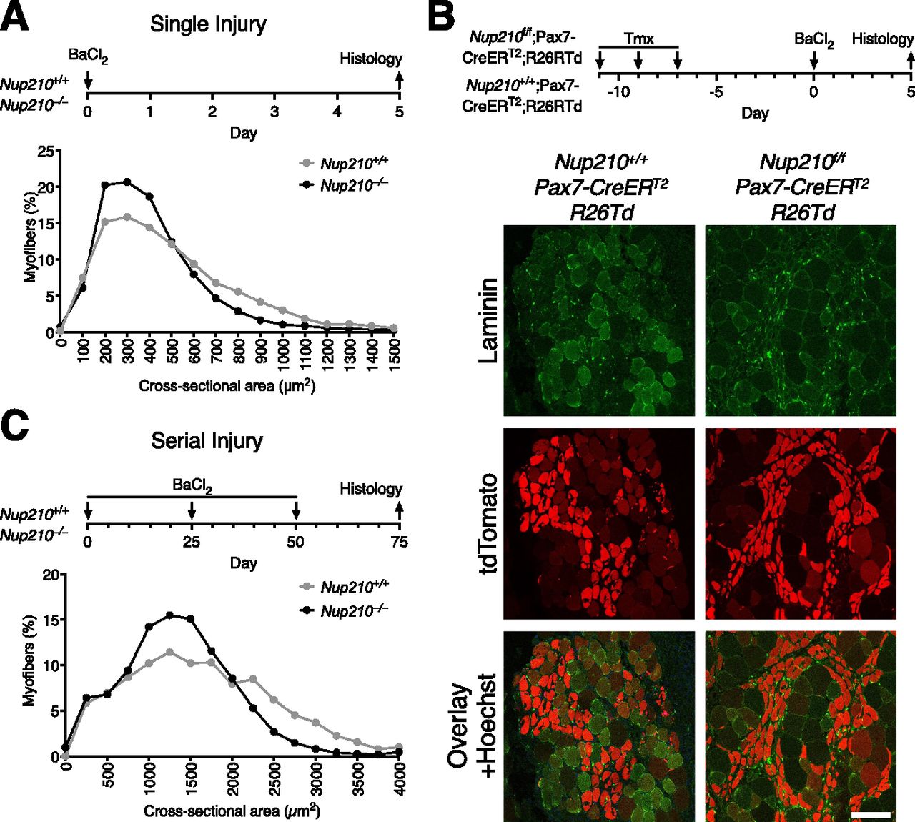
    Figure 3. Nup210 ablation results in delayed muscle regeneration.

    (A) Young (6–8 wk old) Nup210+/+ and Nup210−/− mice were subjected to BaCl2-induced muscle injury and muscle regeneration was analyzed 5 d later. Top: Schematic illustration of experimental approach. Bottom: Quantification of myofiber cross-sectional area distribution. n = 3, data are binned in 250 μm2 bins and are plotted as mean. (B) Nup210+/+/Pax7-CreERT2 and Nup210f/f/Pax7-CreERT2 mice carrying a Cre-inducible tdTomato reporter (R26Td) were treated with tamoxifen before being subjected to BaCl2 muscle injury. The tdTomato-positive myofibers were analyzed by immunofluorescence in muscle sections. Laminin was used as counterstain to detect muscle fibers. Top: Schematic representation of experimental approach. Bottom: Representative immunofluorescence images from TA sections. Scale bar, 100 μm. (C) Young Nup210+/+ and Nup210−/− mice were subjected to BaCl2-induced serial muscle injury (three injuries total), and muscle regeneration was analyzed 25 d after last injury. Top: Schematic illustration of experimental approach. Bottom: Quantification of myofiber cross-sectional area distribution. n = 3, data are binned in 250-μm2 bins and are plotted as mean.

  • Figure 4.
    • Download figure
    • Open in new tab
    • Download powerpoint
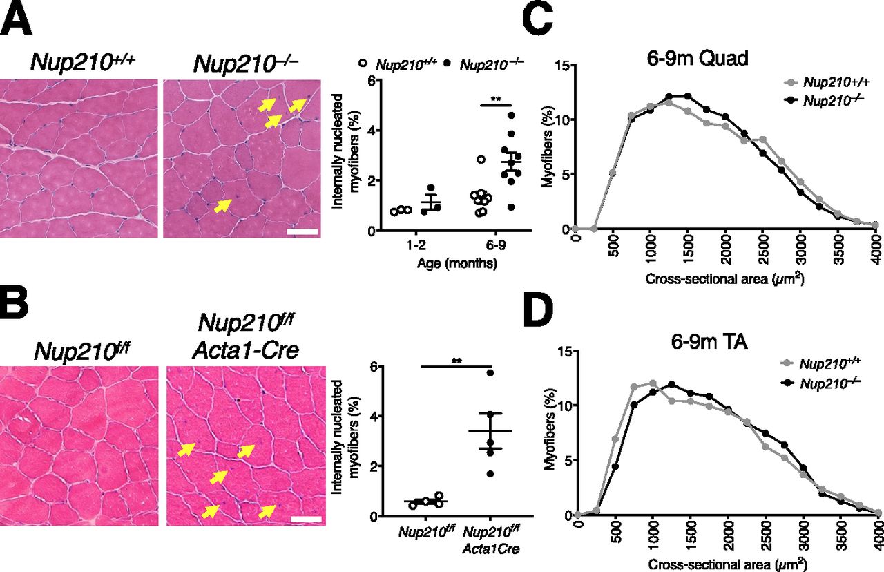
    Figure 4. Nup210 deficiency in muscle results in increased number of centrally nucleated myofibers.

    (A) Representative H&E images (left) and quantification of centrally nucleated myofibers (right) of quadricep muscle from 6- to 9-mo-old Nup210+/+ and Nup210−/− mice (n = 3–9). Arrows denote centrally nucleated fibers. Scale bar 50 μm. (B) Representative H&E images (left) and whole section quantification of centrally nucleated myofibers (right) of quadricep muscle from 6- to 9-mo-old Nup210f/f and Nup210f/f/Acta1-Cre mice (n = 4–5). Arrows denote centrally nucleated fibers. Scale bar 50 μm. (C) Quantification of quadricep myofiber cross-sectional area isolated from 6- to 9-mo-old Nup210+/+ and Nup210−/− mice (n = 3). Data are binned in 250 μm2 bins. (D) Quantification of myofiber cross-sectional area distribution in TA isolated from 6- to 9-mo-old Nup210+/+ and Nup210−/− mice (n = 4). (A, B) Data are binned in 250 μm2 bins. **, P ≤ 0.01 by two-way ANOVA with Sidak’s multiple comparisons test (A), and unpaired t tests in (B). Data are plotted as mean ± SEM in (A, B) and as mean in (C, D).

  • Figure S3.
    • Download figure
    • Open in new tab
    • Download powerpoint
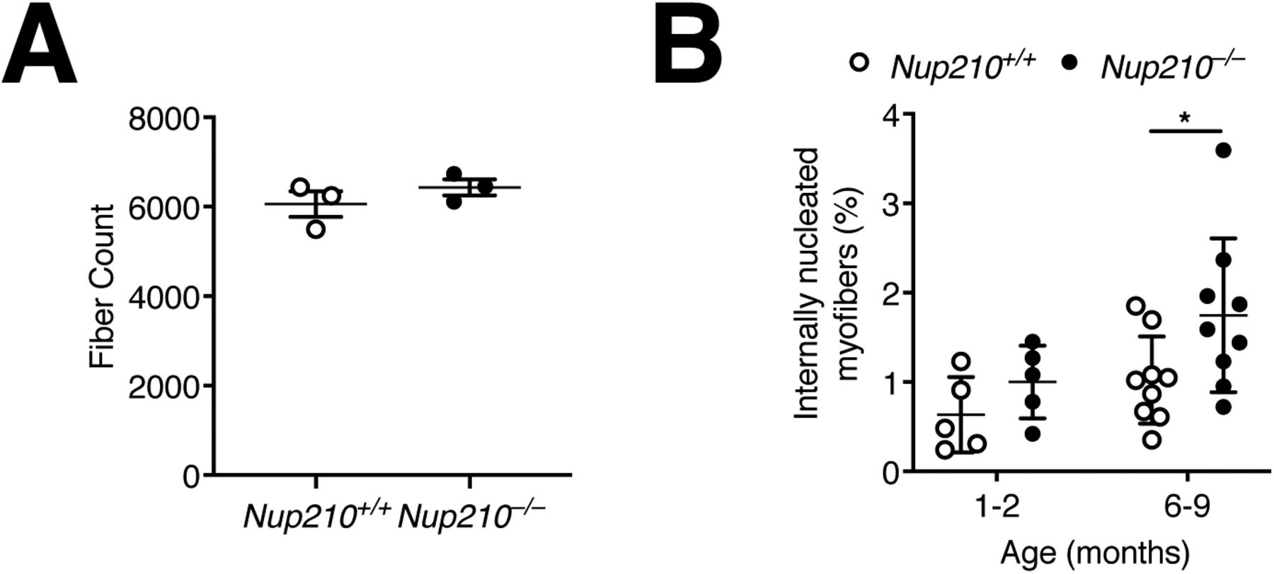
    Figure S3. Evaluation of Quad and TA muscle.

    (A) Quantification of total fiber count for quadricep muscles from 6- to 9-mo-old Nup210+/+ and Nup210−/− mice (n = 3). (B) Quantification of centrally nucleated myofibers in TA muscle from Nup210+/+ and Nup210−/− mice (n = 5–9).

  • Figure 5.
    • Download figure
    • Open in new tab
    • Download powerpoint
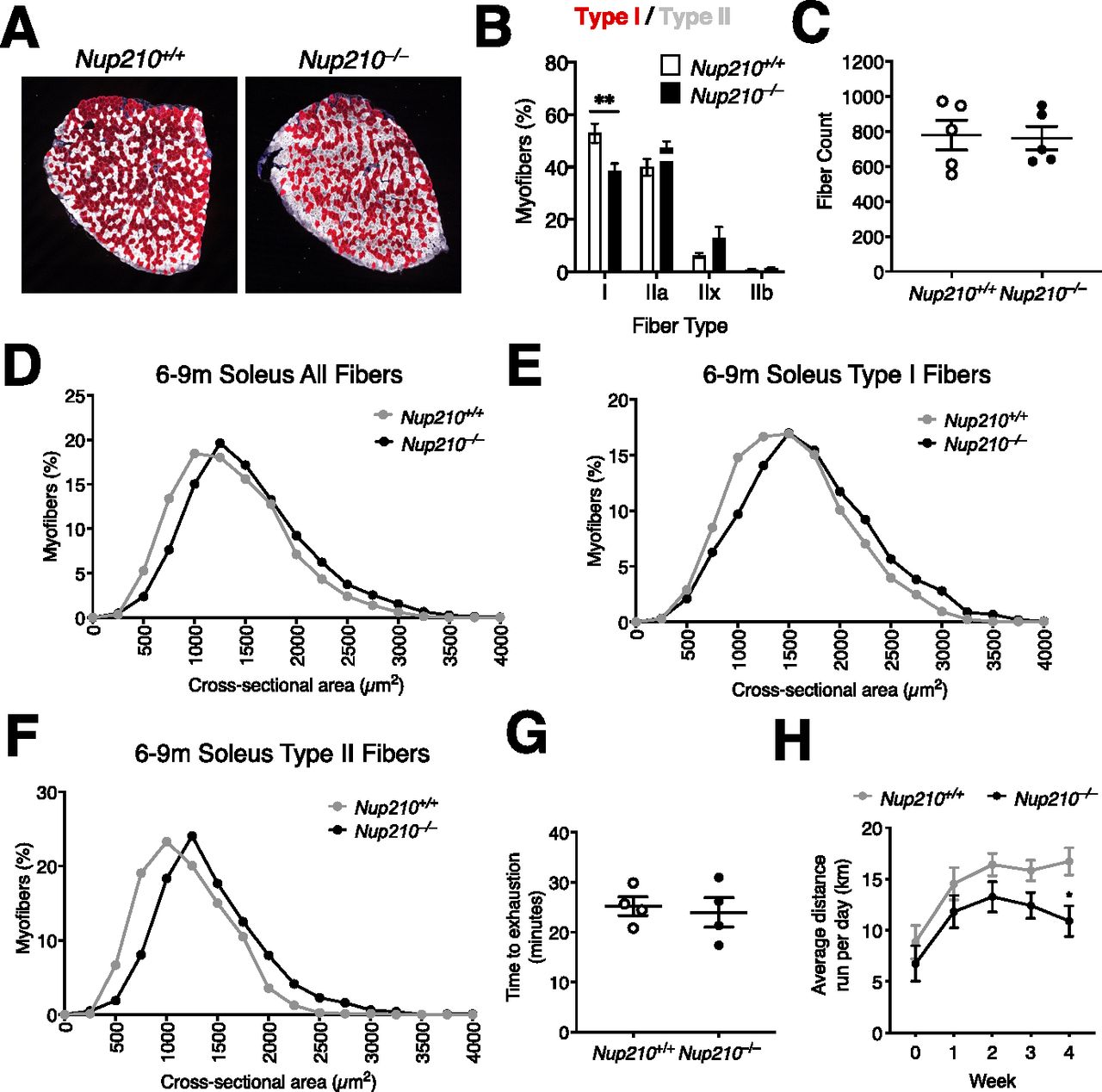
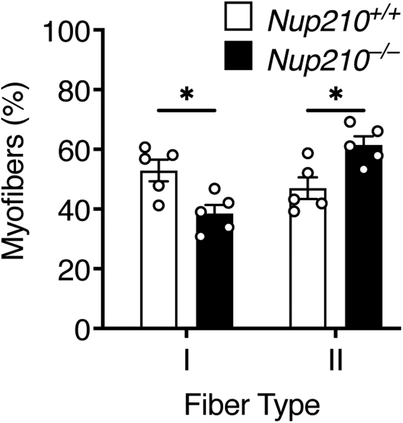
    Figure 5. Nup210 is required for the maintenance of normal skeletal muscle fiber type distribution.

    (A) Representative immunofluorescence images showing Type I (red) and II (white) fibers in soleus of 6- to 9-mo-old Nup210+/+ and Nup210−/− mice. (B) Quantification of fiber type distribution in soleus of 6- to 9-mo-old Nup210+/+ and Nup210−/− mice (n = 5). (C) Quantification of total fiber count for soleus muscles from 6- to 9-mo-old mice (n = 5). (D, E, F) Quantification of myofiber cross-sectional area for all fibers (D), Type I fibers (E), and Type II (IIa, IIx, and IIb) fibers (F) in soleus of 6- to 9-mo-old Nup210+/+ and Nup210−/− mice (n = 5). (G) Nup210+/+ and Nup210−/− mice were subjected to treadmill exercise and the time to exhaustion was quantified. (H) Nup210+/+ and Nup210−/− mice were subjected to 4 wk of voluntary running. The average distance ran per day each week was quantified and plotted. Data are plotted as mean ± SEM in (B), (C, G, H) and mean in (D, E, F). **P ≤ 0.01 by two-way ANOVA with Sidak’s multiple comparisons test.

  • Figure S4.
    • Download figure
    • Open in new tab
    • Download powerpoint
    Figure S4. Quantification of fiber type distribution in soleus.

    Quantification of fiber type distribution in soleus of 6- to 9-mo-old Nup210+/+ and Nup210−/− mice (n = 5). *P ≤ 0.05 by two-way ANOVA with Sidak’s multiple comparisons test.

  • Figure 6.
    • Download figure
    • Open in new tab
    • Download powerpoint
    Figure 6. Nup210 ablation affects the expression of cell adhesion and immune genes.

    (A) Volcano plot of RNA-seq transcriptome data for Nup210 knockout quad muscle relative to control muscle. Significantly up- and down-regulated genes are reported as red and blue dots, respectively (q < 0.05). Genes not changing expression are represented as gray dots. (B) Ingenuity Pathway Analysis of genes deregulated in Nup210 knockout quad muscle (>1.5 fold, q < 0.05). The top 20 pathways are shown. Green dots mark cell adhesion/cytoskeletal-related pathways, red dots show immune-related pathways, yellow dots show pathways associated with both. (C) Upstream transcriptional regulators predicted to be altered in Nup210 knockout muscle by Ingenuity Pathway Analysis. Only pathways with at least 10 target genes affected were included in the analysis. The top 20 pathways are shown. Mef2C is shown in blue. (D) Expression levels for Mef2C muscle structural target genes from the RNAseq data from. Data are plotted as mean ± SEM.

Supplementary Materials

  • Figures
  • Table S1 List of the differentially expressed genes in Nup210+/+ and Nup210−/− quad muscle.

  • Table S2 List of the upstream transcriptional regulators and their target genes predicted altered by Ingenuity Pathway Analysis in Nup210−/− quad muscles.

  • Supplemental Data 1.

    Image J macro.

PreviousNext
Back to top
Download PDF
Article Alerts
Sign In to Email Alerts with your Email Address
Email Article

Thank you for your interest in spreading the word on Life Science Alliance.

NOTE: We only request your email address so that the person you are recommending the page to knows that you wanted them to see it, and that it is not junk mail. We do not capture any email address.

Enter multiple addresses on separate lines or separate them with commas.
Loss of Nup210 results in muscle repair delays and age-associated alterations in muscle integrity
(Your Name) has sent you a message from Life Science Alliance
(Your Name) thought you would like to see the Life Science Alliance web site.
CAPTCHA
This question is for testing whether or not you are a human visitor and to prevent automated spam submissions.
Citation Tools
Nup210 in muscle physiology
Stephen Sakuma, Ethan YS Zhu, Marcela Raices, Pan Zhang, Rabi Murad, Maximiliano A D’Angelo
Life Science Alliance Dec 2021, 5 (3) e202101216; DOI: 10.26508/lsa.202101216

Citation Manager Formats

  • BibTeX
  • Bookends
  • EasyBib
  • EndNote (tagged)
  • EndNote 8 (xml)
  • Medlars
  • Mendeley
  • Papers
  • RefWorks Tagged
  • Ref Manager
  • RIS
  • Zotero
Share
Nup210 in muscle physiology
Stephen Sakuma, Ethan YS Zhu, Marcela Raices, Pan Zhang, Rabi Murad, Maximiliano A D’Angelo
Life Science Alliance Dec 2021, 5 (3) e202101216; DOI: 10.26508/lsa.202101216
del.icio.us logo Digg logo Reddit logo Twitter logo CiteULike logo Facebook logo Google logo Mendeley logo
  • Tweet Widget
  • Facebook Like
Issue Cover

In this Issue

Volume 5, No. 3
March 2022
  • Table of Contents
  • Cover (PDF)
  • About the Cover
  • Masthead (PDF)
Advertisement

Jump to section

  • Article
    • Abstract
    • Introduction
    • Results
    • Discussion
    • Materials and Methods
    • Data Availability
    • Acknowledgements
    • References
  • Figures & Data
  • Info
  • Metrics
  • Reviewer Comments
  • PDF

Subjects

  • Molecular Biology
  • Development
  • Cell Biology

Related Articles

  • No related articles found.

Cited By...

  • No citing articles found.
  • Google Scholar

More in this TOC Section

  • HDAC2, HDAC3, and SIRT1 activity during Mitosis
  • FKBP4 controls 5-FU sensitivity in COAD
  • Aporrectodea caliginosa adaption and acclimatisation
Show more Research Article

Similar Articles

EMBO Press LogoRockefeller University Press LogoCold Spring Harbor Logo

Content

  • Home
  • Newest Articles
  • Current Issue
  • Archive
  • Subject Collections

For Authors

  • Submit a Manuscript
  • Author Guidelines
  • License, copyright, Fee

Other Services

  • Alerts
  • Twitter
  • RSS Feeds

More Information

  • Editors & Staff
  • Reviewer Guidelines
  • Feedback
  • Licensing and Reuse
  • Privacy Policy

ISSN: 2575-1077
© 2022 Life Science Alliance LLC

Life Science Alliance is registered as a trademark in the U.S. Patent and Trade Mark Office and in the European Union Intellectual Property Office.