Advertisement

Main menu

  • Home
  • Articles
    • Newest Articles
    • Current Issue
    • Methods & Resources
    • Author Interviews
    • Archive
    • Subjects
  • Collections
  • Submit
    • Submit a Manuscript
    • Author Guidelines
    • License, Copyright, Fee
    • FAQ
    • Why submit
  • About
    • About Us
    • Editors & Staff
    • Board Members
    • Licensing and Reuse
    • Reviewer Guidelines
    • Privacy Policy
    • Advertise
    • Contact Us
    • LSA LLC
  • Alerts
  • Other Publications
    • EMBO Press
    • The EMBO Journal
    • EMBO reports
    • EMBO Molecular Medicine
    • Molecular Systems Biology
    • Rockefeller University Press
    • Journal of Cell Biology
    • Journal of Experimental Medicine
    • Journal of General Physiology
    • Journal of Human Immunity
    • Cold Spring Harbor Laboratory Press
    • Genes & Development
    • Genome Research

User menu

  • My alerts

Search

  • Advanced search
Life Science Alliance
  • Other Publications
    • EMBO Press
    • The EMBO Journal
    • EMBO reports
    • EMBO Molecular Medicine
    • Molecular Systems Biology
    • Rockefeller University Press
    • Journal of Cell Biology
    • Journal of Experimental Medicine
    • Journal of General Physiology
    • Journal of Human Immunity
    • Cold Spring Harbor Laboratory Press
    • Genes & Development
    • Genome Research
  • My alerts
Life Science Alliance

Advanced Search

  • Home
  • Articles
    • Newest Articles
    • Current Issue
    • Methods & Resources
    • Author Interviews
    • Archive
    • Subjects
  • Collections
  • Submit
    • Submit a Manuscript
    • Author Guidelines
    • License, Copyright, Fee
    • FAQ
    • Why submit
  • About
    • About Us
    • Editors & Staff
    • Board Members
    • Licensing and Reuse
    • Reviewer Guidelines
    • Privacy Policy
    • Advertise
    • Contact Us
    • LSA LLC
  • Alerts
  • Follow LSA on Bluesky
  • Follow lsa Template on Twitter
Review
Open Access

Building in vitro models of the brain to understand the role of APOE in Alzheimer’s disease

View ORCID ProfileRebecca L Pinals, View ORCID ProfileLi-Huei Tsai  Correspondence email
Rebecca L Pinals
1Picower Institute for Learning and Memory, Massachusetts Institute of Technology, Cambridge, MA, USA
2Department of Brain and Cognitive Sciences, Massachusetts Institute of Technology, Cambridge, MA, USA
Roles: Conceptualization, Investigation, Visualization, Writing—original draft, Writing—review and editing
  • Find this author on Google Scholar
  • Find this author on PubMed
  • Search for this author on this site
  • ORCID record for Rebecca L Pinals
Li-Huei Tsai
1Picower Institute for Learning and Memory, Massachusetts Institute of Technology, Cambridge, MA, USA
2Department of Brain and Cognitive Sciences, Massachusetts Institute of Technology, Cambridge, MA, USA
3Broad Institute of Harvard and MIT, Cambridge, MA, USA
Roles: Conceptualization, Resources, Supervision, Funding acquisition, Project administration, Writing—review and editing
  • Find this author on Google Scholar
  • Find this author on PubMed
  • Search for this author on this site
  • ORCID record for Li-Huei Tsai
  • For correspondence: lhtsai@mit.edu
Published 27 September 2022. DOI: 10.26508/lsa.202201542
  • Article
  • Figures & Data
  • Info
  • Metrics
  • PDF
Loading

This article has a correction. Please see:

  • Correction: Building in vitro models of the brain to understand the role of APOE in Alzheimer’s disease - December 1,2022.

Abstract

Alzheimer’s disease (AD) is a devastating, complex, and incurable disease that represents an increasingly problematic global health issue. The etiology of sporadic AD that accounts for a vast majority of cases remains poorly understood, with no effective therapeutic interventions. Genetic studies have identified AD risk genes including the most prominent, APOE, of which the ɛ4 allele increases risk in a dose-dependent manner. A breakthrough discovery enabled the creation of human induced pluripotent stem cells (hiPSCs) that can be differentiated into various brain cell types, facilitating AD research in genetically human models. Herein, we provide a brief background on AD in the context of APOE susceptibility and feature work employing hiPSC-derived brain cell and tissue models to interrogate the contribution of APOE in driving AD pathology. Such models have delivered crucial insights into cellular mechanisms and cell type–specific roles underlying the perturbed biological functions that trigger pathogenic cascades and propagate neurodegeneration. Collectively, hiPSC-based models are envisioned to be an impactful platform for uncovering fundamental AD understanding, with high translational value toward AD drug discovery and testing.

Introduction

Alzheimer’s disease (AD) persists as a debilitating and widespread neurodegenerative disorder, with over 55 million people worldwide currently living with AD or a related form of dementia (Alzheimer’s Disease International, 2020; World Health Organization, 2022). AD is characterized by progressive cognitive and functional decline, in parallel with brain cell dysfunction and death (Alzheimer’s & Dementia, nd; Montine et al, 2012; Knopman et al, 2021). Early-onset familial AD begins to manifest in individuals within the range of ∼30–60 yr of age, in comparison to late-onset sporadic AD that typically develops later in life at ≥65 yr of age (Lambert et al, 2013; National Institute on Aging, 2019). Disease-causing mutations leading to familial AD have become well-established, although this form of AD constitutes only 1–5% of all cases (Reitz & Mayeux, 2014). Work in the 1990s identified the central role of amyloid-β (Aß) in familial AD arising from mutations or duplications in the genes APP, PSEN1, and PSEN2 (Goate et al, 1991; Levy-Lahad et al, 1995; Rogaev et al, 1995; Sherrington et al, 1995; Tanzi & Bertram, 2005). In general, the Aß peptide is released from neurons via sequential proteolytic processing of the membrane-immobilized amyloid precursor protein (APP) by secretase enzymes (Haass et al, 2012). In the amyloidogenic pathway, β-secretase first cleaves APP at the ectodomain, followed by γ-secretase at the intramembrane site, liberating Aß peptides including Aß-40 and Aß-42 (among other peptide lengths). This contrasts with the physiologically normal pathway in which α- then γ-secretases consecutively cleave APP, shedding the shorter Aß-40 species. The genetic modifications underlying familial AD alter the structures of APP (encoded by APP; including near the secretase cleavage sites) and the γ-secretase complex (the catalytic subunit of which is encoded by PSEN1 and PSEN2). As a result, there is elevated generation of the Aß-42 species, which is more prone to aggregate into neurotoxic plaques. This sequence of findings became formative work toward the neuron-centric amyloid hypothesis of AD, whereby accumulation of Aß peptide aggregates in the brain is postulated to drive other AD pathologies, including neurofibrillary tangles of hyperphosphorylated tau (p-tau) protein inside of neurons and, ultimately, neurodegeneration (Hardy & Higgins, 1992; Hardy & Selkoe, 2002). Although a relative ratio of longer to shorter Aß peptides (often the Aß-42/40 ratio) has become a more widely accepted AD biomarker (Tanzi & Bertram, 2005; Selkoe & Hardy, 2016; Hampel et al, 2021), it is worth noting that not all familial AD-causing mutations lead to increased relative or absolute Aß-42 production, including many PSEN1 mutations that impair net γ-secretase activity and thus reduce production of both Aß species (Sun et al, 2017). More broadly, the linear causal structure of the amyloid hypothesis, with the consequent use of Aß species as biomarkers, has suffered from heightened criticism because of failing AD drugs and contradictory findings (Herrup, 2015; Makin, 2018; Panza et al, 2019; Rabinovici, 2021).

Sporadic AD accounts for over 95% of all cases, yet the exact mechanism by which this form of AD arises is still unknown (Reitz & Mayeux, 2014). Based on the understanding of familial AD, research has historically explored the formation of amyloid plaques and tau tangles as key pathological features shared by both AD forms (Tanzi & Bertram, 2005; Serrano-Pozo et al, 2011; Knopman et al, 2021). The cascade of neurodegenerative effects associated with amyloid aggregation suggests that reducing Aß load in the brain could slow or halt cognitive decline. Despite intense efforts in drug development targeting these pathological hallmarks by means of anti-amyloid antibodies and secretase inhibitors, there is no cure for AD; current therapeutic strategies provide only modest relief or yield favorable biomarker changes in the absence of a clinical response (Huang & Mucke, 2012; Canter et al, 2016; Karran & De Strooper, 2022).

In contrast to the recognized genetic changes underlying familial AD, sporadic AD is seemingly driven by a multifactorial combination of genetic and environmental influences. Age remains the most significant risk factor for developing AD (Knopman et al, 2021). Sporadic AD carries an estimated heritability over 50% (Sims et al, 2020), with genome-wide association studies (GWAS) continuing to reveal key genetic loci that modify risk (Lambert et al, 2013; Jansen et al, 2019; Kunkle et al, 2019; Wightman et al, 2021). In particular, the importance of APOE ɛ4 was first identified several decades ago and is now accepted to represent the single largest genetic determinant of AD (Corder et al, 1993; Saunders et al, 1993; Strittmatter et al, 1993; Knopman et al, 2021). Even with APOE displaying only partial penetrance, the imparted risk is significant because the ɛ4 allele is observed at relatively high frequency in the human population. GWAS analyses have impelled a shift to recognize the involvement of multiple genetic factors across different brain cell types in driving AD. However, the interplay of these genetic nodes and corresponding cell type–specific roles require further study (De Strooper & Karran, 2016).

Disentangling the complex causes of AD relies on the development and use of experimental models that recapitulate essential facets of the human brain in the healthy versus diseased state. Animal models have served as the standard platform for the study of AD and other human diseases, offering an integrated system (i.e., connected nervous to other systems, with an immune component) that can undergo controlled manipulation (Elder et al, 2010; Götz et al, 2018). Knowledge of disease-causing mutations facilitates development of animal models, as has been the case for the less common but more genetically tractable familial form of AD. For example, transgenic mouse models have provided a route to study familial AD by overexpression of human genes carrying disease-causative mutations that promote amyloid aggregation (Hsiao et al, 1996; Sasaguri et al, 2017; Götz et al, 2018). More recently, targeted gene-editing to add humanized, pathogenic mutations to endogenous risk-factor loci (e.g., APP and APOE) has rendered more physiologically relevant mouse models (Saito et al, 2014; Sasaguri et al, 2017; Götz et al, 2018; Scearce-Levie et al, 2020). Yet, fundamental biological differences exist between animal and human systems that hinder modeling of complex, human-specific neurodegenerative diseases (Drummond & Wisniewski, 2017; Sasaguri et al, 2017; Wan et al, 2020). Postmortem brain tissues from human donors capture the relevant biology, though only present a static endpoint. Consequently, such tissues do not provide a dynamic model for tracking changes before onset and during the disease nor for experimental interventions that could alter the course of disease (Serrano-Pozo et al, 2011; Lovett et al, 2020). Human cells can be extracted and grown in culture; however, such cells are difficult to isolate from the brain and lack the relevant microenvironment of a three-dimensional tissue (Abud et al, 2017; Lovett et al, 2020). Recent efforts have leveraged advances in stem-cell biology to build in vitro models of the human brain. In 2007, Yamanaka and his team described groundbreaking work in which human induced pluripotent stem cells (hiPSCs) can be derived from more readily accessible patient skin cells and reprogrammed to an embryonic-like, pluripotent state (Takahashi et al, 2007). This paradigm was soon extended to somatic cells from other donor tissues, including peripheral blood cells (Loh et al, 2010; Seki et al, 2010; Staerk et al, 2010). The hiPSCs can then be differentiated into various cell types, such as those of the brain. Accordingly, hiPSC technology has enabled the modeling of various aspects of human brain tissue in the context of Alzheimer’s disease (Penney et al, 2020; Blanchard et al, 2022; Bubnys & Tsai, 2022). Such developments are promising and crucial toward deconvoluting cell-specific roles and tissue-level features as a function of genetic and environmental factors driving AD.

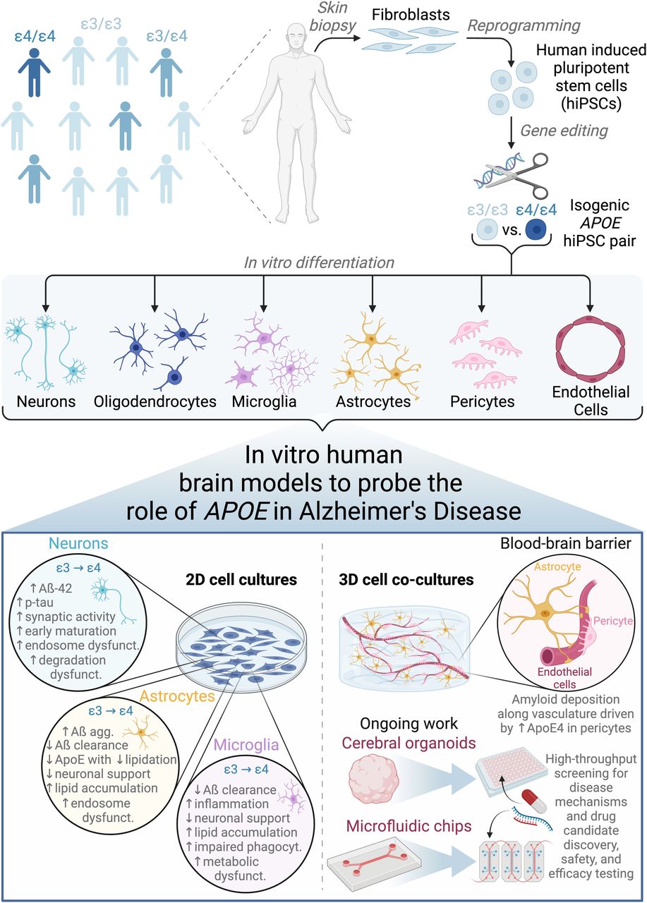
In this review, we provide an overview of hiPSC-derived brain cellular and tissue models, highlighting recent work that employs these models to understand the role of the APOE ε4 genetic risk factor in AD (Fig 1). We begin with a brief background on how the APOE ε4 genotype is implicated in AD. Next, we describe foundational work in hiPSC-based brain cell modeling and then focus on findings from hiPSC-based AD models. We feature new work integrating multiple cell types and/or three-dimensional brain tissue culture systems to model AD, including cerebral organoids and engineered tissues, and conclude with outstanding challenges the field faces.

Figure 1.
  • Download figure
  • Open in new tab
  • Download powerpoint
Figure 1. Employing human induced pluripotent stem cell (hiPSC)–based cellular and tissue models to deconvolute the function of APOE in Alzheimer’s disease.

hiPSCs are derived from human patients of varying genetic background, gene-edited to create isogenic pairs, and differentiated into various cell types of the brain. hiPSC-based cell cultures can be formulated in conventional 2D monoculture or novel 3D co-culture geometries, the latter of which better recapitulates facets of human brain structure and function. Cell type–specific findings as detailed in the main text are summarized from 2D culture studies. Ongoing work will expand the use of cerebral organoids to modeling more diverse cell types beyond neurons and astrocytes and implement perfusable vasculature in microfluidic chip-based models of the blood–brain barrier. Such 3D co-culture models will be advantageous to both fundamental mechanistic studies to understand AD and translation into high-throughput therapeutic discovery and testing pipelines. Figure was created with BioRender.com.

APOE in Alzheimer’s disease

The significance of the APOE gene in governing AD risk was initially recognized in the 1990s, with a series of pioneering studies providing the crucial genotype-to-pathology association and evidence of the physical protein-to-biomarker interaction (Corder et al, 1993; Saunders et al, 1993; Strittmatter et al, 1993). APOE encodes the protein apolipoprotein E (ApoE). Three common forms of the APOE gene exist across the human population: APOE ε2, ε3, and ε4 (Holtzman et al, 2012). The APOE ɛ4 genotype has become well established as the primary genetic risk factor for developing AD through a series of independent studies and datasets across the globe (Farrer et al, 1997; Lambert et al, 2013; Yamazaki et al, 2019). Although the ε4 allele increases risk of developing AD, the ε2 allele is protective (Corder et al, 1993; Saunders et al, 1993; Holtzman et al, 2012; Reiman et al, 2020). APOE confers susceptibility in a dose-dependent manner: relative to an individual with the most common APOE ε3/ε3 background, individuals heterozygous for APOE ε4 (ε4/ε3) are subjected to a 2–4 times greater risk of developing AD and individuals homozygous for APOE ε4 (ε4/ε4) rise to an 8–15 times greater chance of developing AD (Corder et al, 1993; Farrer et al, 1997; Genin et al, 2011; Holtzman et al, 2012). In contrast, a single APOE ε2 allele (together with the common ε3) decreases the odds ratio to ∼0.6 (Corder et al, 1994; Farrer et al, 1997; Holtzman et al, 2012; Reiman et al, 2020). Moreover, the age of AD onset scales inversely with APOE ε4 allele dose, whereby each additional ε4 allele shifts the individual toward a younger age of disease manifestation (estimated 2–5 or 5–10 yr earlier for one or two copies of the ε4 allele, respectively, relative to the lower risk group) (Corder et al, 1993; van der Lee et al, 2018; Yamazaki et al, 2019). For context of the gene prevalence, anywhere from 9 to 25% of humans carry at least one copy of APOE ε4, with this allele frequency varying widely among population groups (Farrer et al, 1997; Bertram et al, 2007; National Institute on Aging, 2019; Yamazaki et al, 2019). However, one or two copies of ε4 is neither necessary nor sufficient to cause AD.

Following the recognition of APOE as a principal genetic determinant in AD, the encoded protein ApoE has become the subject of intense investigation. Yet, the exact functional connection between the polymorphic protein and ensuing AD pathologies remains elusive. ApoE is a 34-kD protein that is broadly involved in lipid metabolism, existing distinctly in the periphery and in the brain (with highest expression in the liver, followed by the brain) (Mahley, 1988; Kim et al, 2009; Holtzman et al, 2012). Within the brain, ApoE represents the most abundantly produced apolipoprotein type and is primarily made by astrocytes under physiological conditions, with lesser contributions from mural cells of the vasculature, damage-associated or neurodegenerative disease-associated microglia, and stressed neurons (Boyles et al, 1985; Pitas et al, 1987; Xu et al, 2006; Casey et al, 2015; Gosselin et al, 2017; Keren-Shaul et al, 2017; Wadhwani et al, 2019; Mahan et al, 2022). ApoE is a component of the high-density lipoprotein–like particles unique to the brain, which adopt a more discoidal morphology compared with those outside the brain (Pitas et al, 1987; LaDu et al, 1998; Holtzman et al, 2012). Similar to its role in the periphery, ApoE in the brain serves as a ligand in receptor-mediated endocytosis of these lipoprotein particles, facilitating transport of phospholipids and cholesterol to neurons (Kim et al, 2009; Holtzman et al, 2012; Yamazaki et al, 2019). Interestingly, ApoE has been found within plaques in human and transgenic mouse brains (Namba et al, 1991; Wisniewski & Frangione, 1992) and has been demonstrated to bind to the Aß peptide (Strittmatter et al, 1993), albeit in acellular experiments with synthetic proteins at above-physiological concentrations.

The three common APOE genetic variants result in distinct amino acid substitutions within the coding sequence of the protein: ApoE ε2 (ApoE2) has cysteines at amino acid positions 112 and 158, ApoE ε3 (ApoE3) has cysteine at position 112 and arginine at position 158, and ApoE ε4 (ApoE4) has arginines at positions 112 and 158. These single–amino acid substitutions strikingly alter the ApoE protein structure, specifically in the receptor-binding domain of ApoE2 and the lipid-binding domain of ApoE4. These conformational differences modify the corresponding protein function: the ApoE2 isoform is severely deficient in its ability to bind to the LDL receptor (<2% receptor binding activity in comparison to ApoE3), and the ApoE4 isoform exhibits a lower lipidation state and lower binding affinity to Aß (Ruiz et al, 2005; Kim et al, 2009; Mahley et al, 2009; Holtzman et al, 2012). Expression of APOE ε4 is reported to cause increased Aß aggregation within and impaired clearance out of the brain, in addition to other processes including synaptic dysfunction and neuroinflammation (Kim et al, 2009; Castellano et al, 2011; Holtzman et al, 2012; Knopman et al, 2021). ApoE thus maintains a clear biological association to AD, yet the multifaceted mechanism by which ApoE4 contributes to AD pathogenesis requires further study.

As APOE ε4 has become firmly entrenched as the strongest genetic factor predisposing individuals to sporadic AD, GWAS studies have expanded in attempt to identify other genes with such significant effects on AD risk. Despite a growing list of such genetic variants, the field increasingly recognizes that these other genes most likely operate interactively with both each other and nongenetic factors, further complicating the story (Kunkle et al, 2019; Mathys et al, 2019; Knopman et al, 2021). Harboring individual risk genes may only confer a minor heritable AD risk but become problematic when existing in certain combinations of multiple, common polymorphisms and/or with a single, rarer genetic variant.

The ability to test the functional consequences of predicted risk factor combinations has been a critical step toward understanding individual genetic contributions. This effort has been enabled by the development and use of appropriate AD models, together with the emergence of larger genomic datasets and more advanced characterization methods necessary to assess functional outputs of the systems under study. A host of animal models exist to recapitulate various aspects of AD, including a growing list of transgenic and genetically modified mice (Drummond & Wisniewski, 2017; Götz et al, 2018). Although these animal models can exhibit certain phenotypes similar to those of human AD patients, the underlying mechanisms are often quite disparate (Götz et al, 2018; Scearce-Levie et al, 2020; D’Avanzo et al, 2015). For example, a popular mouse model (5xFAD) produces high levels of Aß-42 by overexpression of human APP (with three AD-associated mutations) and PSEN1 (with two AD-associated mutations). Although this model develops some pathological AD phenotypes, it has suffered from poor clinical translation. Notably, a third of putative AD risk genes identified in humans lack adequate mouse orthologs, and of particular importance, the APOE polymorphism does not exist in rodents (Mancuso et al, 2019). As will be described, recent work with hiPSC-based models has underscored that human and rodent glia differ significantly in terms of morphology, function, and gene expression profiles (Preman et al, 2021), particularly in the lack of APOE ε4-driven lipid metabolic dysregulation pathways now generally accepted to contribute to AD (TCW et al, 2022). As such, there is a growing movement in the field to take advantage of hiPSC-based models to examine the fundamental disease mechanisms occurring at the molecular and cellular scales within a genetically human background.

Pluripotent stem cell-based models of neurological disease

The scientific breakthrough of generating human iPSCs from somatic cells was first described in 2007, wherein adult human dermal fibroblasts obtained from simple skin biopsies were reprogrammed into stem cells (Takahashi et al, 2007). From this initial discovery, there are now methodologies to differentiate hiPSCs into individual cell types of widely varying identities and organ-like cellular aggregates known as organoids. Herein, we will focus on brain-centric hiPSC-based models. Published protocols exist to derive all major cell types of the brain from hiPSCs: neurons (of different subtypes) (Yeo et al, 2007; Chambers et al, 2012; Zhang et al, 2013), oligodendrocytes (Hu et al, 2009; Wang et al, 2013; Douvaras et al, 2014), microglia (Muffat et al, 2016; Abud et al, 2017; Guttikonda et al, 2021), astrocytes (Shaltouki et al, 2013; TCW et al, 2017), pericytes (Patsch et al, 2015; Kumar et al, 2017), and endothelial cells (Lippmann et al, 2012; Patsch et al, 2015; Qian et al, 2017; Lu et al, 2021b). These differentiation protocols continue to be refined, resulting in brain cells that more accurately represent the requisite genetic expression profiles, functions, and morphologies and at higher yield and purity (Anderson et al, 2021; Lu et al, 2021a). In vivo chimeric models established by transplantation of hiPSC-derived cells into mouse brains has provided another route for producing particular cell populations and for studying neurodegenerative disease, wherein organismal integration provides cell type heterogeneity and an extracellular environment that can drive biologically relevant cell identity and AD phenotypes (Espuny-Camacho et al, 2017; Hasselmann et al, 2019; Najm et al, 2020). Rather than differentiating individual cell types, organoids are three-dimensional models that leverage early developmental programs to drive hiPSCs into self-organized tissue, often with numerous cell types present (Clevers, 2016; Kim et al, 2020; Hofer & Lutolf, 2021). Foundational work by Lancaster et al (2013) described the creation of an in vitro model of the human brain, termed cerebral organoid, and its application to model neurodevelopment and neurological disorders (Lancaster et al, 2013). Namely, the authors generated cerebral organoids to model microcephaly and determined that premature neuronal differentiation underlies the disease phenotype. This work was crucial as a proof-of-principle demonstration for modeling human diseases using patient-derived hiPSCs, showing that key features of the highly complex human brain, such as regional organization, can be emulated in a simplified organoid context.

Numerous groups have translated these hiPSC-based models to study AD over the past decade. A study that conducted neuronal differentiation of hiPSCs from patients with familial AD, sporadic AD, and control individuals highlighted the utility of this stem cell technology in recapitulating some AD-relevant phenotypes, including elevated levels of active kinase GSK-3ß that can phosphorylate tau and the accumulation of early endosomes in neurons (Israel et al, 2012). These findings have been elaborated upon with an orthogonal approach of using three-dimensionally differentiated neuronal cells originating from immortalized human neural stem cells containing familial AD mutations (Choi et al, 2014). Organoids and other three-dimensional neural tissues grown from familial AD patient–derived hiPSCs have been shown to spontaneously develop key pathological features of AD (Bubnys & Tsai, 2022), including accumulation of amyloid plaque– and tau tangle–like structures (Raja et al, 2016; Gonzalez et al, 2018; Jorfi et al, 2018), endosome abnormalities (Raja et al, 2016), and hyperexcitability (Ghatak et al, 2019). Importantly, these AD phenotypes arose in hiPSC-derived cultures in a matter of weeks to months, rather than decades for the disease to manifest in patients. In addition, these model systems supported drug response studies with secretase inhibitors, which limit the production of toxic Aß species (Israel et al, 2012; Choi et al, 2014; Raja et al, 2016; Jorfi et al, 2018). More detailed findings from hiPSC-based familial AD models have recently been reviewed elsewhere (Lee et al, 2020; Penney et al, 2020; Raman et al, 2020). Such advances are promising toward extending this framework to model sporadic AD with more diverse brain cell types present.

Comparison of hiPSC-derived cells sourced from healthy versus diseased individuals continues to be an important route for building hiPSC-based models of the brain. More recently, the CRISPR/Cas9 gene-editing system (among others) has been employed, allowing introduction of mutations into healthy hiPSCs or, conversely, correction of mutations (Ran et al, 2013; Doudna & Charpentier, 2014; Paquet et al, 2016). Accordingly, individual genetic contributions to AD risk can be deconvoluted within otherwise genetically identical (i.e., isogenic) sets of hiPSC-based cellular and tissue models. In the context of AD, this gene-editing approach has been implemented by generating panels of isogenic hiPSCs harboring familial (Konttinen et al, 2019; Kwart et al, 2019; Schrauben et al, 2020) and sporadic AD mutations, with examples of the latter detailed in the following section.

Although hiPSC-based cultures are powerful in vitro models that capture features of brain development and dysfunction, we also must acknowledge their limitations before discussing conclusions ascertained from them. For two-dimensional cell culture systems, the simplified monolayer geometry often results in monomorphic cell populations unable to capture the cell-level heterogeneity and tissue-level architectural complexity inherent in the brain (D’Avanzo et al, 2015; Grenier et al, 2020; Lovett et al, 2020; Blanchard et al, 2022). Likewise, such systems inherently lack a three-dimensional microenvironment that supports the cellular interactions and spatial context necessary to model extracellular dynamics, such as protein aggregation events (D’Avanzo et al, 2015; Grenier et al, 2020; Lovett et al, 2020). Cerebral organoids present a more relevant interstitial environment, but they often lack control and consistency in composition and spatial structuring (Di Lullo & Kriegstein, 2017; Gonzalez et al, 2018; Grenier et al, 2020; Hofer & Lutolf, 2021). Moreover, organoids frequently suffer from necrotic cores because of the lack of vascularization to locally deliver the oxygen and nutrients necessary to sustain growth (Giandomenico & Lancaster, 2017; Mansour et al, 2018; Grenier et al, 2020). The absence of blood vessels is problematic in light of the key role that vascular pathology plays in the two most common neurodegenerative diseases: AD and vascular dementia (Blanchard et al, 2020; Grenier et al, 2020). More generally, there are intrinsic drawbacks in current hiPSC-based models achieving sufficient tissue maturity and cellular diversity (Camp et al, 2015; Di Lullo & Kriegstein, 2017; Bhaduri et al, 2020; Grenier et al, 2020), in addition to losing epigenetic modifications through the reprogramming process (Maherali et al, 2007; Nashun et al, 2015), all of which are important considerations to fully mimic neurological disease states. Strategies are being developed to address each of these shortcomings, such as engrafting absent cell types including microglia, spatial patterning of signals and/or cells to control tissue architecture, bioengineering to introduce infiltrating structures for nutrient delivery, induced aging via targeted protein expression, and avoiding epigenetic erasure by bypassing the hiPSC stage with direct cell reprogramming (Vierbuchen et al, 2010; Miller et al, 2013; Quadrato et al, 2016; Di Lullo & Kriegstein, 2017; Soliman et al, 2017; Lovett et al, 2020; Garreta et al, 2021; Hofer & Lutolf, 2021). Finally, systematic studies implementing these strategies in concert with characterization by emergent technologies, ranging from transcriptomics to high-resolution imaging, will be critical in understanding and subsequently reducing organoid batch-to-batch variability (Quadrato et al, 2016; Di Lullo & Kriegstein, 2017; Garreta et al, 2021; Hofer & Lutolf, 2021). Overall, the hiPSC approach has undergone noteworthy growth with actionable improvements in modeling human neurological disease over the past 15 yr, and the results from applying such models have been proven immediately useful in deepening our understanding of cellular mechanisms driving AD pathologies.

Modeling APOE ε4 risk in Alzheimer’s disease using hiPSC-derived cells

hiPSC-based model systems provide a platform to scrutinize cell type–specific functions that contribute to sporadic AD pathologies in a genotype-dependent manner. The amyloid hypothesis puts forth a neuron-centric view of AD etiology, where neurons do play an essential role as the main producers of Aß and are highly vulnerable to damage (De Strooper & Karran, 2016). However, the combination of hiPSC-based models and more refined characterization methods, including transcriptomic profiling, has enabled the field to study and appreciate the profoundly interconnected roles of other brain cell types, together with neurons, in AD onset and progression (Lambert et al, 2013; De Strooper & Karran, 2016). These findings are summarized in Fig 1.

Neurons

We begin by considering APOE-dependent outcomes in the context of hiPSC-derived neurons. In general, APOE ε4 neurons produce more Aß-42 and have higher p-tau levels in comparison to APOE ε3 neurons (Duan et al, 2014; Lin et al, 2018; Wang et al, 2018; Wadhwani et al, 2019; Lee et al, 2021). This finding on amyloid extends to hiPSC-derived APOE ε4 neurons, generating more Aß aggregates upon transplantation into human APOE ε4- (as compared with APOE ε3-) knockin mice models (Najm et al, 2020). The field has reached some consensus that APOE ε4 represents a gain of toxic function rather than a loss of function, where APOE-deficient neurons display similar Aß and p-tau pathological phenotypes to those expressing APOE ε3 (Shi et al, 2017; Wang et al, 2018). Notably, the heightened Aß production was only observed in APOE ε4 human, not mouse, neurons, highlighting the species difference in APOE isoform–dependent Aß metabolism (Wang et al, 2018). Although Wang et al (2018) established that a small-molecule ApoE4-structure corrector could resolve these AD-related neuronal phenotypes, treatment at the ApoE protein level has yet to be realized in the clinical space (Wang et al, 2018). Transcriptomic analysis of isogenic APOE ε3 versus ε4 neurons (derived from a non–AD-affected individual and gene-edited ε3 to ε4) has revealed broad changes in expression of genes involving synaptic function in neurons (Lin et al, 2018). Specifically, APOE ε4 neurons in culture exhibit early maturation, elevated synaptic activity, and an increase in both the number of synapses and early endosomes, with a corresponding increase in secretion of the more aggregation-prone Aß-42 peptide (Lin et al, 2018; Meyer et al, 2019). Conversion of APOE ε4 to ε3 in hiPSCs from a sporadic AD patient attenuated many of these AD-related phenotypes in the differentiated neurons (Lin et al, 2018). In contrast, transcriptomic analysis of hiPSC-derived mixed cortical cultures in a different study revealed a lack of APOE-dependent differentially expressed genes related to neuronal maturation (TCW et al, 2022). Studies have also uncovered diverging effects dependent on the neuron subtype, demonstrating hyperexcitable glutamatergic neurons versus degeneration of GABAergic interneurons in culture (Lin et al, 2018; Wang et al, 2018). APOE ε4 in hiPSC-derived neurons has additionally been demonstrated to cause defective degradation pathways of autophagy and mitophagy (Fang et al, 2019). Taken together, APOE ε4 neurons suffer from increased Aß secretion and modulated Aß processing pathways, elevated p-tau levels, altered maturation resulting in augmented synaptic activity and increased electrical excitability, and endosomal and mitochondrial dysfunctions.

Glia

Glial cells, which encompass astrocytes, microglia, and oligodendrocytes, provide critical metabolic, immune, and physical support to the brain. Framed by the amyloid hypothesis, APOE ε4–expressing hiPSC-derived glia have been repeatedly shown to develop trafficking defects that perturb cerebral Aß peptide oligomerization and effective clearance (Fernandez et al, 2019; Knopman et al, 2021; de Leeuw et al, 2022). From a combination of transcriptomics and cell culture experiments, APOE ε4 astrocytes demonstrate impaired Aß uptake compared with isogenic APOE ε3 astrocytes, aligning to the expected result of net higher extracellular Aß-42 concentration (Lin et al, 2018). Alternatively, astrocytic trafficking defects arising from APOE ε4 can also disrupt endocytosis in an Aß-independent manner. Further study of trafficking in hiPSC-derived astrocytes has established a compensatory functional connection between APOE ε4 and another AD risk factor, PICALM: although APOE ε4 expression was shown to cause defects in early endosomes that disrupted endocytic trafficking in astrocytes, increasing expression of PICALM was able to rescue the system (Narayan et al, 2020). Mechanistically, APOE ε4 astrocytes produce significantly less ApoE protein than ε3 (Lin et al, 2018), and this protein remains in a hypolipidated state (Zhao et al, 2017), in agreement with previous studies in human tissue and mouse models (Mooijaart et al, 2006; Shi et al, 2017). These ApoE4-containing lipoprotein particles, in turn, possess diminished binding efficiency to clear Aß (Kim et al, 2009). Some evidence suggests that ApoE isoform-dependent effects on amyloid clearance chiefly stem from competition for the same receptor-mediated removal pathways from the brain rather than direct interaction (Holtzman et al, 2012; Verghese et al, 2013).

Recent work addressing APOE-based AD risk has consistently demonstrated the dysregulation of key lipid pathways in hiPSC-derived APOE ε4 glia. As a primary function of ApoE is the transport of cholesterol, substantial effort has been dedicated to deciphering the cholesterol connection to AD. Lipid metabolism perturbed in APOE ε4 astrocytes at the transcriptomic level has been validated in culture, with APOE ε4 astrocytes exhibiting an accumulation of cholesterol both intracellularly and extracellularly in the media, suggesting dysregulated cholesterol metabolism (Lin et al, 2018). The work of TCW et al (2022) mainly corroborates these findings, where hiPSC-based APOE ε4 astrocytes and microglia feature elevated cholesterol synthesis and accumulation, similarly validated through transcriptomic profiling with corresponding in vitro experiments (TCW et al, 2022). The authors suggest a mechanism in which lysosomes sequester the elevated free cholesterol away from the endoplasmic reticulum, causing the cell to falsely sense low intracellular cholesterol concentration. In turn, this miscommunication induces the glial cell to up-regulate de novo cholesterol biosynthesis and decrease cholesterol efflux. Importantly, these effects are only seen in human, not mouse, glial cells, underscoring the utility of hiPSC-based models (TCW et al, 2022). This putative cholesterol sequestration mechanism is supported by another study that applied proteomic and lipidomic analyses to characterize APOE genotype-dependent changes in hiPSC-based astrocytes (de Leeuw et al, 2022). However, the reduced cholesterol efflux observed by de Leeuw et al (2022) and TCW et al (2022) renders the increased cholesterol level in the media measured by Lin et al (2018) counter-intuitive, pointing to the complex metabolic dysregulations occurring in APOE ε4 astrocytes that require further study. Overall, each of these conclusions highlights the disruption of net cholesterol flux. The apparent distinctions likely arise from experimental differences in parameters such as incubation timings, cell media compositions, and methods of quantification. Bulk media measurements grant a valuable view into cholesterol load that neighboring cells may experience but only a snapshot of net accumulation that is a sum of dynamic processes including biosynthesis, efflux, influx, and turnover. hiPSC donor-specific differences and the number of hiPSC lines under study introduce the added factor of genetic heterogeneity between individuals.

In addition to cholesterol, hiPSC-based APOE ε4 astrocytes demonstrate broad lipid imbalances, including accumulation of unsaturated triacylglycerides within intracellular lipid droplets (Sienski et al, 2021). Such imbalances cause the astrocytes to be more sensitive to nutritional conditions or exogenous lipid stress. Promoting phospholipid synthesis via choline supplementation of culture medium can avert such lipid droplet accumulation and restore lipid homeostasis. These findings support the manipulation of glial lipid metabolism through exogenous supplementation (i.e., dietary changes) as a therapeutic strategy to alleviate APOE ε4-associated disease risk.

Building from these findings ascertained from astrocytes alone, another APOE ε4-induced feature that can be modeled with hiPSC systems is the disrupted metabolic coupling between neurons and astrocytes. From previous animal work, toxic fatty acids produced during periods of neuronal hyperactivity are shunted to astrocytes via lipoprotein particles of which ApoE is a constituent (Liu et al, 2015, 2017; Fernandez et al, 2019; Ioannou et al, 2019). Astrocytes subsequently store these fatty acids in lipid droplets. However, APOE ε4 both reduces the transport efficiency from neurons to astrocytes and diminishes the proficiency of astrocytes degrading neuronal lipids, resulting in compromised neurotrophic support (Qi et al, 2021). Similarly, hiPSC-derived APOE ε4 astrocytes in co-culture with neurons are less effective in supporting neuronal survival and synaptogenesis, thus jeopardizing neuronal health (Zhao et al, 2017). Another study finds that APOE ε4 astrocytes oversupply cholesterol to neurons, resulting in more neuronal lipid rafts to which APP and its processing secretases localize, culminating in higher Aß generation from neurons (Lee et al, 2021) (in agreement with a study done in mouse cells [Wang et al, 2021]). Here, as noted regarding astrocyte monocultures above, perturbations in net cholesterol flux can negatively impact surrounding cells and may implicate combined effects of cholesterol efflux, influx, and turnover. Of note, the faulty lipid transport capabilities of ApoE4 are exacerbated in the aging brain, in comparison with the young APOE ε4 carrier brain that seemingly has compensatory mechanisms to cope with deficient neurotrophic support from astrocytes (Fernandez et al, 2019). Adapting hiPSC-based cultures to better capture these aging effects and ApoE4-mediated disruption of this neuron-supportive function will provide a clearer picture of the nature and consequences of ApoE4-mediated lipid dysregulation.

ApoE is primarily regarded as a lipid transport protein originating from astrocytes, yet the expanding transcriptomic analyses of human tissue samples have identified many AD-driven changes in gene expression within microglia (Mathys et al, 2019; Bellenguez et al, 2022). In particular, transcriptomic analysis of the prefrontal cortex from AD patients has revealed a concomitant up-regulation of APOE in microglia and down-regulation in astrocytes, emphasizing the cell type–specific effects of the gene (Mathys et al, 2019). The recent addition of protocols to derive microglia from hiPSCs has now enabled in vitro modeling of this transcriptomic data. Studying hiPSC-derived microglia is particularly advantageous because of their highly reactive nature and difficulty to transfect that limits the successful application of viral techniques (Maes et al, 2019; Victor et al, 2022). Advances in understanding microglial roles in AD will benefit from recent developments in chimeric models that entail grafting, for example, iPSC hematopoietic progenitors onto humanized, immune-deficient mice, resulting in differentiation into microglia that acquire appropriate human microglial gene signatures and responsive behaviors (Hasselmann et al, 2019). Such models acknowledge the profoundly sensitive nature of microglia to their local environment and are able to correct for the transcriptomic deficiencies that microglia develop in isolation in vitro (Hasselmann et al, 2019; Mancuso et al, 2019; Svoboda et al, 2019; Xu et al, 2020; Claes et al, 2021).

APOE ε4 hiPSC-based microglia are reported to exhibit inflammatory gene activation and associated phenotypes, adopting distinct morphologies with impaired phagocytosis of extracellular Aß aggregates (Lin et al, 2018). Another study on hiPSC-derived microglia determined that the APOE ε4 genotype compromised phagocytosis, reduced migration, increased proinflammatory cytokine secretion, and led to defective glycolytic and mitochondrial metabolism (Konttinen et al, 2019). Collectively, glial activation often arises as a consequence of the APOE ε4 genotype, revealed by analysis of transcriptomic changes together with measurement of secreted proinflammatory chemokines and cytokines (Lin et al, 2018; de Leeuw et al, 2022; TCW et al, 2022). Inflammation is often considered a nonspecific hallmark of neurodegeneration (Krasemann et al, 2017; Butovsky & Weiner, 2018) and in the case of AD, is seemingly connected with and induced by other pathways, such as lipid dyshomeostasis. Indeed, the metabolic shift associated with activation and inflammation includes the accumulation of neutral lipids and lipid droplets, reminiscent of the baseline lipid state in APOE ε4 cells (Sienski et al, 2021; Victor et al, 2022).

Although some work has been done with microglia in co-culture systems, there remains a need for more hiPSC-based studies that elucidate the crosstalk between microglia and other brain cell types. Recently, Victor et al (2022) investigated the cellular interactions between hiPSC-derived neurons and microglia as a function of the APOE genotype (Victor et al, 2022). Interestingly, soluble signaling from neurons provoked APOE ε4 microglia to enter a unique metabolic program, leading to the accumulation of neutral lipid droplets because of impaired lipid catabolism, in conjunction with decreased uptake of extracellular fatty acids because of the already saturated intracellular lipid machinery. In turn, this response shifted microglia away from their prototypical immune surveillance functions and weakened the neuron-microglia coupling required for microglia to adequately respond to modulations in neuronal activity, to the extent of microglia even disrupting coordinated neuronal activity. This cascade ultimately resulted in an intensified pro-inflammatory response, in line with APOE ε4 expression in microglia generally being associated with inflammation (Lin et al, 2018; Fernandez et al, 2019; Yamazaki et al, 2019). In this study, neurons were found to express APOE ε4, as expected in hiPSC-derived cells that are often in a stressed state. Toward therapeutic intervention, pharmacological blocking of lipid synthesis in APOE ε4 microglia was able to remediate these intracellular lipid droplets and restore microglial homeostasis.

Finally, the roles of oligodendrocyte dysfunction and myelin degeneration in AD pathology are becoming increasingly appreciated (Akay et al, 2021; Blanchard et al, 2022). Transcriptomic analysis of prefrontal cortex tissue has identified oligodendrocytes as one of the most altered cell types in AD (Mathys et al, 2019; Lau et al, 2020). The advent of protocols to derive oligodendrocyte precursor cells from hiPSCs offers an avenue to hiPSC-based models of this cell type (Douvaras et al, 2014; Penney et al, 2020; Akay et al, 2021). Model systems have been developed to exemplify in vitro myelination with neuronal co-cultures or artificial axons, as reviewed elsewhere (Blanchard et al, 2022), providing promising future directions to the study of myelin in the context of AD-relevant risk factors such as APOE ε4.

Three-dimensional co-culture systems

Some findings as outlined above have been built upon to capture sporadic AD in organoid culture. This is exemplified by Lin et al (2018), in which the study was extended to model the APOE ε4–dependent defects in organoids containing neurons and astrocytes. In line with the neuron monoculture results (after 6 wk), APOE ε4 organoids exhibited more extracellular Aß accumulation and elevated tau phosphorylation (after 6 mo in culture; in comparison, the corresponding fAD organoid model requires the shorter time course of 2–3 mo to display a similar phenotype [Raja et al, 2016]). Crucially, this demonstrates that APOE ε4 alone is sufficient to cause AD hallmarks in cerebral organoids. Also using organoids, these findings have subsequently been validated and provided with a molecular mechanism implicating impaired function of the transcriptional regulator REST (Meyer et al, 2019). REST serves as a key repressor of neuronal differentiation that is normally induced by aging yet was found via gene network analysis to exhibit a loss of function in both sporadic AD and APOE ε4 neural cells (Meyer et al, 2019). Reduced REST function arises from its decreased nuclear localization and altered chromatin binding, with associated nuclear lamina disruption (Meyer et al, 2019). In turn, neuronal maturation processes are up-regulated, resulting in the previously described phenotype of premature neuronal differentiation, reduced progenitor cell renewal, accelerated synapse formation, and heightened electrical excitability (Meyer et al, 2019). Of note, accelerated differentiation was not reversed by inhibiting Aß generation and appeared before increased levels of tau phosphorylation; therefore, REST dysfunction may precede the canonical amyloid and tau pathologies (Meyer et al, 2019). Taken together, reduced REST function leading to a depleted progenitor pool and disrupted neural circuit formation may contribute to AD onset. A related study using AD patient hiPSC-derived cerebral organoids discovered that although APOE ε4 likely leads to early neuronal maturation, it also exacerbates synaptic loss in mature cerebral organoids (week 12) (Zhao et al, 2020). As such, enhanced differentiation and maturation of neurons in the early stages of development is posited to induce a corresponding mechanistic exhaustion and depleted cognitive reserve that accelerates neurodegeneration in the late disease stages.

Blood–brain barrier cells

The blood–brain barrier (BBB) is comprised of endothelial cells, pericytes, and astrocytes that play an indispensable role in nutrient and oxygen delivery to and waste removal from the brain. BBB dysfunction and breakdown is observed across many neurodegenerative diseases, including AD, with a dependency on APOE isoform (Bell et al, 2012; Montagne et al, 2020). Blanchard et al (2020) have newly developed a three-dimensional, in vitro BBB (iBBB) composed of hiPSC-derived endothelial cells, pericytes, and astrocytes to model the effect of APOE ε4 on cerebral amyloid angiopathy, a condition in AD where amyloid deposits along the brain vasculature (Blanchard et al, 2020). Upon exposure to conditioned media from familial AD neuronal culture as the source of Aß, APOE ε4 iBBB cultures exhibited significantly higher amyloid accumulation along the blood vessels compared with risk-neutral APOE ε3 cultures. Based on a combinatorial cell-type screen with complementary transcriptomic analysis, it was determined that up-regulated APOE ε4 expression by pericytes was the critical component necessary for the amyloid angiopathy phenotype to occur. Further analysis revealed that dysregulation of nuclear factor of activated T cells (NFAT)–calcineurin signaling mediated the up-regulation of ApoE4 in pericytes, therefore increasing amyloid deposition and BBB disruption and providing a potential therapeutic target. Looking forward, microfluidic-based co-culture platforms are excitingly moving toward fully hiPSC-derived cell models of the BBB with functioning blood vessels, offering a promising route to probe AD pathology as a function of the APOE ε4 genotype (Campisi et al, 2018; Shin et al, 2019; Hajal et al, 2022). More broadly, developing a fully hiPSC-derived functional brain tissue with an integrated BBB is of high interest for studying the interplay between these vascular cells and other brain cell types.

Outstanding challenges and future directions

Alzheimer’s disease continues to be a global health problem with severe psychological, social, and economic implications. A disease-altering treatment has yet to be realized. The repeated failures of hundreds of clinical trials over several decades to demonstrate efficacy in human AD patients has spurred the movement to develop more predictive disease models (Gonzalez et al, 2018; Penney et al, 2020). Although no single model has yet to holistically capture the complex AD etiology, advances in this platform-development space have proven useful (Lovett et al, 2020; Penney et al, 2020; Blanchard et al, 2022; Bubnys & Tsai, 2022). Animal models have enabled key contributions to understanding AD, though evolutionary differences render sole reliance on these systems difficult. The capacity to reprogram human fibroblasts into stem cells has begun to revolutionize the study of human disease. Recently developed hiPSC-based brain models provide an avenue to study the mechanisms underlying AD pathologies and drug responses in treating such pathologies in a genetically human background. In contrast to most other AD models, hiPSC systems do not necessitate exogenous overexpression of proteins to induce disease pathologies. Mounting evidence supports the use of hiPSC technology, together with human postmortem tissues and animal models, to build consensus within the field on the connection between genetic susceptibilities and consequent molecular mechanisms and cellular contributions.

Most of the hiPSC work modeling sporadic AD to-date has involved single- or few-cell-type cultures to derive fundamental understanding of the APOE risk factor function. Technologies merging multiple cell types and physiological features present in the actual human brain will be key in building next-generation in vitro platforms to study neurodegenerative disease. Moreover, this will enable the expansion from probing simplified genetic factors to more complex polygenic and/or environmental factors in driving AD risk (Cairns et al, 2020). Broadly, the study of APOE and other AD risk factors requires robustly validated hiPSC-derived brain models that accurately and reproducibly express relevant pathologies, potentially requiring multiple complementary models tailored to best address different biological questions (Blanchard et al, 2022). However, as we move closer to recapitulating the necessary biological complexity of the human brain, it is increasingly important to incorporate the ethical considerations of these models into the research itself (Farahany et al, 2018; Sawai et al, 2019; Garreta et al, 2021). Such models can serve as testing platforms toward the ultimate goal of rational interventions to treat or ideally prevent AD. Some work has established the translatability of highly uniform and homogeneous cerebral organoid models into high-throughput array screening platforms, with applications in discovering novel drug targets and testing candidate drugs to assess effective therapeutic intervention (Gonzalez et al, 2018; Park et al, 2021).

In the future, such hiPSC-based brain cell cultures will benefit from several technological developments, including integration of sensors to monitor dynamic molecular changes (Acarón Ledesma et al, 2019), engineering the extracellular milieu to support longer term culture of various cell types (Bretherton & DeForest, 2021; Hofer & Lutolf, 2021), and implementing spatiotemporal control to tune cell culture conditions in real time (Karimi et al, 2016; Lovett et al, 2020). Brain model platforms will be further enhanced by biological advances in incorporating an in vivo analogous immune component and perfusable vasculature (Park et al, 2018; Hajal et al, 2021; Blanchard et al, 2022). We anticipate hiPSC-based brain co-culture models to open avenues for intervention by revolutionizing AD drug development and testing toward a future cure.

Acknowledgements

We thank Drs. A Bubnys, J Penney, MB Victor, and JM Bonner for critically reading the manuscript. RL Pinals acknowledges support from the Schmidt Science Fellows program, in partnership with the Rhodes Trust. We acknowledge the support of the National Institutes of Health grants UH3NS115064 and RF1AG062377.

Author Contributions

  • RL Pinals: conceptualization, investigation, visualization, and writing—original draft, review, and editing.

  • L-H Tsai: conceptualization, resources, supervision, project administration, and writing—review and editing.

Conflict of Interest Statement

L-H Tsai filed a patent application on the iBBB technology described in Blanchard et al (2020) (PCT/US2020/014572). The authors declare no other conflict of interests.

  • Received May 31, 2022.
  • Revision received September 13, 2022.
  • Accepted September 14, 2022.
  • © 2022 Pinals and Tsai
Creative Commons logoCreative Commons logohttps://creativecommons.org/licenses/by/4.0/

This article is available under a Creative Commons License (Attribution 4.0 International, as described at https://creativecommons.org/licenses/by/4.0/).

References

  1. ↵
    1. Abud EM,
    2. Ramirez RN,
    3. Martinez ES,
    4. Healy LM,
    5. Nguyen CHH,
    6. Newman SA,
    7. Yeromin AV,
    8. Scarfone VM,
    9. Marsh SE,
    10. Fimbres C, et al.
    (2017) iPSC-derived human microglia-like cells to study neurological diseases. Neuron 94: 278–293.e9. doi:10.1016/j.neuron.2017.03.042
    OpenUrlCrossRefPubMed
  2. ↵
    1. Acarón Ledesma H,
    2. Li X,
    3. Carvalho-de-Souza JL,
    4. Wei W,
    5. Bezanilla F,
    6. Tian B
    (2019) An atlas of nano-enabled neural interfaces. Nat Nanotechnol 14: 645–657. doi:10.1038/s41565-019-0487-x
    OpenUrlCrossRef
  3. ↵
    1. Akay LA,
    2. Effenberger AH,
    3. Tsai L-H
    (2021) Cell of all trades: Oligodendrocyte precursor cells in synaptic, vascular, and immune function. Genes Dev 35: 180–198. doi:10.1101/gad.344218.120
    OpenUrlAbstract/FREE Full Text
  4. ↵
    1. Alzheimer’s Disease International
    (2020) Dementia Statistics. https://www.alzint.org/about/dementia-facts-figures/dementia-statistics/
  5. ↵
    1. Alzheimer’s and Dementia
    (nd) Alzheimer’s disease and dementia. https://www.alz.org/alzheimer_s_dementia
  6. ↵
    1. Anderson NC,
    2. Chen P-F,
    3. Meganathan K,
    4. Afshar Saber W,
    5. Petersen AJ,
    6. Bhattacharyya A,
    7. Kroll KL,
    8. Sahin M
    (2021) Balancing serendipity and reproducibility: Pluripotent stem cells as experimental systems for intellectual and developmental disorders. Stem Cell Rep 16: 1446–1457. doi:10.1016/j.stemcr.2021.03.025
    OpenUrlCrossRef
  7. ↵
    1. Bell RD,
    2. Winkler EA,
    3. Singh I,
    4. Sagare AP,
    5. Deane R,
    6. Wu Z,
    7. Holtzman DM,
    8. Betsholtz C,
    9. Armulik A,
    10. Sallstrom J, et al.
    (2012) Apolipoprotein E controls cerebrovascular integrity via cyclophilin A. Nature 485: 512–516. doi:10.1038/nature11087
    OpenUrlCrossRefPubMed
  8. ↵
    1. Bellenguez C,
    2. Küçükali F,
    3. Jansen IE,
    4. Kleineidam L,
    5. Moreno-Grau S,
    6. Amin N,
    7. Naj AC,
    8. Campos-Martin R,
    9. Grenier-Boley B,
    10. Andrade V, et al.
    (2022) New insights into the genetic etiology of Alzheimer’s disease and related dementias. Nat Genet 54: 412–436. doi:10.1038/s41588-022-01024-z
    OpenUrlCrossRef
  9. ↵
    1. Bertram L,
    2. McQueen MB,
    3. Mullin K,
    4. Blacker D,
    5. Tanzi RE
    (2007) Systematic meta-analyses of Alzheimer disease genetic association studies: The AlzGene database. Nat Genet 39: 17–23. doi:10.1038/ng1934
    OpenUrlCrossRefPubMed
  10. ↵
    1. Bhaduri A,
    2. Andrews MG,
    3. Mancia Leon W,
    4. Jung D,
    5. Shin D,
    6. Allen D,
    7. Jung D,
    8. Schmunk G,
    9. Haeussler M,
    10. Salma J, et al.
    (2020) Cell stress in cortical organoids impairs molecular subtype specification. Nature 578: 142–148. doi:10.1038/s41586-020-1962-0
    OpenUrlCrossRef
  11. ↵
    1. Blanchard JW,
    2. Bula M,
    3. Davila-Velderrain J,
    4. Akay LA,
    5. Zhu L,
    6. Frank A,
    7. Victor MB,
    8. Bonner JM,
    9. Mathys H,
    10. Lin Y-T, et al.
    (2020) Reconstruction of the human blood–brain barrier in vitro reveals a pathogenic mechanism of APOE4 in pericytes. Nat Med 26: 952–963. doi:10.1038/s41591-020-0886-4
    OpenUrlCrossRefPubMed
  12. ↵
    1. Blanchard JW,
    2. Victor MB,
    3. Tsai L-H
    (2022) Dissecting the complexities of Alzheimer disease with in vitro models of the human brain. Nat Rev Neurol 18: 25–39. doi:10.1038/s41582-021-00578-6
    OpenUrlCrossRef
  13. ↵
    1. Boyles JK,
    2. Pitas RE,
    3. Wilson E,
    4. Mahley RW,
    5. Taylor JM
    (1985) Apolipoprotein E associated with astrocytic glia of the central nervous system and with nonmyelinating glia of the peripheral nervous system. J Clin Invest 76: 1501–1513. doi:10.1172/jci112130
    OpenUrlCrossRefPubMed
  14. ↵
    1. Bretherton RC,
    2. DeForest CA
    (2021) The art of engineering biomimetic cellular microenvironments. ACS Biomater Sci Eng 7: 3997–4008. doi:10.1021/acsbiomaterials.0c01549
    OpenUrlCrossRef
  15. ↵
    1. Bubnys A,
    2. Tsai L-H
    (2022) Harnessing cerebral organoids for Alzheimer’s disease research. Curr Opin Neurobiol 72: 120–130. doi:10.1016/j.conb.2021.10.003
    OpenUrlCrossRef
  16. ↵
    1. Butovsky O,
    2. Weiner HL
    (2018) Microglial signatures and their role in health and disease. Nat Rev Neurosci 19: 622–635. doi:10.1038/s41583-018-0057-5
    OpenUrlCrossRefPubMed
  17. ↵
    1. Cairns DM,
    2. Rouleau N,
    3. Parker RN,
    4. Walsh KG,
    5. Gehrke L,
    6. Kaplan DL
    (2020) A 3D human brain–like tissue model of herpes-induced Alzheimer’s disease. Sci Adv 6: eaay8828. doi:10.1126/sciadv.aay8828
    OpenUrlFREE Full Text
  18. ↵
    1. Camp JG,
    2. Badsha F,
    3. Florio M,
    4. Kanton S,
    5. Gerber T,
    6. Wilsch-Bräuninger M,
    7. Lewitus E,
    8. Sykes A,
    9. Hevers W,
    10. Lancaster M, et al.
    (2015) Human cerebral organoids recapitulate gene expression programs of fetal neocortex development. Proc Natl Acad Sci U S A 112: 15672–15677. doi:10.1073/pnas.1520760112
    OpenUrlAbstract/FREE Full Text
  19. ↵
    1. Campisi M,
    2. Shin Y,
    3. Osaki T,
    4. Hajal C,
    5. Chiono V,
    6. Kamm RD
    (2018) 3D self-organized microvascular model of the human blood-brain barrier with endothelial cells, pericytes and astrocytes. Biomaterials 180: 117–129. doi:10.1016/j.biomaterials.2018.07.014
    OpenUrlCrossRef
  20. ↵
    1. Canter RG,
    2. Penney J,
    3. Tsai L-H
    (2016) The road to restoring neural circuits for the treatment of Alzheimer’s disease. Nature 539: 187–196. doi:10.1038/nature20412
    OpenUrlCrossRefPubMed
  21. ↵
    1. Casey CS,
    2. Atagi Y,
    3. Yamazaki Y,
    4. Shinohara M,
    5. Tachibana M,
    6. Fu Y,
    7. Bu G,
    8. Kanekiyo T
    (2015) Apolipoprotein E inhibits cerebrovascular pericyte mobility through a RhoA protein-mediated pathway. J Biol Chem 290: 14208–14217. doi:10.1074/jbc.M114.625251
    OpenUrlAbstract/FREE Full Text
  22. ↵
    1. Castellano JM,
    2. Kim J,
    3. Stewart FR,
    4. Jiang H,
    5. DeMattos RB,
    6. Patterson BW,
    7. Fagan AM,
    8. Morris JC,
    9. Mawuenyega KG,
    10. Cruchaga C, et al.
    (2011) Human apoE isoforms differentially regulate brain amyloid-β peptide clearance. Sci Transl Med 3: 89ra57. doi:10.1126/scitranslmed.3002156
    OpenUrlAbstract/FREE Full Text
  23. ↵
    1. Chambers SM,
    2. Qi Y,
    3. Mica Y,
    4. Lee G,
    5. Zhang X-J,
    6. Niu L,
    7. Bilsland J,
    8. Cao L,
    9. Stevens E,
    10. Whiting P, et al.
    (2012) Combined small-molecule inhibition accelerates developmental timing and converts human pluripotent stem cells into nociceptors. Nat Biotechnol 30: 715–720. doi:10.1038/nbt.2249
    OpenUrlCrossRefPubMed
  24. ↵
    1. Choi SH,
    2. Kim YH,
    3. Hebisch M,
    4. Sliwinski C,
    5. Lee S,
    6. D’Avanzo C,
    7. Chen H,
    8. Hooli B,
    9. Asselin C,
    10. Muffat J, et al.
    (2014) A three-dimensional human neural cell culture model of Alzheimer’s disease. Nature 515: 274–278. doi:10.1038/nature13800
    OpenUrlCrossRefPubMed
  25. ↵
    1. Claes C,
    2. Danhash EP,
    3. Hasselmann J,
    4. Chadarevian JP,
    5. Shabestari SK,
    6. England WE,
    7. Lim TE,
    8. Hidalgo JLS,
    9. Spitale RC,
    10. Davtyan H, et al.
    (2021) Plaque-associated human microglia accumulate lipid droplets in a chimeric model of Alzheimer’s disease. Mol Neurodegener 16: 50. doi:10.1186/s13024-021-00473-0
    OpenUrlCrossRef
  26. ↵
    1. Clevers H
    (2016) Modeling development and disease with organoids. Cell 165: 1586–1597. doi:10.1016/j.cell.2016.05.082
    OpenUrlCrossRef
  27. ↵
    1. Corder EH,
    2. Saunders AM,
    3. Risch NJ,
    4. Strittmatter WJ,
    5. Schmechel DE,
    6. Gaskell PC,
    7. Rimmler JB,
    8. Locke PA,
    9. Conneally PM,
    10. Schmader KE, et al.
    (1994) Protective effect of apolipoprotein E type 2 allele for late onset Alzheimer disease. Nat Genet 7: 180–184. doi:10.1038/ng0694-180
    OpenUrlCrossRefPubMed
  28. ↵
    1. Corder EH,
    2. Saunders AM,
    3. Strittmatter WJ,
    4. Schmechel DE,
    5. Gaskell PC,
    6. Small GW,
    7. Roses AD,
    8. Haines JL,
    9. Pericak-Vance MA
    (1993) Gene dose of apolipoprotein E type 4 allele and the risk of Alzheimer’s disease in late onset families. Science 261: 921–923. doi:10.1126/science.8346443
    OpenUrlAbstract/FREE Full Text
  29. ↵
    1. D’Avanzo C,
    2. Aronson J,
    3. Kim YH,
    4. Choi SH,
    5. Tanzi RE,
    6. Kim DY
    (2015) Alzheimer’s in 3D culture: Challenges and perspectives. BioEssays 37: 1139–1148. doi:10.1002/bies.201500063
    OpenUrlCrossRef
  30. ↵
    1. de Leeuw SM,
    2. Kirschner AWT,
    3. Lindner K,
    4. Rust R,
    5. Budny V,
    6. Wolski WE,
    7. Gavin A-C,
    8. Nitsch RM,
    9. Tackenberg C
    (2022) APOE2, E3, and E4 differentially modulate cellular homeostasis, cholesterol metabolism, and inflammatory response in isogenic iPSC-derived astrocytes. Stem Cell Rep 17: 110–126. doi:10.1016/j.stemcr.2021.11.007
    OpenUrlCrossRef
  31. ↵
    1. De Strooper B,
    2. Karran E
    (2016) The cellular phase of Alzheimer’s disease. Cell 164: 603–615. doi:10.1016/j.cell.2015.12.056
    OpenUrlCrossRefPubMed
  32. ↵
    1. Di Lullo E,
    2. Kriegstein AR
    (2017) The use of brain organoids to investigate neural development and disease. Nat Rev Neurosci 18: 573–584. doi:10.1038/nrn.2017.107
    OpenUrlCrossRefPubMed
  33. ↵
    1. Doudna JA,
    2. Charpentier E
    (2014) Genome editing. The new frontier of genome engineering with CRISPR-Cas9. Science 346: 1258096. doi:10.1126/science.1258096
    OpenUrlAbstract/FREE Full Text
  34. ↵
    1. Douvaras P,
    2. Wang J,
    3. Zimmer M,
    4. Hanchuk S,
    5. O’Bara MA,
    6. Sadiq S,
    7. Sim FJ,
    8. Goldman J,
    9. Fossati V
    (2014) Efficient generation of myelinating oligodendrocytes from primary progressive multiple sclerosis patients by induced pluripotent stem cells. Stem Cell Rep 3: 250–259. doi:10.1016/j.stemcr.2014.06.012
    OpenUrlCrossRefPubMed
  35. ↵
    1. Drummond E,
    2. Wisniewski T
    (2017) Alzheimer’s disease: Experimental models and reality. Acta Neuropathol 133: 155–175. doi:10.1007/s00401-016-1662-x
    OpenUrlCrossRefPubMed
  36. ↵
    1. Duan L,
    2. Bhattacharyya BJ,
    3. Belmadani A,
    4. Pan L,
    5. Miller RJ,
    6. Kessler JA
    (2014) Stem cell derived basal forebrain cholinergic neurons from Alzheimer’s disease patients are more susceptible to cell death. Mol Neurodegener 9: 3. doi:10.1186/1750-1326-9-3
    OpenUrlCrossRefPubMed
  37. ↵
    1. Elder GA,
    2. Gama Sosa MA,
    3. De Gasperi R
    (2010) Transgenic mouse models of Alzheimer’s disease. Mt Sinai J Med 77: 69–81. doi:10.1002/msj.20159
    OpenUrlCrossRefPubMed
  38. ↵
    1. Espuny-Camacho I,
    2. Arranz AM,
    3. Fiers M,
    4. Snellinx A,
    5. Ando K,
    6. Munck S,
    7. Bonnefont J,
    8. Lambot L,
    9. Corthout N,
    10. Omodho L, et al.
    (2017) Hallmarks of Alzheimer’s disease in stem-cell-derived human neurons transplanted into mouse brain. Neuron 93: 1066–1081.e8. doi:10.1016/j.neuron.2017.02.001
    OpenUrlCrossRefPubMed
  39. ↵
    1. Fang EF,
    2. Hou Y,
    3. Palikaras K,
    4. Adriaanse BA,
    5. Kerr JS,
    6. Yang B,
    7. Lautrup S,
    8. Hasan-Olive MM,
    9. Caponio D,
    10. Dan X, et al.
    (2019) Mitophagy inhibits amyloid-β and tau pathology and reverses cognitive deficits in models of Alzheimer’s disease. Nat Neurosci 22: 401–412. doi:10.1038/s41593-018-0332-9
    OpenUrlCrossRefPubMed
  40. ↵
    1. Farahany NA,
    2. Greely HT,
    3. Hyman S,
    4. Koch C,
    5. Grady C,
    6. Pașca SP,
    7. Sestan N,
    8. Arlotta P,
    9. Bernat JL,
    10. Ting J, et al.
    (2018) The ethics of experimenting with human brain tissue. Nature 556: 429–432. doi:10.1038/d41586-018-04813-x
    OpenUrlCrossRefPubMed
  41. ↵
    1. Farrer LA,
    2. Cupples LA,
    3. Haines JL,
    4. Hyman B,
    5. Kukull WA,
    6. Mayeux R,
    7. Myers RH,
    8. Pericak-Vance MA,
    9. Risch N,
    10. van Duijn CM
    (1997) Effects of age, sex, and ethnicity on the association between apolipoprotein E genotype and alzheimer disease: A meta-analysis. JAMA 278: 1349–1356. doi:10.1001/jama.1997.03550160069041
    OpenUrlCrossRefPubMed
  42. ↵
    1. Fernandez CG,
    2. Hamby ME,
    3. McReynolds ML,
    4. Ray WJ
    (2019) The role of APOE4 in disrupting the homeostatic functions of astrocytes and microglia in aging and Alzheimer’s disease. Front Aging Neurosci 11: 14. doi:10.3389/fnagi.2019.00014
    OpenUrlCrossRef
  43. ↵
    1. Garreta E,
    2. Kamm RD,
    3. Chuva de Sousa Lopes SM,
    4. Lancaster MA,
    5. Weiss R,
    6. Trepat X,
    7. Hyun I,
    8. Montserrat N
    (2021) Rethinking organoid technology through bioengineering. Nat Mater 20: 145–155. doi:10.1038/s41563-020-00804-4
    OpenUrlCrossRef
  44. ↵
    1. Genin E,
    2. Hannequin D,
    3. Wallon D,
    4. Sleegers K,
    5. Hiltunen M,
    6. Combarros O,
    7. Bullido MJ,
    8. Engelborghs S,
    9. De Deyn P,
    10. Berr C, et al.
    (2011) APOE and alzheimer disease: A major gene with semi-dominant inheritance. Mol Psychiatry 16: 903–907. doi:10.1038/mp.2011.52
    OpenUrlCrossRefPubMed
  45. ↵
    1. Ghatak S,
    2. Dolatabadi N,
    3. Trudler D,
    4. Zhang X,
    5. Wu Y,
    6. Mohata M,
    7. Ambasudhan R,
    8. Talantova M,
    9. Lipton SA
    (2019) Mechanisms of hyperexcitability in Alzheimer’s disease hiPSC-derived neurons and cerebral organoids vs isogenic controls. Elife 8: e50333. doi:10.7554/elife.50333
    OpenUrlCrossRef
  46. ↵
    1. Giandomenico SL,
    2. Lancaster MA
    (2017) Probing human brain evolution and development in organoids. Curr Opin Cell Biol 44: 36–43. doi:10.1016/j.ceb.2017.01.001
    OpenUrlCrossRef
  47. ↵
    1. Goate A,
    2. Chartier-Harlin M-C,
    3. Mullan M,
    4. Brown J,
    5. Crawford F,
    6. Fidani L,
    7. Giuffra L,
    8. Haynes A,
    9. Irving N,
    10. James L, et al.
    (1991) Segregation of a missense mutation in the amyloid precursor protein gene with familial Alzheimer’s disease. Nature 349: 704–706. doi:10.1038/349704a0
    OpenUrlCrossRefPubMed
  48. ↵
    1. Gonzalez C,
    2. Armijo E,
    3. Bravo-Alegria J,
    4. Becerra-Calixto A,
    5. Mays CE,
    6. Soto C
    (2018) Modeling amyloid beta and tau pathology in human cerebral organoids. Mol Psychiatry 23: 2363–2374. doi:10.1038/s41380-018-0229-8
    OpenUrlCrossRefPubMed
  49. ↵
    1. Gosselin D,
    2. Skola D,
    3. Coufal NG,
    4. Holtman IR,
    5. Schlachetzki JCM,
    6. Sajti E,
    7. Jaeger BN,
    8. O’Connor C,
    9. Fitzpatrick C,
    10. Pasillas MP, et al.
    (2017) An environment-dependent transcriptional network specifies human microglia identity. Science 356: eaal3222. doi:10.1126/science.aal3222
    OpenUrlAbstract/FREE Full Text
  50. ↵
    1. Götz J,
    2. Bodea L-G,
    3. Goedert M
    (2018) Rodent models for Alzheimer disease. Nat Rev Neurosci 19: 583–598. doi:10.1038/s41583-018-0054-8
    OpenUrlCrossRefPubMed
  51. ↵
    1. Grenier K,
    2. Kao J,
    3. Diamandis P
    (2020) Three-dimensional modeling of human neurodegeneration: Brain organoids coming of age. Mol Psychiatry 25: 254–274. doi:10.1038/s41380-019-0500-7
    OpenUrlCrossRef
  52. ↵
    1. Guttikonda SR,
    2. Sikkema L,
    3. Tchieu J,
    4. Saurat N,
    5. Walsh RM,
    6. Harschnitz O,
    7. Ciceri G,
    8. Sneeboer M,
    9. Mazutis L,
    10. Setty M, et al.
    (2021) Fully defined human pluripotent stem cell-derived microglia and tri-culture system model C3 production in Alzheimer’s disease. Nat Neurosci 24: 343–354. doi:10.1038/s41593-020-00796-z
    OpenUrlCrossRef
  53. ↵
    1. Haass C,
    2. Kaether C,
    3. Thinakaran G,
    4. Sisodia S
    (2012) Trafficking and proteolytic processing of APP. Cold Spring Harb Perspect Med 2: a006270. doi:10.1101/cshperspect.a006270
    OpenUrlAbstract/FREE Full Text
  54. ↵
    1. Hajal C,
    2. Le Roi B,
    3. Kamm RD,
    4. Maoz BM
    (2021) Biology and models of the blood–brain barrier. Annu Rev Biomed Eng 23: 359–384. doi:10.1146/annurev-bioeng-082120-042814
    OpenUrlCrossRef
  55. ↵
    1. Hajal C,
    2. Offeddu GS,
    3. Shin Y,
    4. Zhang S,
    5. Morozova O,
    6. Hickman D,
    7. Knutson CG,
    8. Kamm RD
    (2022) Engineered human blood–brain barrier microfluidic model for vascular permeability analyses. Nat Protoc 17: 95–128. doi:10.1038/s41596-021-00635-w
    OpenUrlCrossRef
  56. ↵
    1. Hampel H,
    2. Hardy J,
    3. Blennow K,
    4. Chen C,
    5. Perry G,
    6. Kim SH,
    7. Villemagne VL,
    8. Aisen P,
    9. Vendruscolo M,
    10. Iwatsubo T, et al.
    (2021) The amyloid-β pathway in Alzheimer’s disease. Mol Psychiatry 26: 5481–5503. doi:10.1038/s41380-021-01249-0
    OpenUrlCrossRef
  57. ↵
    1. Hardy J,
    2. Selkoe DJ
    (2002) The amyloid hypothesis of Alzheimer’s disease: Progress and problems on the road to therapeutics. Science 297: 353–356. doi:10.1126/science.1072994
    OpenUrlAbstract/FREE Full Text
  58. ↵
    1. Hardy JA,
    2. Higgins GA
    (1992) Alzheimer’s disease: The amyloid cascade hypothesis. Science 256: 184–185. doi:10.1126/science.1566067
    OpenUrlFREE Full Text
  59. ↵
    1. Hasselmann J,
    2. Coburn MA,
    3. England W,
    4. Figueroa Velez DX,
    5. Kiani Shabestari S,
    6. Tu CH,
    7. McQuade A,
    8. Kolahdouzan M,
    9. Echeverria K,
    10. Claes C, et al.
    (2019) Development of a chimeric model to study and manipulate human microglia in vivo. Neuron 103: 1016–1033.e10. doi:10.1016/j.neuron.2019.07.002
    OpenUrlCrossRefPubMed
  60. ↵
    1. Herrup K
    (2015) The case for rejecting the amyloid cascade hypothesis. Nat Neurosci 18: 794–799. doi:10.1038/nn.4017
    OpenUrlCrossRefPubMed
  61. ↵
    1. Hofer M,
    2. Lutolf MP
    (2021) Engineering organoids. Nat Rev Mater 6: 402–420. doi:10.1038/s41578-021-00279-y
    OpenUrlCrossRef
  62. ↵
    1. Holtzman DM,
    2. Herz J,
    3. Bu G
    (2012) Apolipoprotein E and apolipoprotein E receptors: Normal biology and roles in alzheimer disease. Cold Spring Harb Perspect Med 2: a006312. doi:10.1101/cshperspect.a006312
    OpenUrlAbstract/FREE Full Text
  63. ↵
    1. Hsiao K,
    2. Chapman P,
    3. Nilsen S,
    4. Eckman C,
    5. Harigaya Y,
    6. Younkin S,
    7. Yang F,
    8. Cole G
    (1996) Correlative memory deficits, aβ elevation, and amyloid plaques in transgenic mice. Science 274: 99–103. doi:10.1126/science.274.5284.99
    OpenUrlAbstract/FREE Full Text
  64. ↵
    1. Hu B-Y,
    2. Du Z-W,
    3. Zhang S-C
    (2009) Differentiation of human oligodendrocytes from pluripotent stem cells. Nat Protoc 4: 1614–1622. doi:10.1038/nprot.2009.186
    OpenUrlCrossRefPubMed
  65. ↵
    1. Huang Y,
    2. Mucke L
    (2012) Alzheimer mechanisms and therapeutic strategies. Cell 148: 1204–1222. doi:10.1016/j.cell.2012.02.040
    OpenUrlCrossRefPubMed
  66. ↵
    1. Ioannou MS,
    2. Jackson J,
    3. Sheu S-H,
    4. Chang C-L,
    5. Weigel AV,
    6. Liu H,
    7. Pasolli HA,
    8. Xu CS,
    9. Pang S,
    10. Matthies D, et al.
    (2019) Neuron-astrocyte metabolic coupling protects against activity-induced fatty acid toxicity. Cell 177: 1522–1535.e14. doi:10.1016/j.cell.2019.04.001
    OpenUrlCrossRefPubMed
  67. ↵
    1. Israel MA,
    2. Yuan SH,
    3. Bardy C,
    4. Reyna SM,
    5. Mu Y,
    6. Herrera C,
    7. Hefferan MP,
    8. Van Gorp S,
    9. Nazor KL,
    10. Boscolo FS, et al.
    (2012) Probing sporadic and familial Alzheimer’s disease using induced pluripotent stem cells. Nature 482: 216–220. doi:10.1038/nature10821
    OpenUrlCrossRefPubMed
  68. ↵
    1. Jansen IE,
    2. Savage JE,
    3. Watanabe K,
    4. Bryois J,
    5. Williams DM,
    6. Steinberg S,
    7. Sealock J,
    8. Karlsson IK,
    9. Hägg S,
    10. Athanasiu L, et al.
    (2019) Genome-wide meta-analysis identifies new loci and functional pathways influencing Alzheimer’s disease risk. Nat Genet 51: 404–413. doi:10.1038/s41588-018-0311-9
    OpenUrlCrossRefPubMed
  69. ↵
    1. Jorfi M,
    2. D’Avanzo C,
    3. Tanzi RE,
    4. Kim DY,
    5. Irimia D
    (2018) Human neurospheroid arrays for in vitro studies of Alzheimer’s disease. Sci Rep 8: 2450. doi:10.1038/s41598-018-20436-8
    OpenUrlCrossRef
  70. ↵
    1. Karimi M,
    2. Bahrami S,
    3. Mirshekari H,
    4. Basri SMM,
    5. Nik AB,
    6. Aref AR,
    7. Akbari M,
    8. Hamblin MR
    (2016) Microfluidic systems for stem cell-based neural tissue engineering. Lab Chip 16: 2551–2571. doi:10.1039/c6lc00489j
    OpenUrlCrossRef
  71. ↵
    1. Karran E,
    2. De Strooper B
    (2022) The amyloid hypothesis in alzheimer disease: New insights from new therapeutics. Nat Rev Drug Discov 21: 306–318. doi:10.1038/s41573-022-00391-w
    OpenUrlCrossRef
  72. ↵
    1. Keren-Shaul H,
    2. Spinrad A,
    3. Weiner A,
    4. Matcovitch-Natan O,
    5. Dvir-Szternfeld R,
    6. Ulland TK,
    7. David E,
    8. Baruch K,
    9. Lara-Astaiso D,
    10. Toth B, et al.
    (2017) A unique microglia type Associated with restricting development of Alzheimer’s disease. Cell 169: 1276–1290.e17. doi:10.1016/j.cell.2017.05.018
    OpenUrlCrossRefPubMed
  73. ↵
    1. Kim J,
    2. Basak JM,
    3. Holtzman DM
    (2009) The role of apolipoprotein E in Alzheimer’s disease. Neuron 63: 287–303. doi:10.1016/j.neuron.2009.06.026
    OpenUrlCrossRefPubMed
  74. ↵
    1. Kim J,
    2. Koo B-K,
    3. Knoblich JA
    (2020) Human organoids: Model systems for human biology and medicine. Nat Rev Mol Cell Biol 21: 571–584. doi:10.1038/s41580-020-0259-3
    OpenUrlCrossRef
  75. ↵
    1. Knopman DS,
    2. Amieva H,
    3. Petersen RC,
    4. Chételat G,
    5. Holtzman DM,
    6. Hyman BT,
    7. Nixon RA,
    8. Jones DT
    (2021) Alzheimer disease. Nat Rev Dis Primers 7: 33. doi:10.1038/s41572-021-00269-y
    OpenUrlCrossRefPubMed
  76. ↵
    1. Konttinen H,
    2. Cabral-da-Silva MEC,
    3. Ohtonen S,
    4. Wojciechowski S,
    5. Shakirzyanova A,
    6. Caligola S,
    7. Giugno R,
    8. Ishchenko Y,
    9. Hernández D,
    10. Fazaludeen MF, et al.
    (2019) PSEN1ΔE9, APPswe, and APOE4 confer disparate phenotypes in human iPSC-derived microglia. Stem Cell Rep 13: 669–683. doi:10.1016/j.stemcr.2019.08.004
    OpenUrlCrossRef
  77. ↵
    1. Krasemann S,
    2. Madore C,
    3. Cialic R,
    4. Baufeld C,
    5. Calcagno N,
    6. El Fatimy R,
    7. Beckers L,
    8. O’Loughlin E,
    9. Xu Y,
    10. Fanek Z, et al.
    (2017) The TREM2-APOE pathway drives the transcriptional phenotype of dysfunctional microglia in neurodegenerative diseases. Immunity 47: 566–581.e9. doi:10.1016/j.immuni.2017.08.008
    OpenUrlCrossRefPubMed
  78. ↵
    1. Kumar A,
    2. D’Souza SS,
    3. Moskvin OV,
    4. Toh H,
    5. Wang B,
    6. Zhang J,
    7. Swanson S,
    8. Guo L-W,
    9. Thomson JA,
    10. Slukvin II
    (2017) Specification and diversification of pericytes and smooth muscle cells from mesenchymoangioblasts. Cell Rep 19: 1902–1916. doi:10.1016/j.celrep.2017.05.019
    OpenUrlCrossRef
  79. ↵
    1. Kunkle BW,
    2. Grenier-Boley B,
    3. Sims R,
    4. Bis JC,
    5. Damotte V,
    6. Naj AC,
    7. Boland A,
    8. Vronskaya M,
    9. van der Lee SJ,
    10. Amlie-Wolf A, et al.
    (2019) Genetic meta-analysis of diagnosed Alzheimer’s disease identifies new risk loci and implicates Aβ, tau, immunity and lipid processing. Nat Genet 51: 414–430. doi:10.1038/s41588-019-0358-2
    OpenUrlCrossRefPubMed
  80. ↵
    1. Kwart D,
    2. Gregg A,
    3. Scheckel C,
    4. Murphy EA,
    5. Paquet D,
    6. Duffield M,
    7. Fak J,
    8. Olsen O,
    9. Darnell RB,
    10. Tessier-Lavigne M
    (2019) A large panel of isogenic APP and PSEN1 mutant human iPSC neurons reveals shared endosomal abnormalities mediated by APP β-CTFs, not aβ. Neuron 104: 256–270.e5. doi:10.1016/j.neuron.2019.07.010
    OpenUrlCrossRefPubMed
  81. ↵
    1. LaDu MJ,
    2. Gilligan SM,
    3. Lukens JR,
    4. Cabana VG,
    5. Reardon CA,
    6. Van Eldik LJ,
    7. Holtzman DM
    (1998) Nascent astrocyte particles differ from lipoproteins in CSF. J Neurochem 70: 2070–2081. doi:10.1046/j.1471-4159.1998.70052070.x
    OpenUrlCrossRefPubMed
  82. ↵
    1. Lambert J-C,
    2. Ibrahim-Verbaas CA,
    3. Harold D,
    4. Naj AC,
    5. Sims R,
    6. Bellenguez C,
    7. DeStafano AL,
    8. Bis JC,
    9. Beecham GW,
    10. Grenier-Boley B, et al.
    (2013) Meta-analysis of 74, 046 individuals identifies 11 new susceptibility loci for Alzheimer’s disease. Nat Genet 45: 1452–1458. doi:10.1038/ng.2802
    OpenUrlCrossRefPubMed
  83. ↵
    1. Lancaster MA,
    2. Renner M,
    3. Martin C-A,
    4. Wenzel D,
    5. Bicknell LS,
    6. Hurles ME,
    7. Homfray T,
    8. Penninger JM,
    9. Jackson AP,
    10. Knoblich JA
    (2013) Cerebral organoids model human brain development and microcephaly. Nature 501: 373–379. doi:10.1038/nature12517
    OpenUrlCrossRefPubMed
  84. ↵
    1. Lau S-F,
    2. Cao H,
    3. Fu AKY,
    4. Ip NY
    (2020) Single-nucleus transcriptome analysis reveals dysregulation of angiogenic endothelial cells and neuroprotective glia in Alzheimer’s disease. Proc Natl Acad Sci U S A 117: 25800–25809. doi:10.1073/pnas.2008762117
    OpenUrlAbstract/FREE Full Text
  85. ↵
    1. Lee C,
    2. Willerth SM,
    3. Nygaard HB
    (2020) The use of patient-derived induced pluripotent stem cells for Alzheimer’s disease modeling. Prog Neurobiol 192: 101804. doi:10.1016/j.pneurobio.2020.101804
    OpenUrlCrossRef
  86. ↵
    1. Lee S-I,
    2. Jeong W,
    3. Lim H,
    4. Cho S,
    5. Lee H,
    6. Jang Y,
    7. Cho J,
    8. Bae S,
    9. Lin Y-T,
    10. Tsai L-H, et al.
    (2021) APOE4-carrying human astrocytes oversupply cholesterol to promote neuronal lipid raft expansion and Aβ generation. Stem Cell Rep 16: 2128–2137. doi:10.1016/j.stemcr.2021.07.017
    OpenUrlCrossRef
  87. ↵
    1. Levy-Lahad E,
    2. Wasco W,
    3. Poorkaj P,
    4. Romano DM,
    5. Oshima J,
    6. Pettingell WH,
    7. Yu CE,
    8. Jondro PD,
    9. Schmidt SD,
    10. Wang K, et al.
    (1995) Candidate gene for the chromosome 1 familial Alzheimer’s disease locus. Science 269: 973–977. doi:10.1126/science.7638622
    OpenUrlAbstract/FREE Full Text
  88. ↵
    1. Lin Y-T,
    2. Seo J,
    3. Gao F,
    4. Feldman HM,
    5. Wen H-L,
    6. Penney J,
    7. Cam HP,
    8. Gjoneska E,
    9. Raja WK,
    10. Cheng J, et al.
    (2018) APOE4 causes widespread molecular and cellular alterations associated with Alzheimer’s disease phenotypes in human iPSC-derived brain cell types. Neuron 98: 1141–1154.e7. doi:10.1016/j.neuron.2018.05.008
    OpenUrlCrossRefPubMed
  89. ↵
    1. Lippmann ES,
    2. Azarin SM,
    3. Kay JE,
    4. Nessler RA,
    5. Wilson HK,
    6. Al-Ahmad A,
    7. Palecek SP,
    8. Shusta EV
    (2012) Derivation of blood-brain barrier endothelial cells from human pluripotent stem cells. Nat Biotechnol 30: 783–791. doi:10.1038/nbt.2247
    OpenUrlCrossRefPubMed
  90. ↵
    1. Liu L,
    2. MacKenzie KR,
    3. Putluri N,
    4. Maletić-Savatić M,
    5. Bellen HJ
    (2017) The glia-neuron lactate shuttle and elevated ROS promote lipid synthesis in neurons and lipid droplet accumulation in glia via APOE/D. Cell Metab 26: 719–737.e6. doi:10.1016/j.cmet.2017.08.024
    OpenUrlCrossRefPubMed
  91. ↵
    1. Liu L,
    2. Zhang K,
    3. Sandoval H,
    4. Yamamoto S,
    5. Jaiswal M,
    6. Sanz E,
    7. Li Z,
    8. Hui J,
    9. Graham BH,
    10. Quintana A, et al.
    (2015) Glial lipid droplets and ROS induced by mitochondrial defects promote neurodegeneration. Cell 160: 177–190. doi:10.1016/j.cell.2014.12.019
    OpenUrlCrossRefPubMed
  92. ↵
    1. Loh Y-H,
    2. Hartung O,
    3. Li H,
    4. Guo C,
    5. Sahalie JM,
    6. Manos PD,
    7. Urbach A,
    8. Heffner GC,
    9. Grskovic M,
    10. Vigneault F, et al.
    (2010) Reprogramming of T Cells from human peripheral blood. Cell Stem Cell 7: 15–19. doi:10.1016/j.stem.2010.06.004
    OpenUrlCrossRefPubMed
  93. ↵
    1. Lovett ML,
    2. Nieland TJF,
    3. Dingle YL,
    4. Kaplan DL
    (2020) Innovations in 3D tissue models of human brain physiology and diseases. Adv Funct Mater 30: 1909146. doi:10.1002/adfm.201909146
    OpenUrlCrossRef
  94. ↵
    1. Lu TM,
    2. Barcia Durán JG,
    3. Houghton S,
    4. Rafii S,
    5. Redmond D,
    6. Lis R
    (2021a) Human induced pluripotent stem cell-derived brain endothelial cells: Current controversies. Front Physiol 12: 642812. doi:10.3389/fphys.2021.642812
    OpenUrlCrossRef
  95. ↵
    1. Lu TM,
    2. Houghton S,
    3. Magdeldin T,
    4. Durán JGB,
    5. Minotti AP,
    6. Snead A,
    7. Sproul A,
    8. Nguyen D-HT,
    9. Xiang J,
    10. Fine HA, et al.
    (2021b) Pluripotent stem cell-derived epithelium misidentified as brain microvascular endothelium requires ETS factors to acquire vascular fate. Proc Natl Acad Sci U S A 118: e2016950118. doi:10.1073/pnas.2016950118
    OpenUrlAbstract/FREE Full Text
  96. ↵
    1. Maes ME,
    2. Colombo G,
    3. Schulz R,
    4. Siegert S
    (2019) Targeting microglia with lentivirus and AAV: Recent advances and remaining challenges. Neurosci Lett 707: 134310. doi:10.1016/j.neulet.2019.134310
    OpenUrlCrossRef
  97. ↵
    1. Mahan TE,
    2. Wang C,
    3. Bao X,
    4. Choudhury A,
    5. Ulrich JD,
    6. Holtzman DM
    (2022) Selective reduction of astrocyte apoE3 and apoE4 strongly reduces Aβ accumulation and plaque-related pathology in a mouse model of amyloidosis. Mol Neurodegener 17: 13. doi:10.1186/s13024-022-00516-0
    OpenUrlCrossRef
  98. ↵
    1. Maherali N,
    2. Sridharan R,
    3. Xie W,
    4. Utikal J,
    5. Eminli S,
    6. Arnold K,
    7. Stadtfeld M,
    8. Yachechko R,
    9. Tchieu J,
    10. Jaenisch R, et al.
    (2007) Directly reprogrammed fibroblasts show global epigenetic remodeling and widespread tissue contribution. Cell Stem Cell 1: 55–70. doi:10.1016/j.stem.2007.05.014
    OpenUrlCrossRefPubMed
  99. ↵
    1. Mahley RW
    (1988) Apolipoprotein E: Cholesterol transport protein with expanding role in cell biology. Science 240: 622–630. doi:10.1126/science.3283935
    OpenUrlAbstract/FREE Full Text
  100. ↵
    1. Mahley RW,
    2. Weisgraber KH,
    3. Huang Y
    (2009) Apolipoprotein E: Structure determines function, from atherosclerosis to Alzheimer’s disease to AIDS. J Lipid Res 50: S183–S188. doi:10.1194/jlr.r800069-jlr200
    OpenUrlAbstract/FREE Full Text
  101. ↵
    1. Makin S
    (2018) The amyloid hypothesis on trial. Nature 559: S4–S7. doi:10.1038/d41586-018-05719-4
    OpenUrlCrossRef
  102. ↵
    1. Mancuso R,
    2. Van Den Daele J,
    3. Fattorelli N,
    4. Wolfs L,
    5. Balusu S,
    6. Burton O,
    7. Liston A,
    8. Sierksma A,
    9. Fourne Y,
    10. Poovathingal S, et al.
    (2019) Stem-cell-derived human microglia transplanted in mouse brain to study human disease. Nat Neurosci 22: 2111–2116. doi:10.1038/s41593-019-0525-x
    OpenUrlCrossRefPubMed
  103. ↵
    1. Mansour AA,
    2. Gonçalves JT,
    3. Bloyd CW,
    4. Li H,
    5. Fernandes S,
    6. Quang D,
    7. Johnston S,
    8. Parylak SL,
    9. Jin X,
    10. Gage FH
    (2018) An in vivo model of functional and vascularized human brain organoids. Nat Biotechnol 36: 432–441. doi:10.1038/nbt.4127
    OpenUrlCrossRefPubMed
  104. ↵
    1. Mathys H,
    2. Davila-Velderrain J,
    3. Peng Z,
    4. Gao F,
    5. Mohammadi S,
    6. Young JZ,
    7. Menon M,
    8. He L,
    9. Abdurrob F,
    10. Jiang X, et al.
    (2019) Single-cell transcriptomic analysis of Alzheimer’s disease. Nature 570: 332–337. doi:10.1038/s41586-019-1195-2
    OpenUrlCrossRefPubMed
  105. ↵
    1. Meyer K,
    2. Feldman HM,
    3. Lu T,
    4. Drake D,
    5. Lim ET,
    6. Ling K-H,
    7. Bishop NA,
    8. Pan Y,
    9. Seo J,
    10. Lin Y-T, et al.
    (2019) REST and neural gene network dysregulation in iPSC models of Alzheimer’s disease. Cell Rep 26: 1112–1127.e9. doi:10.1016/j.celrep.2019.01.023
    OpenUrlCrossRefPubMed
  106. ↵
    1. Miller JD,
    2. Ganat YM,
    3. Kishinevsky S,
    4. Bowman RL,
    5. Liu B,
    6. Tu EY,
    7. Mandal PK,
    8. Vera E,
    9. Shim J-w,
    10. Kriks S, et al.
    (2013) Human iPSC-based modeling of late-onset disease via progerin-induced aging. Cell Stem Cell 13: 691–705. doi:10.1016/j.stem.2013.11.006
    OpenUrlCrossRefPubMed
  107. ↵
    1. Montagne A,
    2. Nation DA,
    3. Sagare AP,
    4. Barisano G,
    5. Sweeney MD,
    6. Chakhoyan A,
    7. Pachicano M,
    8. Joe E,
    9. Nelson AR,
    10. D’Orazio LM, et al.
    (2020) APOE4 leads to blood–brain barrier dysfunction predicting cognitive decline. Nature 581: 71–76. doi:10.1038/s41586-020-2247-3
    OpenUrlCrossRefPubMed
  108. ↵
    1. Montine TJ,
    2. Phelps CH,
    3. Beach TG,
    4. Bigio EH,
    5. Cairns NJ,
    6. Dickson DW,
    7. Duyckaerts C,
    8. Frosch MP,
    9. Masliah E,
    10. Mirra SS, et al.
    (2012) National institute on aging–Alzheimer’s association guidelines for the neuropathologic assessment of Alzheimer’s disease: A practical approach. Acta Neuropathol 123: 1–11. doi:10.1007/s00401-011-0910-3
    OpenUrlCrossRefPubMed
  109. ↵
    1. Mooijaart SP,
    2. Berbée JFP,
    3. van Heemst D,
    4. Havekes LM,
    5. de Craen AJM,
    6. Slagboom PE,
    7. Rensen PCN,
    8. Westendorp RGJ
    (2006) ApoE plasma levels and risk of cardiovascular mortality in old age. PLoS Med 3: e176. doi:10.1371/journal.pmed.0030176
    OpenUrlCrossRefPubMed
  110. ↵
    1. Muffat J,
    2. Li Y,
    3. Yuan B,
    4. Mitalipova M,
    5. Omer A,
    6. Corcoran S,
    7. Bakiasi G,
    8. Tsai L-H,
    9. Aubourg P,
    10. Ransohoff RM, et al.
    (2016) Efficient derivation of microglia-like cells from human pluripotent stem cells. Nat Med 22: 1358–1367. doi:10.1038/nm.4189
    OpenUrlCrossRefPubMed
  111. ↵
    1. Najm R,
    2. Zalocusky KA,
    3. Zilberter M,
    4. Yoon SY,
    5. Hao Y,
    6. Koutsodendris N,
    7. Nelson M,
    8. Rao A,
    9. Taubes A,
    10. Jones EA, et al.
    (2020) In vivo chimeric Alzheimer’s disease modeling of apolipoprotein E4 toxicity in human neurons. Cell Rep 32: 107962. doi:10.1016/j.celrep.2020.107962
    OpenUrlCrossRef
  112. ↵
    1. Namba Y,
    2. Tomonaga M,
    3. Kawasaki H,
    4. Otomo E,
    5. Ikeda K
    (1991) Apolipoprotein E immunoreactivity in cerebral amyloid deposits and neurofibrillary tangles in Alzheimer’s disease and kuru plaque amyloid in Creutzfeldt-Jakob disease. Brain Res 541: 163–166. doi:10.1016/0006-8993(91)91092-f
    OpenUrlCrossRefPubMed
  113. ↵
    1. Narayan P,
    2. Sienski G,
    3. Bonner JM,
    4. Lin Y-T,
    5. Seo J,
    6. Baru V,
    7. Haque A,
    8. Milo B,
    9. Akay LA,
    10. Graziosi A, et al.
    (2020) PICALM rescues endocytic defects caused by the Alzheimer’s disease risk factor APOE4. Cell Rep 33: 108224. doi:10.1016/j.celrep.2020.108224
    OpenUrlCrossRef
  114. ↵
    1. Nashun B,
    2. Hill PW,
    3. Hajkova P
    (2015) Reprogramming of cell fate: Epigenetic memory and the erasure of memories past. EMBO J 34: 1296–1308. doi:10.15252/embj.201490649
    OpenUrlAbstract/FREE Full Text
  115. ↵
    1. National Institute on Aging
    (2019) Alzheimer’s Disease Genetics Fact Sheet. https://www.nia.nih.gov/health/alzheimers-disease-genetics-fact-sheet
  116. ↵
    1. Panza F,
    2. Lozupone M,
    3. Logroscino G,
    4. Imbimbo BP
    (2019) A critical appraisal of amyloid-β-targeting therapies for Alzheimer disease. Nat Rev Neurol 15: 73–88. doi:10.1038/s41582-018-0116-6
    OpenUrlCrossRefPubMed
  117. ↵
    1. Paquet D,
    2. Kwart D,
    3. Chen A,
    4. Sproul A,
    5. Jacob S,
    6. Teo S,
    7. Olsen KM,
    8. Gregg A,
    9. Noggle S,
    10. Tessier-Lavigne M
    (2016) Efficient introduction of specific homozygous and heterozygous mutations using CRISPR/Cas9. Nature 533: 125–129. doi:10.1038/nature17664
    OpenUrlCrossRefPubMed
  118. ↵
    1. Park J,
    2. Wetzel I,
    3. Marriott I,
    4. Dréau D,
    5. D’Avanzo C,
    6. Kim DY,
    7. Tanzi RE,
    8. Cho H
    (2018) A 3D human triculture system modeling neurodegeneration and neuroinflammation in Alzheimer’s disease. Nat Neurosci 21: 941–951. doi:10.1038/s41593-018-0175-4
    OpenUrlCrossRefPubMed
  119. ↵
    1. Park J-C,
    2. Jang S-Y,
    3. Lee D,
    4. Lee J,
    5. Kang U,
    6. Chang H,
    7. Kim HJ,
    8. Han S-H,
    9. Seo J,
    10. Choi M, et al.
    (2021) A logical network-based drug-screening platform for Alzheimer’s disease representing pathological features of human brain organoids. Nat Commun 12: 280. doi:10.1038/s41467-020-20440-5
    OpenUrlCrossRefPubMed
  120. ↵
    1. Patsch C,
    2. Challet-Meylan L,
    3. Thoma EC,
    4. Urich E,
    5. Heckel T,
    6. O’Sullivan JF,
    7. Grainger SJ,
    8. Kapp FG,
    9. Sun L,
    10. Christensen K, et al.
    (2015) Generation of vascular endothelial and smooth muscle cells from human pluripotent stem cells. Nat Cell Biol 17: 994–1003. doi:10.1038/ncb3205
    OpenUrlCrossRefPubMed
  121. ↵
    1. Penney J,
    2. Ralvenius WT,
    3. Tsai L-H
    (2020) Modeling Alzheimer’s disease with iPSC-derived brain cells. Mol Psychiatry 25: 148–167. doi:10.1038/s41380-019-0468-3
    OpenUrlCrossRefPubMed
  122. ↵
    1. Pitas RE,
    2. Boyles JK,
    3. Lee SH,
    4. Foss D,
    5. Mahley RW
    (1987) Astrocytes synthesize apolipoprotein E and metabolize apolipoprotein E-containing lipoproteins. Biochim Biophys Acta 917: 148–161. doi:10.1016/0005-2760(87)90295-5
    OpenUrlCrossRefPubMed
  123. ↵
    1. Preman P,
    2. TCW J,
    3. Calafate S,
    4. Snellinx A,
    5. Alfonso-Triguero M,
    6. Corthout N,
    7. Munck S,
    8. Thal DR,
    9. Goate AM,
    10. De Strooper B, et al.
    (2021) Human iPSC-derived astrocytes transplanted into the mouse brain undergo morphological changes in response to amyloid-β plaques. Mol Neurodegener 16: 68. doi:10.1186/s13024-021-00487-8
    OpenUrlCrossRef
  124. ↵
    1. Qi G,
    2. Mi Y,
    3. Shi X,
    4. Gu H,
    5. Brinton RD,
    6. Yin F
    (2021) ApoE4 impairs neuron-astrocyte coupling of fatty acid metabolism. Cell Rep 34: 108572. doi:10.1016/j.celrep.2020.108572
    OpenUrlCrossRefPubMed
  125. ↵
    1. Qian T,
    2. Maguire SE,
    3. Canfield SG,
    4. Bao X,
    5. Olson WR,
    6. Shusta EV,
    7. Palecek SP
    (2017) Directed differentiation of human pluripotent stem cells to blood-brain barrier endothelial cells. Sci Adv 3: e1701679. doi:10.1126/sciadv.1701679
    OpenUrlFREE Full Text
  126. ↵
    1. Quadrato G,
    2. Brown J,
    3. Arlotta P
    (2016) The promises and challenges of human brain organoids as models of neuropsychiatric disease. Nat Med 22: 1220–1228. doi:10.1038/nm.4214
    OpenUrlCrossRef
  127. ↵
    1. Rabinovici GD
    (2021) Controversy and progress in Alzheimer’s disease — FDA approval of aducanumab. N Engl J Med 385: 771–774. doi:10.1056/nejmp2111320
    OpenUrlCrossRefPubMed
  128. ↵
    1. Raja WK,
    2. Mungenast AE,
    3. Lin Y-T,
    4. Ko T,
    5. Abdurrob F,
    6. Seo J,
    7. Tsai L-H
    (2016) Self-organizing 3D human neural tissue derived from induced pluripotent stem cells recapitulate Alzheimer’s disease phenotypes. PLoS One 11: e0161969. doi:10.1371/journal.pone.0161969
    OpenUrlCrossRefPubMed
  129. ↵
    1. Raman S,
    2. Brookhouser N,
    3. Brafman DA
    (2020) Using human induced pluripotent stem cells (hiPSCs) to investigate the mechanisms by which Apolipoprotein E (APOE) contributes to Alzheimer’s disease (AD) risk. Neurobiol Dis 138: 104788. doi:10.1016/j.nbd.2020.104788
    OpenUrlCrossRef
  130. ↵
    1. Ran FA,
    2. Hsu PD,
    3. Wright J,
    4. Agarwala V,
    5. Scott DA,
    6. Zhang F
    (2013) Genome engineering using the CRISPR-Cas9 system. Nat Protoc 8: 2281–2308. doi:10.1038/nprot.2013.143
    OpenUrlCrossRefPubMed
  131. ↵
    1. Reiman EM,
    2. Arboleda-Velasquez JF,
    3. Quiroz YT,
    4. Huentelman MJ,
    5. Beach TG,
    6. Caselli RJ,
    7. Chen Y,
    8. Su Y,
    9. Myers AJ,
    10. Hardy J, et al.
    (2020) Exceptionally low likelihood of Alzheimer’s dementia in APOE2 homozygotes from a 5, 000-person neuropathological study. Nat Commun 11: 667. doi:10.1038/s41467-019-14279-8
    OpenUrlCrossRef
  132. ↵
    1. Reitz C,
    2. Mayeux R
    (2014) Alzheimer disease: Epidemiology, diagnostic criteria, risk factors and biomarkers. Biochem Pharmacol 88: 640–651. doi:10.1016/j.bcp.2013.12.024
    OpenUrlCrossRefPubMed
  133. ↵
    1. Rogaev EI,
    2. Sherrington R,
    3. Rogaeva EA,
    4. Levesque G,
    5. Ikeda M,
    6. Liang Y,
    7. Chi H,
    8. Lin C,
    9. Holman K,
    10. Tsuda T, et al.
    (1995) Familial Alzheimer’s disease in kindreds with missense mutations in a gene on chromosome 1 related to the Alzheimer’s disease type 3 gene. Nature 376: 775–778. doi:10.1038/376775a0
    OpenUrlCrossRefPubMed
  134. ↵
    1. Ruiz J,
    2. Kouiavskaia D,
    3. Migliorini M,
    4. Robinson S,
    5. Saenko EL,
    6. Gorlatova N,
    7. Li D,
    8. Lawrence D,
    9. Hyman BT,
    10. Weisgraber KH, et al.
    (2005) The apoE isoform binding properties of the VLDL receptor reveal marked differences from LRP and the LDL receptor. J Lipid Res 46: 1721–1731. doi:10.1194/jlr.m500114-jlr200
    OpenUrlAbstract/FREE Full Text
  135. ↵
    1. Saito T,
    2. Matsuba Y,
    3. Mihira N,
    4. Takano J,
    5. Nilsson P,
    6. Itohara S,
    7. Iwata N,
    8. Saido TC
    (2014) Single App knock-in mouse models of Alzheimer’s disease. Nat Neurosci 17: 661–663. doi:10.1038/nn.3697
    OpenUrlCrossRefPubMed
  136. ↵
    1. Sasaguri H,
    2. Nilsson P,
    3. Hashimoto S,
    4. Nagata K,
    5. Saito T,
    6. De Strooper B,
    7. Hardy J,
    8. Vassar R,
    9. Winblad B,
    10. Saido TC
    (2017) APP mouse models for Alzheimer’s disease preclinical studies. EMBO J 36: 2473–2487. doi:10.15252/embj.201797397
    OpenUrlAbstract/FREE Full Text
  137. ↵
    1. Saunders AM,
    2. Strittmatter WJ,
    3. Schmechel D,
    4. George-Hyslop PH,
    5. Pericak-Vance MA,
    6. Joo SH,
    7. Rosi BL,
    8. Gusella JF,
    9. Crapper-MacLachlan DR,
    10. Alberts MJ, et al.
    (1993) Association of apolipoprotein E allele ϵ4 with late-onset familial and sporadic Alzheimer’s disease. Neurology 43: 1467. doi:10.1212/wnl.43.8.1467
    OpenUrlCrossRefPubMed
  138. ↵
    1. Sawai T,
    2. Sakaguchi H,
    3. Thomas E,
    4. Takahashi J,
    5. Fujita M
    (2019) The ethics of cerebral organoid research: Being conscious of consciousness. Stem Cell Rep 13: 440–447. doi:10.1016/j.stemcr.2019.08.003
    OpenUrlCrossRef
  139. ↵
    1. Scearce-Levie K,
    2. Sanchez PE,
    3. Lewcock JW
    (2020) Leveraging preclinical models for the development of Alzheimer disease therapeutics. Nat Rev Drug Discov 19: 447–462. doi:10.1038/s41573-020-0065-9
    OpenUrlCrossRef
  140. ↵
    1. Schrauben M,
    2. Dempster E,
    3. Lunnon K
    (2020) Applying gene-editing technology to elucidate the functional consequence of genetic and epigenetic variation in Alzheimer’s disease. Brain Pathol 30: 992–1004. doi:10.1111/bpa.12881
    OpenUrlCrossRef
  141. ↵
    1. Seki T,
    2. Yuasa S,
    3. Oda M,
    4. Egashira T,
    5. Yae K,
    6. Kusumoto D,
    7. Nakata H,
    8. Tohyama S,
    9. Hashimoto H,
    10. Kodaira M, et al.
    (2010) Generation of induced pluripotent stem cells from human terminally differentiated circulating T cells. Cell Stem Cell 7: 11–14. doi:10.1016/j.stem.2010.06.003
    OpenUrlCrossRefPubMed
  142. ↵
    1. Selkoe DJ,
    2. Hardy J
    (2016) The amyloid hypothesis of Alzheimer’s disease at 25 years. EMBO Mol Med 8: 595–608. doi:10.15252/emmm.201606210
    OpenUrlAbstract/FREE Full Text
  143. ↵
    1. Serrano-Pozo A,
    2. Frosch MP,
    3. Masliah E,
    4. Hyman BT
    (2011) Neuropathological alterations in alzheimer disease. Cold Spring Harb Perspect Med 1: a006189. doi:10.1101/cshperspect.a006189
    OpenUrlAbstract/FREE Full Text
  144. ↵
    1. Shaltouki A,
    2. Peng J,
    3. Liu Q,
    4. Rao MS,
    5. Zeng X
    (2013) Efficient generation of astrocytes from human pluripotent stem cells in defined conditions. Stem Cells 31: 941–952. doi:10.1002/stem.1334
    OpenUrlCrossRefPubMed
  145. ↵
    1. Sherrington R,
    2. Rogaev EI,
    3. Liang Y,
    4. Rogaeva EA,
    5. Levesque G,
    6. Ikeda M,
    7. Chi H,
    8. Lin C,
    9. Li G,
    10. Holman K, et al.
    (1995) Cloning of a gene bearing missense mutations in early-onset familial Alzheimer’s disease. Nature 375: 754–760. doi:10.1038/375754a0
    OpenUrlCrossRefPubMed
  146. ↵
    1. Shi Y,
    2. Yamada K,
    3. Liddelow SA,
    4. Smith ST,
    5. Zhao L,
    6. Luo W,
    7. Tsai RM,
    8. Spina S,
    9. Grinberg LT,
    10. Rojas JC, et al.
    (2017) ApoE4 markedly exacerbates tau-mediated neurodegeneration in a mouse model of tauopathy. Nature 549: 523–527. doi:10.1038/nature24016
    OpenUrlCrossRefPubMed
  147. ↵
    1. Shin Y,
    2. Choi SH,
    3. Kim E,
    4. Bylykbashi E,
    5. Kim JA,
    6. Chung S,
    7. Kim DY,
    8. Kamm RD,
    9. Tanzi RE
    (2019) Blood-brain barrier dysfunction in a 3D in vitro model of Alzheimer’s disease. Adv Sci (Weinh) 6: 1900962. doi:10.1002/advs.201900962
    OpenUrlCrossRef
  148. ↵
    1. Sienski G,
    2. Narayan P,
    3. Bonner JM,
    4. Kory N,
    5. Boland S,
    6. Arczewska AA,
    7. Ralvenius WT,
    8. Akay L,
    9. Lockshin E,
    10. He L, et al.
    (2021) APOE4 disrupts intracellular lipid homeostasis in human iPSC-derived glia. Sci Transl Med 13: eaaz4564. doi:10.1126/scitranslmed.aaz4564
    OpenUrlFREE Full Text
  149. ↵
    1. Sims R,
    2. Hill M,
    3. Williams J
    (2020) The multiplex model of the genetics of Alzheimer’s disease. Nat Neurosci 23: 311–322. doi:10.1038/s41593-020-0599-5
    OpenUrlCrossRefPubMed
  150. ↵
    1. Soliman MA,
    2. Aboharb F,
    3. Zeltner N,
    4. Studer L
    (2017) Pluripotent stem cells in neuropsychiatric disorders. Mol Psychiatry 22: 1241–1249. doi:10.1038/mp.2017.40
    OpenUrlCrossRefPubMed
  151. ↵
    1. Staerk J,
    2. Dawlaty MM,
    3. Gao Q,
    4. Maetzel D,
    5. Hanna J,
    6. Sommer CA,
    7. Mostoslavsky G,
    8. Jaenisch R
    (2010) Reprogramming of human peripheral blood cells to induced pluripotent stem cells. Cell Stem Cell 7: 20–24. doi:10.1016/j.stem.2010.06.002
    OpenUrlCrossRefPubMed
  152. ↵
    1. Strittmatter WJ,
    2. Saunders AM,
    3. Schmechel D,
    4. Pericak-Vance M,
    5. Enghild J,
    6. Salvesen GS,
    7. Roses AD
    (1993) Apolipoprotein E: High-avidity binding to beta-amyloid and increased frequency of type 4 allele in late-onset familial alzheimer disease. Proc Natl Acad Sci U S A 90: 1977–1981. doi:10.1073/pnas.90.5.1977
    OpenUrlAbstract/FREE Full Text
  153. ↵
    1. Sun L,
    2. Zhou R,
    3. Yang G,
    4. Shi Y
    (2017) Analysis of 138 pathogenic mutations in presenilin-1 on the in vitro production of Aβ42 and Aβ40 peptides by γ-secretase. Proc Natl Acad Sci U S A 114: E476–E485. doi:10.1073/pnas.1618657114
    OpenUrlAbstract/FREE Full Text
  154. ↵
    1. Svoboda DS,
    2. Barrasa MI,
    3. Shu J,
    4. Rietjens R,
    5. Zhang S,
    6. Mitalipova M,
    7. Berube P,
    8. Fu D,
    9. Shultz LD,
    10. Bell GW, et al.
    (2019) Human iPSC-derived microglia assume a primary microglia-like state after transplantation into the neonatal mouse brain. Proc Natl Acad Sci U S A 116: 25293–25303. doi:10.1073/pnas.1913541116
    OpenUrlAbstract/FREE Full Text
  155. ↵
    1. Takahashi K,
    2. Tanabe K,
    3. Ohnuki M,
    4. Narita M,
    5. Ichisaka T,
    6. Tomoda K,
    7. Yamanaka S
    (2007) Induction of pluripotent stem cells from adult human fibroblasts by defined factors. Cell 131: 861–872. doi:10.1016/j.cell.2007.11.019
    OpenUrlCrossRefPubMed
  156. ↵
    1. Tanzi RE,
    2. Bertram L
    (2005) Twenty years of the Alzheimer’s disease amyloid hypothesis: A genetic perspective. Cell 120: 545–555. doi:10.1016/j.cell.2005.02.008
    OpenUrlCrossRefPubMed
  157. ↵
    1. TCW J,
    2. Qian L,
    3. Pipalia NH,
    4. Chao MJ,
    5. Liang SA,
    6. Shi Y,
    7. Jain BR,
    8. Bertelsen SE,
    9. Kapoor M,
    10. Marcora E, et al.
    (2022) Cholesterol and matrisome pathways dysregulated in astrocytes and microglia. Cell 185: 2213–2233.e25. doi:10.1016/j.cell.2022.05.017
    OpenUrlCrossRef
  158. ↵
    1. TCW J,
    2. Wang M,
    3. Pimenova AA,
    4. Bowles KR,
    5. Hartley BJ,
    6. Lacin E,
    7. Machlovi SI,
    8. Abdelaal R,
    9. Karch CM,
    10. Phatnani H, et al.
    (2017) An efficient platform for astrocyte differentiation from human induced pluripotent stem cells. Stem Cell Rep 9: 600–614. doi:10.1016/j.stemcr.2017.06.018
    OpenUrlCrossRefPubMed
  159. ↵
    1. van der Lee SJ,
    2. Wolters FJ,
    3. Ikram MK,
    4. Hofman A,
    5. Ikram MA,
    6. Amin N,
    7. van Duijn CM
    (2018) The effect of APOE and other common genetic variants on the onset of Alzheimer’s disease and dementia: A community-based cohort study. Lancet Neurol 17: 434–444. doi:10.1016/s1474-4422(18)30053-x
    OpenUrlCrossRef
  160. ↵
    1. Verghese PB,
    2. Castellano JM,
    3. Garai K,
    4. Wang Y,
    5. Jiang H,
    6. Shah A,
    7. Bu G,
    8. Frieden C,
    9. Holtzman DM
    (2013) ApoE influences amyloid-β (Aβ) clearance despite minimal apoE/Aβ association in physiological conditions. Proc Natl Acad Sci U S A 110: E1807–E1816. doi:10.1073/pnas.1220484110
    OpenUrlAbstract/FREE Full Text
  161. ↵
    1. Victor MB,
    2. Leary N,
    3. Luna X,
    4. Meharena HS,
    5. Scannail AN,
    6. Bozzelli PL,
    7. Samaan G,
    8. Murdock MH,
    9. von Maydell D,
    10. Effenberger AH, et al.
    (2022) Lipid accumulation induced by APOE4 impairs microglial surveillance of neuronal-network activity. Cell Stem Cell 29: 1197–1212.e8. doi:10.1016/j.stem.2022.07.005
    OpenUrlCrossRef
  162. ↵
    1. Vierbuchen T,
    2. Ostermeier A,
    3. Pang ZP,
    4. Kokubu Y,
    5. Südhof TC,
    6. Wernig M
    (2010) Direct conversion of fibroblasts to functional neurons by defined factors. Nature 463: 1035–1041. doi:10.1038/nature08797
    OpenUrlCrossRefPubMed
  163. ↵
    1. Wadhwani AR,
    2. Affaneh A,
    3. Van Gulden S,
    4. Kessler JA
    (2019) Neuronal apolipoprotein E4 increases cell death and phosphorylated tau release in alzheimer disease. Ann Neurol 85: 726–739. doi:10.1002/ana.25455
    OpenUrlCrossRef
  164. ↵
    1. Wan Y-W,
    2. Al-Ouran R,
    3. Mangleburg CG,
    4. Perumal TM,
    5. Lee TV,
    6. Allison K,
    7. Swarup V,
    8. Funk CC,
    9. Gaiteri C,
    10. Allen M, et al.
    (2020) Meta-analysis of the Alzheimer’s disease human brain transcriptome and functional dissection in mouse models. Cell Rep 32: 107908. doi:10.1016/j.celrep.2020.107908
    OpenUrlCrossRef
  165. ↵
    1. Wang C,
    2. Najm R,
    3. Xu Q,
    4. Jeong DE,
    5. Walker D,
    6. Balestra ME,
    7. Yoon SY,
    8. Yuan H,
    9. Li G,
    10. Miller ZA, et al.
    (2018) Gain of toxic apolipoprotein E4 effects in human iPSC-derived neurons is ameliorated by a small-molecule structure corrector. Nat Med 24: 647–657. doi:10.1038/s41591-018-0004-z
    OpenUrlCrossRefPubMed
  166. ↵
    1. Wang H,
    2. Kulas JA,
    3. Wang C,
    4. Holtzman DM,
    5. Ferris HA,
    6. Hansen SB
    (2021) Regulation of beta-amyloid production in neurons by astrocyte-derived cholesterol. Proc Natl Acad Sci U S A 118: e2102191118. doi:10.1073/pnas.2102191118
    OpenUrlAbstract/FREE Full Text
  167. ↵
    1. Wang S,
    2. Bates J,
    3. Li X,
    4. Schanz S,
    5. Chandler-Militello D,
    6. Levine C,
    7. Maherali N,
    8. Studer L,
    9. Hochedlinger K,
    10. Windrem M, et al.
    (2013) Human iPSC-derived oligodendrocyte progenitor cells can myelinate and rescue a mouse model of congenital hypomyelination. Cell Stem Cell 12: 252–264. doi:10.1016/j.stem.2012.12.002
    OpenUrlCrossRefPubMed
  168. ↵
    1. Wightman DP,
    2. Jansen IE,
    3. Savage JE,
    4. Shadrin AA,
    5. Bahrami S,
    6. Holland D,
    7. Rongve A,
    8. Børte S,
    9. Winsvold BS,
    10. Drange OK, et al.
    (2021) A genome-wide association study with 1, 126, 563 individuals identifies new risk loci for Alzheimer’s disease. Nat Genet 53: 1276–1282. doi:10.1038/s41588-021-00921-z
    OpenUrlCrossRef
  169. ↵
    1. Wisniewski T,
    2. Frangione B
    (1992) Apolipoprotein E: A pathological chaperone protein in patients with cerebral and systemic amyloid. Neurosci Lett 135: 235–238. doi:10.1016/0304-3940(92)90444-c
    OpenUrlCrossRefPubMed
  170. ↵
    1. World Health Organization
    (2022) Dementia. https://www.who.int/news-room/fact-sheets/detail/dementia
  171. ↵
    1. Xu Q,
    2. Bernardo A,
    3. Walker D,
    4. Kanegawa T,
    5. Mahley RW,
    6. Huang Y
    (2006) Profile and regulation of apolipoprotein E (ApoE) expression in the CNS in mice with targeting of green fluorescent protein gene to the ApoE locus. J Neurosci 26: 4985–4994. doi:10.1523/jneurosci.5476-05.2006
    OpenUrlAbstract/FREE Full Text
  172. ↵
    1. Xu R,
    2. Li X,
    3. Boreland AJ,
    4. Posyton A,
    5. Kwan K,
    6. Hart RP,
    7. Jiang P
    (2020) Human iPSC-derived mature microglia retain their identity and functionally integrate in the chimeric mouse brain. Nat Commun 11: 1577. doi:10.1038/s41467-020-15411-9
    OpenUrlCrossRef
  173. ↵
    1. Yamazaki Y,
    2. Zhao N,
    3. Caulfield TR,
    4. Liu C-C,
    5. Bu G
    (2019) Apolipoprotein E and alzheimer disease: Pathobiology and targeting strategies. Nat Rev Neurol 15: 501–518. doi:10.1038/s41582-019-0228-7
    OpenUrlCrossRefPubMed
  174. ↵
    1. Yeo GW,
    2. Xu X,
    3. Liang TY,
    4. Muotri AR,
    5. Carson CT,
    6. Coufal NG,
    7. Gage FH
    (2007) Alternative splicing events identified in human embryonic stem cells and neural progenitors. PLoS Comput Biol 3: e196. doi:10.1371/journal.pcbi.0030196
    OpenUrlCrossRef
  175. ↵
    1. Zhang Y,
    2. Pak C,
    3. Han Y,
    4. Ahlenius H,
    5. Zhang Z,
    6. Chanda S,
    7. Marro S,
    8. Patzke C,
    9. Acuna C,
    10. Covy J, et al.
    (2013) Rapid single-step induction of functional neurons from human pluripotent stem cells. Neuron 78: 785–798. doi:10.1016/j.neuron.2013.05.029
    OpenUrlCrossRefPubMed
  176. ↵
    1. Zhao J,
    2. Davis MD,
    3. Martens YA,
    4. Shinohara M,
    5. Graff-Radford NR,
    6. Younkin SG,
    7. Wszolek ZK,
    8. Kanekiyo T,
    9. Bu G
    (2017) APOE ε4/ε4 diminishes neurotrophic function of human iPSC-derived astrocytes. Hum Mol Genet 26: 2690–2700. doi:10.1093/hmg/ddx155
    OpenUrlCrossRefPubMed
  177. ↵
    1. Zhao J,
    2. Fu Y,
    3. Yamazaki Y,
    4. Ren Y,
    5. Davis MD,
    6. Liu C-C,
    7. Lu W,
    8. Wang X,
    9. Chen K,
    10. Cherukuri Y, et al.
    (2020) APOE4 exacerbates synapse loss and neurodegeneration in Alzheimer’s disease patient iPSC-derived cerebral organoids. Nat Commun 11: 5540. doi:10.1038/s41467-020-19264-0
    OpenUrlCrossRef
PreviousNext
Back to top
Download PDF
Email Article

Thank you for your interest in spreading the word on Life Science Alliance.

NOTE: We only request your email address so that the person you are recommending the page to knows that you wanted them to see it, and that it is not junk mail. We do not capture any email address.

Enter multiple addresses on separate lines or separate them with commas.
Building in vitro models of the brain to understand the role of APOE in Alzheimer’s disease
(Your Name) has sent you a message from Life Science Alliance
(Your Name) thought you would like to see the Life Science Alliance web site.
CAPTCHA
This question is for testing whether or not you are a human visitor and to prevent automated spam submissions.
Citation Tools
In vitro brain models to probe APOE in Alzheimer’s disease
Rebecca L Pinals, Li-Huei Tsai
Life Science Alliance Sep 2022, 5 (11) e202201542; DOI: 10.26508/lsa.202201542

Citation Manager Formats

  • BibTeX
  • Bookends
  • EasyBib
  • EndNote (tagged)
  • EndNote 8 (xml)
  • Medlars
  • Mendeley
  • Papers
  • RefWorks Tagged
  • Ref Manager
  • RIS
  • Zotero
Share
In vitro brain models to probe APOE in Alzheimer’s disease
Rebecca L Pinals, Li-Huei Tsai
Life Science Alliance Sep 2022, 5 (11) e202201542; DOI: 10.26508/lsa.202201542
Twitter logo Facebook logo Mendeley logo
  • Tweet Widget
Issue Cover

In this Issue

Volume 5, No. 11
November 2022
  • Table of Contents
  • Cover (PDF)
  • About the Cover
  • Masthead (PDF)
Advertisement

Jump to section

  • Article
    • Abstract
    • Introduction
    • Acknowledgements
    • References
  • Figures & Data
  • Info
  • Metrics
  • PDF

Subjects

  • Neuroscience

Related Articles

  • Pinals, R. L., & Tsai, L. (2023). Correction: Building in vitro models of the brain to understand the role of APOE in Alzheimer’s disease. Life Science Alliance, 6(2), e202201845. Accessed June 14, 2025. https://doi.org/10.26508/lsa.202201845.

Cited By...

  • A{beta} Amyloid Scaffolds the Accumulation of Matrisome and Additional Proteins in Alzheimers Disease
  • Google Scholar

More in this TOC Section

  • Molecular mechanisms of EPI and PE fate determination
  • Stem cell stress
  • Three case studies of computational modelling of pathogens
Show more Review

Similar Articles

EMBO Press LogoRockefeller University Press LogoCold Spring Harbor Logo

Content

  • Home
  • Newest Articles
  • Current Issue
  • Archive
  • Subject Collections

For Authors

  • Submit a Manuscript
  • Author Guidelines
  • License, copyright, Fee

Other Services

  • Alerts
  • Bluesky
  • X/Twitter
  • RSS Feeds

More Information

  • Editors & Staff
  • Reviewer Guidelines
  • Feedback
  • Licensing and Reuse
  • Privacy Policy

ISSN: 2575-1077
© 2025 Life Science Alliance LLC

Life Science Alliance is registered as a trademark in the U.S. Patent and Trade Mark Office and in the European Union Intellectual Property Office.