Advertisement

Main menu

  • Home
  • Articles
    • Newest Articles
    • Current Issue
    • Methods & Resources
    • Archive
    • Subjects
  • Collections
  • Submit
    • Submit a Manuscript
    • Author Guidelines
    • License, Copyright, Fee
    • FAQ
    • Why submit
  • About
    • About Us
    • Editors & Staff
    • Board Members
    • Licensing and Reuse
    • Reviewer Guidelines
    • Privacy Policy
    • Advertise
    • Contact Us
    • LSA LLC
  • Alerts
  • Other Publications
    • EMBO Press
    • The EMBO Journal
    • EMBO reports
    • EMBO Molecular Medicine
    • Molecular Systems Biology
    • Rockefeller University Press
    • Journal of Cell Biology
    • Journal of Experimental Medicine
    • Journal of General Physiology
    • Cold Spring Harbor Laboratory Press
    • Genes & Development
    • Genome Research

User menu

  • My alerts

Search

  • Advanced search
Life Science Alliance
  • Other Publications
    • EMBO Press
    • The EMBO Journal
    • EMBO reports
    • EMBO Molecular Medicine
    • Molecular Systems Biology
    • Rockefeller University Press
    • Journal of Cell Biology
    • Journal of Experimental Medicine
    • Journal of General Physiology
    • Cold Spring Harbor Laboratory Press
    • Genes & Development
    • Genome Research
  • My alerts
Life Science Alliance

Advanced Search

  • Home
  • Articles
    • Newest Articles
    • Current Issue
    • Methods & Resources
    • Archive
    • Subjects
  • Collections
  • Submit
    • Submit a Manuscript
    • Author Guidelines
    • License, Copyright, Fee
    • FAQ
    • Why submit
  • About
    • About Us
    • Editors & Staff
    • Board Members
    • Licensing and Reuse
    • Reviewer Guidelines
    • Privacy Policy
    • Advertise
    • Contact Us
    • LSA LLC
  • Alerts
  • Follow lsa Template on Twitter
Research Article
Transparent Process
Open Access

Structural basis of membrane recognition of Toxoplasma gondii vacuole by Irgb6

View ORCID ProfileYumiko Saijo-Hamano, Aalaa Alrahman Sherif, Ariel Pradipta, Miwa Sasai, View ORCID ProfileNaoki Sakai, Yoshiaki Sakihama, View ORCID ProfileMasahiro Yamamoto, View ORCID ProfileDaron M Standley, View ORCID ProfileRyo Nitta  Correspondence email
Yumiko Saijo-Hamano
1Division of Structural Medicine and Anatomy, Department of Physiology and Cell Biology, Kobe University Graduate School of Medicine, Kobe, Japan
Roles: Conceptualization, Data curation, Formal analysis, Funding acquisition, Validation, Investigation, Visualization, Writing—original draft, review, and editing
  • Find this author on Google Scholar
  • Find this author on PubMed
  • Search for this author on this site
  • ORCID record for Yumiko Saijo-Hamano
Aalaa Alrahman Sherif
2Depertment of Genome Informatics, Research Institute for Microbial Diseases, Osaka University, Osaka, Japan
Roles: Data curation, Formal analysis, Validation, Investigation, Visualization
  • Find this author on Google Scholar
  • Find this author on PubMed
  • Search for this author on this site
Ariel Pradipta
3Department of Immunoparasitology, Research Institute for Microbial Diseases, Osaka University, Osaka, Japan
4Laboratory of Immunoparasitology, World Premier International Immunology Frontier Research Center, Osaka University, Osaka, Japan
Roles: Data curation, Formal analysis, Investigation
  • Find this author on Google Scholar
  • Find this author on PubMed
  • Search for this author on this site
Miwa Sasai
3Department of Immunoparasitology, Research Institute for Microbial Diseases, Osaka University, Osaka, Japan
4Laboratory of Immunoparasitology, World Premier International Immunology Frontier Research Center, Osaka University, Osaka, Japan
5Division of Microbiology and Immunology, Center for Infectious Disease Education and Research, Osaka University, Osaka, Japan
Roles: Data curation, Formal analysis, Investigation
  • Find this author on Google Scholar
  • Find this author on PubMed
  • Search for this author on this site
Naoki Sakai
6RIKEN SPring-8 Center, Hyogo, Japan
Roles: Data curation, Formal analysis, Supervision, Validation, Investigation, Visualization
  • Find this author on Google Scholar
  • Find this author on PubMed
  • Search for this author on this site
  • ORCID record for Naoki Sakai
Yoshiaki Sakihama
1Division of Structural Medicine and Anatomy, Department of Physiology and Cell Biology, Kobe University Graduate School of Medicine, Kobe, Japan
Roles: Data curation, Investigation
  • Find this author on Google Scholar
  • Find this author on PubMed
  • Search for this author on this site
Masahiro Yamamoto
3Department of Immunoparasitology, Research Institute for Microbial Diseases, Osaka University, Osaka, Japan
4Laboratory of Immunoparasitology, World Premier International Immunology Frontier Research Center, Osaka University, Osaka, Japan
5Division of Microbiology and Immunology, Center for Infectious Disease Education and Research, Osaka University, Osaka, Japan
Roles: Conceptualization, Data curation, Formal analysis, Supervision, Funding acquisition, Validation, Investigation, Visualization, Project administration, Writing—original draft, review, and editing
  • Find this author on Google Scholar
  • Find this author on PubMed
  • Search for this author on this site
  • ORCID record for Masahiro Yamamoto
Daron M Standley
2Depertment of Genome Informatics, Research Institute for Microbial Diseases, Osaka University, Osaka, Japan
Roles: Formal analysis, Supervision, Investigation, Project administration, Writing—original draft, review, and editing
  • Find this author on Google Scholar
  • Find this author on PubMed
  • Search for this author on this site
  • ORCID record for Daron M Standley
Ryo Nitta
1Division of Structural Medicine and Anatomy, Department of Physiology and Cell Biology, Kobe University Graduate School of Medicine, Kobe, Japan
Roles: Conceptualization, Formal analysis, Supervision, Funding acquisition, Visualization, Project administration, Writing—original draft, review, and editing
  • Find this author on Google Scholar
  • Find this author on PubMed
  • Search for this author on this site
  • ORCID record for Ryo Nitta
  • For correspondence: ryonitta@med.kobe-u.ac.jp
Published 9 November 2021. DOI: 10.26508/lsa.202101149
  • Article
  • Figures & Data
  • Info
  • Metrics
  • Reviewer Comments
  • PDF
Loading

Article Figures & Data

Figures

  • Supplementary Materials
  • Figure S1.
    • Download figure
    • Open in new tab
    • Download powerpoint
    Figure S1. Purification of Irgb6.

    (A) Size exclusion chromatography analysis of Irgb6. Chromatogram of size exclusion chromatography analysis (top) and SDS–PAGE analysis of peak fractions are shown. (B) Analyses of nucleotide components through the anion column. The incubation of Irgb6 with GTP at 36°C for 30 min induced hydrolysis of GTP into GDP.

  • Figure 1.
    • Download figure
    • Open in new tab
    • Download powerpoint
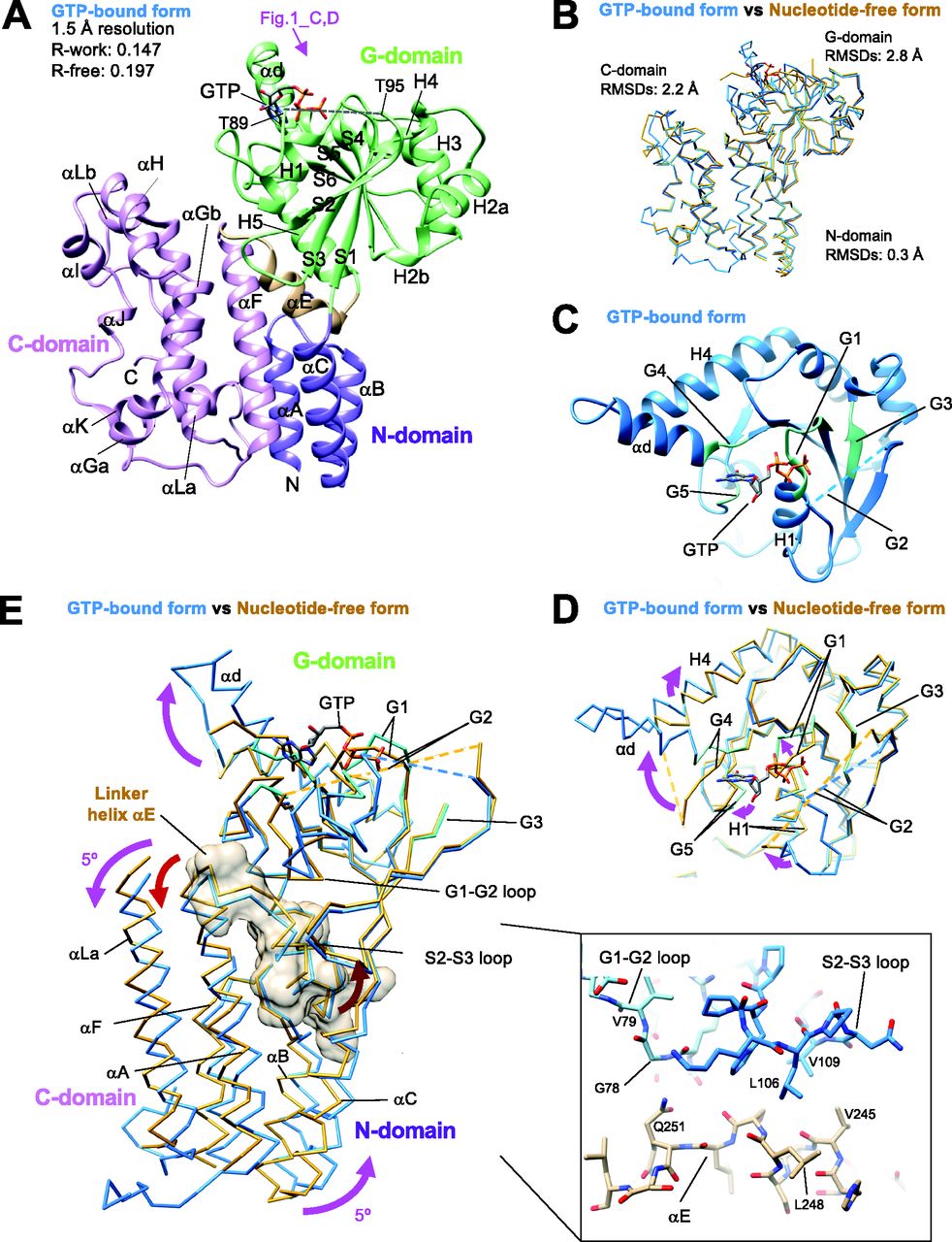
    Figure 1. Crystal structures of Irgb6 with GTP and without any nucleotide.

    (A) Crystal structure of Irgb6 with GTP. (B) Structural comparison between Irgb6 with GTP (blue) and without any nucleotide (yellow-brown), superimposed using all residues to minimize root-mean square deviations. (A, C) Structure around the nucleotide-binding pocket in G-domain observed from the top indicated by pink arrow in panel (A). (D) Conformational change of nucleotide-binding pocket during GTP binding. (E) Conformational change of N- and C-domains during GTP binding. Irgb6 with GTP (blue) and without any nucleotide (yellow-brown) was superimposed on their G-domains to illustrate the relay of conformational changes from the nucleotide-binding pocket. Whole N-domain, main components of G-domain around the nucleotide-binding pocket, linker helix αE, and helices αG and αLa of C-domain are shown. Linker helix αE is also shown with surface model. (Inset) Close-up view of the interactions between switches I-II and the helix αE.

  • Figure S2.
    • Download figure
    • Open in new tab
    • Download powerpoint
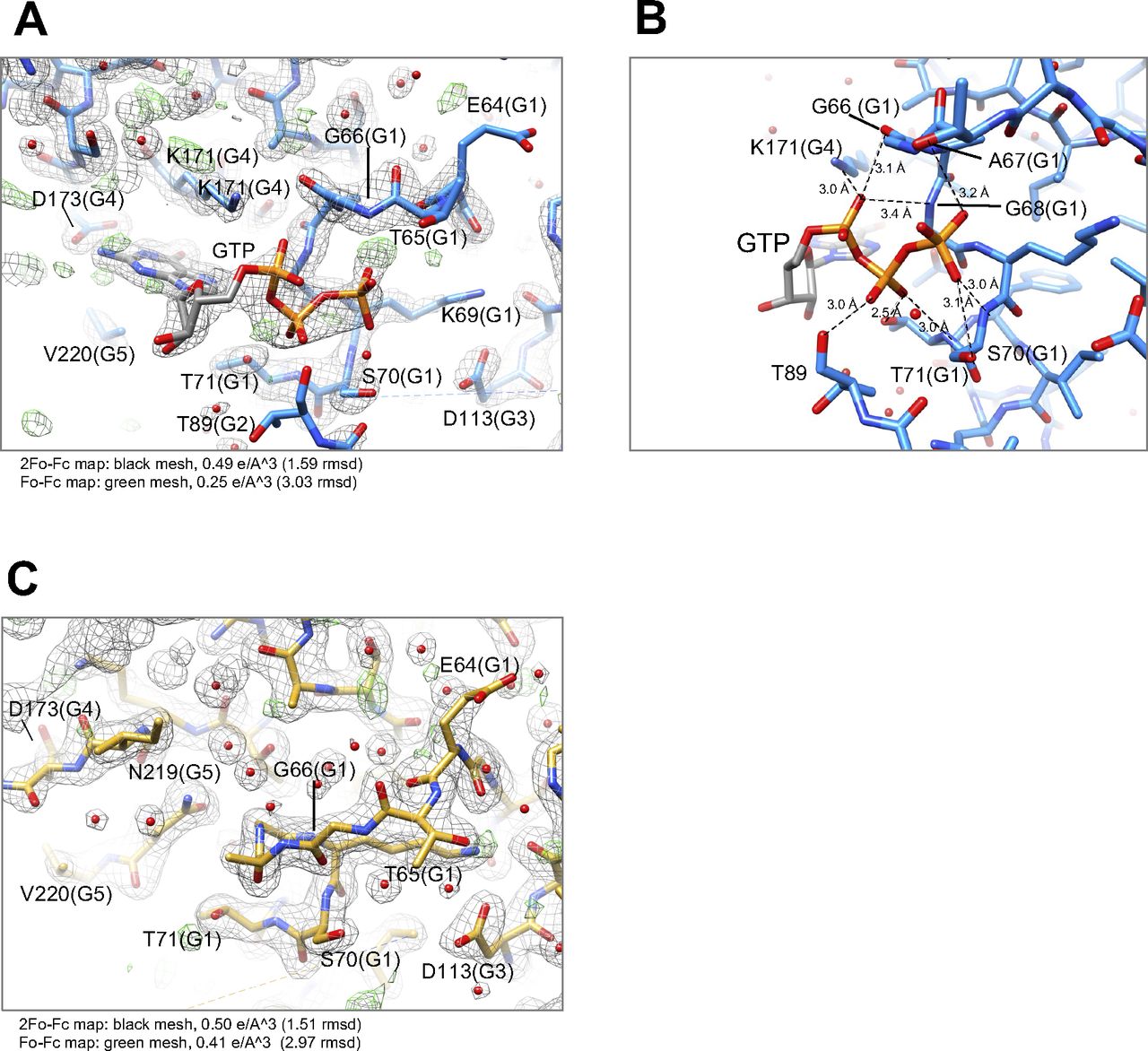
    Figure S2. Detailed structures of nucleotide-binding pocket, sequence alignment, and structural comparison among IRGs.

    (A) and (B) Nucleotide-binding pocket of Irgb6 with GTP. (C) Nucleotide-binding pocket of Irgb6 without any nucleotide.

  • Figure S3.
    • Download figure
    • Open in new tab
    • Download powerpoint
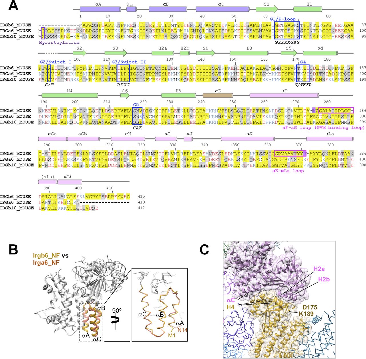
    Figure S3. Sequence alignment and structural comparison among IRGs.

    (A) Amino acid sequence alignment of three mouse IRGs: Irgb6, Irga6, and Irgb10. (B) Structural comparison of N-domains of Irgb6 and Irga6, both in the NF state. (C) Crystal packing environment of Irgb6 in the NF state. The helix H4 was stabilized by the interactions with the neighboring molecule.

  • Figure 2.
    • Download figure
    • Open in new tab
    • Download powerpoint
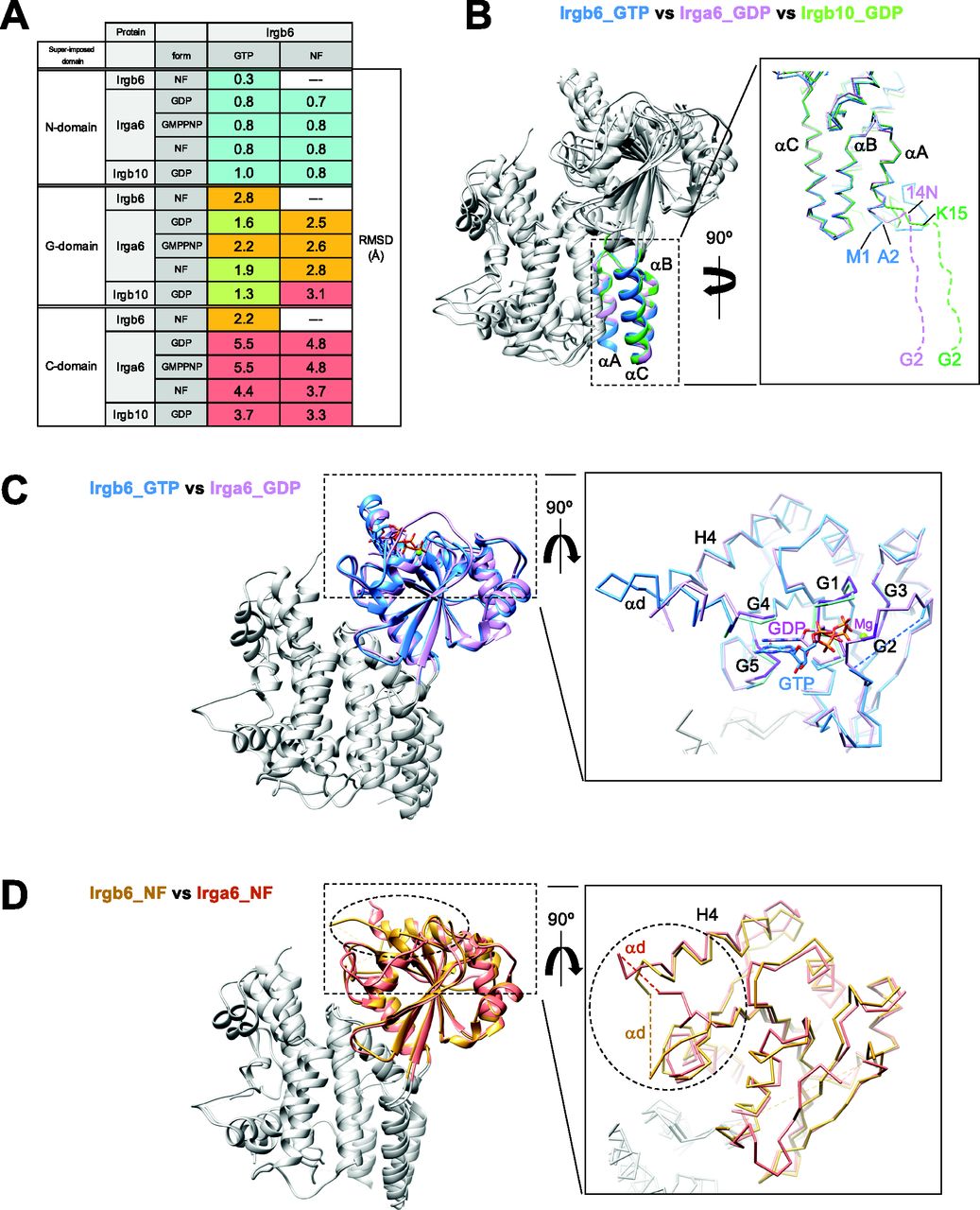
    Figure 2. Structural comparison among Irgb6, Irga6, and Irgb10.

    (A) Root-mean square deviations (RMSDs) among Irgb6, Irga6, and Irgb10, superimposed on their N-, G-, and C-domains. RMSDs≦1: cyan, 1 < RMSDs ≦ 2: yellow-green, 2 < RMSDs ≦ 3: orange, 3 < RMSDs: red. (B) Structural comparison among Irgb6 with GTP (blue), Irga6 with GDP (pink), and Irgb10 with GDP (green) superimposed on their N-domains. (C) Structural comparison between Irgb6 with GTP (blue) and Irga6 with GDP (pink), superimposed on their G-domains. (D) Structural comparison between Irgb6 without any nucleotide (yellow-brown) and Irga6 without any nucleotide (red), superimposed on their G-domains.

  • Figure 3.
    • Download figure
    • Open in new tab
    • Download powerpoint
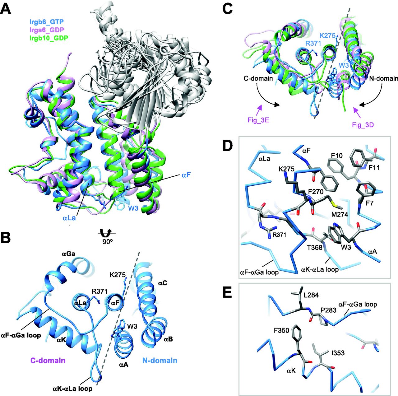
    Figure 3. Unique conformation of C-domain in Irgb6.

    (A) Structural comparison among Irgb6 with GTP (blue), Irga6 with GDP (pink), and Irgb10 with GDP (green) superimposed on their C-domains. (B) Bottom view of Irgb6. Broken line indicates the boundary between N- and C-domains. (C) Bottom view of panel (A) showing the conformational differences among Irgb6 with GTP (blue), Irga6 with GDP (pink), and Irgb10 with GDP (green). (D) Close-up view of the boundary indicates the interaction between the helices αA and αF. (E) Close-up view of the interaction between the αF-αGa loop and the helix αK.

  • Figure S4.
    • Download figure
    • Open in new tab
    • Download powerpoint
    Figure S4. Glide scores of Irgb6 with phospholipids.

    (A) Structures of PI5P, PS, PE, and PC. (B) Glide scores of wild-type Irgb6 protein docking with only phospholipids’ polar head. (C) Glide scores of Irgb6(W3A) mutant protein docking with only phospholipids’ polar head and with phospholipids’ polar head and glycerol backbone. (D) Glide scores of Irgb6(G277D/G285T/G285F) mutant protein docking with only phospholipids’ polar head and with phospholipids’ polar head and glycerol backbone. (E) Glide scores of wild-type Irgb6 protein docking with phospholipids’ polar head and glycerol backbone. (F) Glide scores of wild-type Irgb6 protein docking with phospholipids that have 4C and 16C acyl chains. (G) Glide scores of wild-type Irga6 protein docking with only phospholipids’ polar head. See Fig S4 H for detail. (H) Glide scores of wild-type Irga6 protein docking with only phospholipids’ polar head and with phospholipids’ polar head and glycerol backbone. (I) Glide scores of wild-type Irgb10 protein docking with only phospholipids’ polar head. See Fig S4 J for detail. (J) Glide scores of wild-type Irgb10 protein docking with only phospholipids’ polar head and with phospholipids’ polar head and glycerol backbone.

  • Figure 4.
    • Download figure
    • Open in new tab
    • Download powerpoint
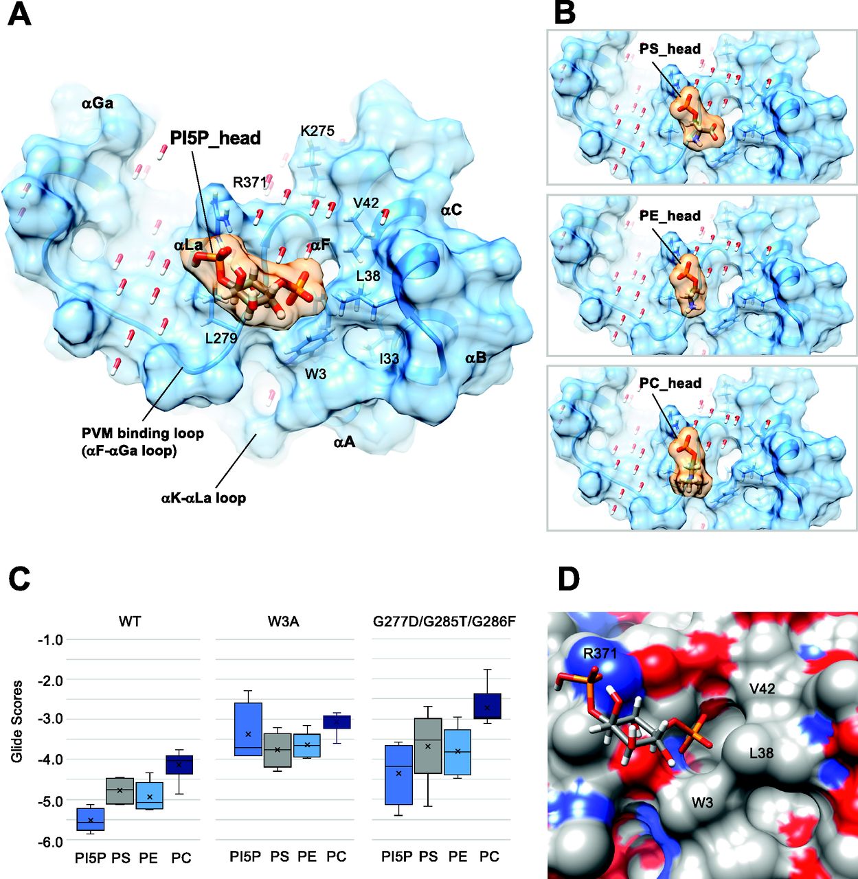
    Figure 4. Docking simulation of phospholipids to the Irgb6.

    (A) Docking of the polar head of PI5P to the GTP-bound Irgb6. (B) Docking of the polar head of PS, PE, and PC to the GTP-bound Irgb6. (C) Glide scores of Irgb6 (wild-type, W3A mutant, and G277D/G285T/G286F mutant) docking with phospholipid polar head groups. See Fig S4 for detail. (D) Surface presentation of the PI5P pocket colored by elements. Blue: nitrogen, red: oxygen, and gray: carbon. The left side of the pocket where the PI5P head docks is covered with the hydrophilic/ionic residues, whereas the right side is covered with the hydrophobic residues (Trp3, Leu38, and Val42).

  • Figure 5.
    • Download figure
    • Open in new tab
    • Download powerpoint
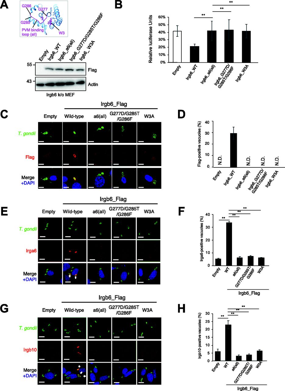
    Figure 5. The membrane-binding region is essential for Irgb6 accumulation on Toxoplasma gondii parasitophorous vacuole membrane.

    (A) Western blot image to detect stably expressed Irgb6 protein after retroviral transfection and puromycin selection. The mutation positions are indicated in the top panel. (B) T. gondii survival rate in the indicated Irgb6 reconstitution in Irgb6-KO MEFs with IFN-γ stimulation relative to those without IFN-γ treatment by luciferase analysis at 24 h post infection. All graphs show the mean ± SEM in three independent experiments. All images are representative of three independent experiments. N.D., not detected; **P < 0.01. T. gondii survival and Irgb6_Flag recruitment comparison between genotypes applied one-way ANOVA (Tukey’s multiple comparisons test). White arrows to indicate recruitment of effector on T. gondii PV. Scale bars on microscope images represent 10 μm. (C) Confocal microscope images to show the localization of Irgb6-Flag (red) to T. gondii PV (green), and DAPI (blue) at 4 h post infection in IFN-γ–treated Irgb6-KO MEFs reconstituted with indicated Irgb6. (D) Recruitment percentages of Irgb6_Flag. (E) Confocal microscope images to show the localization of Irga6 (red) to T. gondii PV (anti-GRA2; green), and DAPI (blue) at 4 h post infection in IFN-γ–treated Irgb6-KO MEFs reconstituted with indicated Irgb6. (F) Recruitment percentages of Irga6. (G) Confocal microscope images to show the localization of Irgb10 (red) to T. gondii PV (anti-GRA2; green), and DAPI (blue) at 4 h post infection in IFN-γ–treated Irgb6-KO MEFs reconstituted with indicated Irgb6. (H) Recruitment percentages of Irgb10. All graphs show the mean ± SEM in three independent experiments. All images are representative of three independent experiments. Recruitment percentages of indicated effectors calculated by counting almost 100 T. gondii PV in one experiment were shown as results of three independent experiments. N.D., not detected; **P < 0.01. T. gondii survival and Irgb6_Flag recruitment comparison between genotypes applied one-way ANOVA (Tukey’s multiple comparisons test). White arrows to indicate recruitment of effector on T. gondii PV. Scale bars on microscope images represent 10 μm.

  • Figure S5.
    • Download figure
    • Open in new tab
    • Download powerpoint
    Figure S5. Localization of Irgb6 mutants was similar to that of wild-type Irgb6.

    (A) Confocal microscope images to show the localization of Irgb6 (red) and KDEL (green), and DAPI (blue) in uninfected Irgb6-KO MEFs reconstituted with indicated Irgb6. All images are representative of three independent experiments. Scale bars on microscope images represent 10 μm. (B) Raw data of Fig 5D. Recruitment percentages of indicated effectors were calculated by counting almost 100 Toxoplasma gondii PV in one experiment. Experiments were repeated three times.

  • Figure 6.
    • Download figure
    • Open in new tab
    • Download powerpoint
    Figure 6. Structural model of parasitophorous vacuole membrane recognition during the GTP binding.

    (A) Structure of Irgb6 in the GTP-bound state. (A, B) Schematic model of conformational change of Irgb6 during GTP binding, shown with the same colors in the panel (A).

  • Figure S6.
    • Download figure
    • Open in new tab
    • Download powerpoint
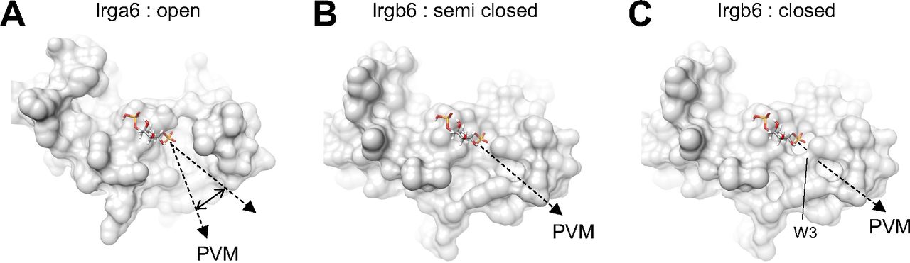
    Figure S6. Structures of parasitophorous vacuole membrane (PVM)-binding pocket.

    (A) PVM-binding site of Irga6 in the active GTP-form (PDB ID: 1TPZ) represents widely open pocket. (B) PVM-binding pocket of Irgb6 in the GTP-bound form (alternative conformation 1) represents semi closed form. (C) PVM-binding pocket of Irgb6 in the GTP-bound form (alternative conformation 2) represents closed form.

Supplementary Materials

  • Figures
  • Table S1 Data collection and refinement statistics.

PreviousNext
Back to top
Download PDF
Email Article

Thank you for your interest in spreading the word on Life Science Alliance.

NOTE: We only request your email address so that the person you are recommending the page to knows that you wanted them to see it, and that it is not junk mail. We do not capture any email address.

Enter multiple addresses on separate lines or separate them with commas.
Structural basis of membrane recognition of Toxoplasma gondii vacuole by Irgb6
(Your Name) has sent you a message from Life Science Alliance
(Your Name) thought you would like to see the Life Science Alliance web site.
CAPTCHA
This question is for testing whether or not you are a human visitor and to prevent automated spam submissions.
Citation Tools
Structural mechanism of PVM recognition by Irgb6
Yumiko Saijo-Hamano, Aalaa Alrahman Sherif, Ariel Pradipta, Miwa Sasai, Naoki Sakai, Yoshiaki Sakihama, Masahiro Yamamoto, Daron M Standley, Ryo Nitta
Life Science Alliance Nov 2021, 5 (1) e202101149; DOI: 10.26508/lsa.202101149

Citation Manager Formats

  • BibTeX
  • Bookends
  • EasyBib
  • EndNote (tagged)
  • EndNote 8 (xml)
  • Medlars
  • Mendeley
  • Papers
  • RefWorks Tagged
  • Ref Manager
  • RIS
  • Zotero
Share
Structural mechanism of PVM recognition by Irgb6
Yumiko Saijo-Hamano, Aalaa Alrahman Sherif, Ariel Pradipta, Miwa Sasai, Naoki Sakai, Yoshiaki Sakihama, Masahiro Yamamoto, Daron M Standley, Ryo Nitta
Life Science Alliance Nov 2021, 5 (1) e202101149; DOI: 10.26508/lsa.202101149
Reddit logo Twitter logo Facebook logo Mendeley logo
  • Tweet Widget
  • Facebook Like
Issue Cover

In this Issue

Volume 5, No. 1
January 2022
  • Table of Contents
  • Cover (PDF)
  • About the Cover
  • Masthead (PDF)
Advertisement

Jump to section

  • Article
    • Abstract
    • Introduction
    • Results
    • Discussion
    • Materials and Methods
    • Data Availability
    • Acknowledgements
    • References
  • Figures & Data
  • Info
  • Metrics
  • Reviewer Comments
  • PDF

Subjects

  • Microbiology, Virology & Host Pathogen Interaction
  • Structural Biology

Related Articles

  • No related articles found.

Cited By...

  • No citing articles found.
  • Google Scholar

More in this TOC Section

  • UPF3A does not repress NMD in murine cells
  • Bats experience age-related hearing loss
  • Regulation of C5aR1 and IL-1b expression
Show more Research Article

Similar Articles

EMBO Press LogoRockefeller University Press LogoCold Spring Harbor Logo

Content

  • Home
  • Newest Articles
  • Current Issue
  • Archive
  • Subject Collections

For Authors

  • Submit a Manuscript
  • Author Guidelines
  • License, copyright, Fee

Other Services

  • Alerts
  • Twitter
  • RSS Feeds

More Information

  • Editors & Staff
  • Reviewer Guidelines
  • Feedback
  • Licensing and Reuse
  • Privacy Policy

ISSN: 2575-1077
© 2023 Life Science Alliance LLC

Life Science Alliance is registered as a trademark in the U.S. Patent and Trade Mark Office and in the European Union Intellectual Property Office.