Advertisement

Main menu

  • Home
  • Articles
    • Newest Articles
    • Current Issue
    • Methods & Resources
    • Archive
    • Subjects
  • Collections
  • Submit
    • Submit a Manuscript
    • Author Guidelines
    • License, Copyright, Fee
    • FAQ
    • Why Submit
  • About
    • About Us
    • Editors & Staff
    • Board Members
    • Licensing and Reuse
    • Reviewer Guidelines
    • Privacy Policy
    • Advertise
    • Contact Us
    • LSA LLC
  • Alerts
  • Other Publications
    • EMBO Press
    • The EMBO Journal
    • EMBO reports
    • EMBO Molecular Medicine
    • Molecular Systems Biology
    • Rockefeller University Press
    • Journal of Cell Biology
    • Journal of Experimental Medicine
    • Journal of General Physiology
    • Cold Spring Harbor Laboratory Press
    • Genes & Development
    • Genome Research

User menu

  • My alerts

Search

  • Advanced search
Life Science Alliance
  • Other Publications
    • EMBO Press
    • The EMBO Journal
    • EMBO reports
    • EMBO Molecular Medicine
    • Molecular Systems Biology
    • Rockefeller University Press
    • Journal of Cell Biology
    • Journal of Experimental Medicine
    • Journal of General Physiology
    • Cold Spring Harbor Laboratory Press
    • Genes & Development
    • Genome Research
  • My alerts
Life Science Alliance

Advanced Search

  • Home
  • Articles
    • Newest Articles
    • Current Issue
    • Methods & Resources
    • Archive
    • Subjects
  • Collections
  • Submit
    • Submit a Manuscript
    • Author Guidelines
    • License, Copyright, Fee
    • FAQ
    • Why Submit
  • About
    • About Us
    • Editors & Staff
    • Board Members
    • Licensing and Reuse
    • Reviewer Guidelines
    • Privacy Policy
    • Advertise
    • Contact Us
    • LSA LLC
  • Alerts
  • Follow lsa Template on Twitter
Research Article
Transparent Process
Open Access

Heterogeneous nuclear ribonucleoprotein E1 binds polycytosine DNA and monitors genome integrity

Bidyut K Mohanty, View ORCID ProfileJoseph AQ Karam, Breege V Howley, Annamarie C Dalton, Simon Grelet, View ORCID ProfileToros Dincman, View ORCID ProfileWilliam S Streitfeld, Je-Hyun Yoon, Lata Balakrishnan, View ORCID ProfileWalter J Chazin, View ORCID ProfileDavid T Long, View ORCID ProfilePhilip H Howe  Correspondence email
Bidyut K Mohanty
1Department of Biochemistry and Molecular Biology, Medical University of South Carolina, Charleston, SC, USA
Roles: Conceptualization, Formal analysis, Investigation, Methodology, Writing—original draft
  • Find this author on Google Scholar
  • Find this author on PubMed
  • Search for this author on this site
Joseph AQ Karam
1Department of Biochemistry and Molecular Biology, Medical University of South Carolina, Charleston, SC, USA
Roles: Conceptualization, Formal analysis, Validation, Investigation, Methodology, Writing—review and editing
  • Find this author on Google Scholar
  • Find this author on PubMed
  • Search for this author on this site
  • ORCID record for Joseph AQ Karam
Breege V Howley
1Department of Biochemistry and Molecular Biology, Medical University of South Carolina, Charleston, SC, USA
Roles: Conceptualization, Investigation, Writing—review and editing
  • Find this author on Google Scholar
  • Find this author on PubMed
  • Search for this author on this site
Annamarie C Dalton
1Department of Biochemistry and Molecular Biology, Medical University of South Carolina, Charleston, SC, USA
Roles: Investigation, Writing—review and editing
  • Find this author on Google Scholar
  • Find this author on PubMed
  • Search for this author on this site
Simon Grelet
1Department of Biochemistry and Molecular Biology, Medical University of South Carolina, Charleston, SC, USA
2Hollings Cancer Center, Medical University of South Carolina, Charleston, SC, USA
Roles: Formal analysis, Investigation, Methodology
  • Find this author on Google Scholar
  • Find this author on PubMed
  • Search for this author on this site
Toros Dincman
3Division of Hematology and Oncology, Department of Medicine, Medical University of South Carolina, Charleston, SC, USA
Roles: Investigation
  • Find this author on Google Scholar
  • Find this author on PubMed
  • Search for this author on this site
  • ORCID record for Toros Dincman
William S Streitfeld
1Department of Biochemistry and Molecular Biology, Medical University of South Carolina, Charleston, SC, USA
Roles: Investigation
  • Find this author on Google Scholar
  • Find this author on PubMed
  • Search for this author on this site
  • ORCID record for William S Streitfeld
Je-Hyun Yoon
1Department of Biochemistry and Molecular Biology, Medical University of South Carolina, Charleston, SC, USA
Roles: Investigation
  • Find this author on Google Scholar
  • Find this author on PubMed
  • Search for this author on this site
Lata Balakrishnan
4Department of Biology, School of Science, Indiana University Purdue University Indianapolis, Indianapolis, IN, USA
Roles: Resources
  • Find this author on Google Scholar
  • Find this author on PubMed
  • Search for this author on this site
Walter J Chazin
5Departments of Biochemistry and Chemistry and Center for Structural Biology, Vanderbilt University, Nashville, TN, USA
Roles: Resources
  • Find this author on Google Scholar
  • Find this author on PubMed
  • Search for this author on this site
  • ORCID record for Walter J Chazin
David T Long
1Department of Biochemistry and Molecular Biology, Medical University of South Carolina, Charleston, SC, USA
Roles: Resources, Writing—original draft, review, and editing
  • Find this author on Google Scholar
  • Find this author on PubMed
  • Search for this author on this site
  • ORCID record for David T Long
Philip H Howe
1Department of Biochemistry and Molecular Biology, Medical University of South Carolina, Charleston, SC, USA
2Hollings Cancer Center, Medical University of South Carolina, Charleston, SC, USA
Roles: Conceptualization, Resources, Supervision, Funding acquisition, Writing—review and editing
  • Find this author on Google Scholar
  • Find this author on PubMed
  • Search for this author on this site
  • ORCID record for Philip H Howe
  • For correspondence: howep@musc.edu
Published 16 July 2021. DOI: 10.26508/lsa.202000995
  • Article
  • Figures & Data
  • Info
  • Metrics
  • Reviewer Comments
  • PDF
Loading

Article Figures & Data

Figures

  • Supplementary Materials
  • Figure 1.
    • Download figure
    • Open in new tab
    • Download powerpoint
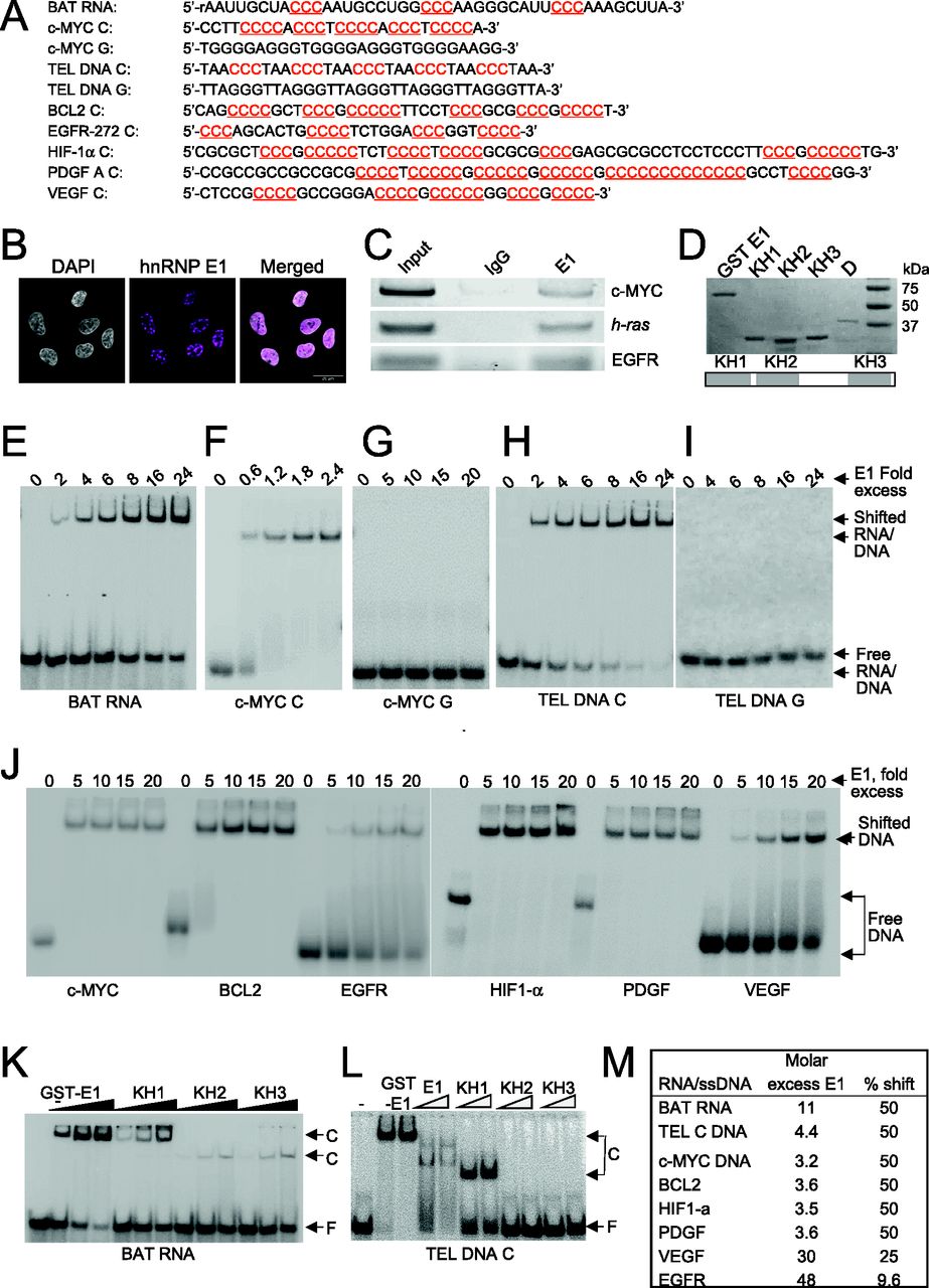
    Figure 1. hnRNP E1 binds polycytosine-rich DNA tracts.

    (A) Oligonucleotides and deoxyoligonucleotides used in RNA electrophoretic mobility shift assay (REMSA)/electrophoretic mobility shift assay (EMSA). (B) Immunofluorescence of hnRNP E1 and DAPI staining showing localization of hnRNP E1. (C) ChIP-PCR products from A549 DNA showing enrichment of hnRNP E1 at promoter regions of c-MYC, h-Ras, and EGFR genes. (D) hnRNP E1 protein and its KH domains. (D) Top. SDS–PAGE gel showing GST fusion proteins of hnRNP E1; KH1, KH2, and KH3; and hnRNP D. Bottom. The three KH domains of hnRNP E1. (E, F, G, H, I, J, K, L) Autoradiograms of representative REMSA/EMSA assays. (E) REMSA showing hnRNP E1 binding to BAT RNA. (F, G, H, I, J) EMSA showing hnRNP E1 binding to c-MYC C (F), c-MYC G (G), TEL DNA C (H), TEL DNA G (I), and c-MYC C, BCL2, EGFR, HIF1-α, PDGF, and VEGF (J). (K) EMSA showing binding of hnRNP E1, KH1, KH2, and KH3 to BAT RNA. (L) EMSA showing binding of hnRNP E1, KH1, KH2, and KH3 to TEL DNA C. (M) Quantification of REMSA and EMSA assay.

  • Figure 2.
    • Download figure
    • Open in new tab
    • Download powerpoint
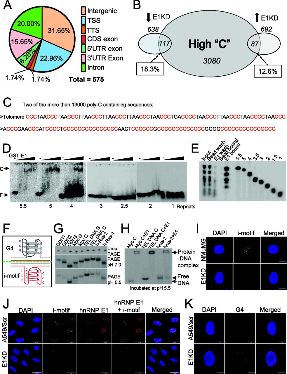
    Figure 2. Genome-wide DNA binding by hnRNP E1 and its cellular function.

    (A) Per cent of regions in the mouse (NMuMG) genome with at least 50% of “C” nucleotide. (B) Per cent (and number) of promoter proximal regions containing high-C sequences. (C) Two poly-C representative sequences from Table S1. (D) Autoradiographs of electrophoretic mobility shift assay showing differential binding of telomeric oligodeoxynucleotides containing one to five repeats of “CCCTAA” to GST-hnRNP E1 protein. (E) PAGE gel showing binding of telomeric oligodeoxynucleotides containing one to five repeats of “CCCTAA” upon loading on GST-hnRNP E1 immobilized on agarose beads. (F) Model showing i-motif and G4 structures. (G) Autoradiograph of urea-PAGE and PAGE (pH 7.5 and 5.5) gels showing fractionation pattern of various oligonucleotides. (H) Autoradiograph of a PAGE gel (run at pH 7.5) showing binding of hnRNP E1 to c-MYC, TEL DNA C and hRas2 deoxynucleotides incubated at pH 5.5. (I) Immunofluorescence of NMuMG control and E1KD cells showing i-motif foci. (J) Immunofluorescence of A549 control and E1KD showing i-motif foci, hnRNP E1 foci, and their colocalization. (K) Immunofluorescence of A549 control and E1KD cells showing G4 foci.

  • Figure S1.
    • Download figure
    • Open in new tab
    • Download powerpoint
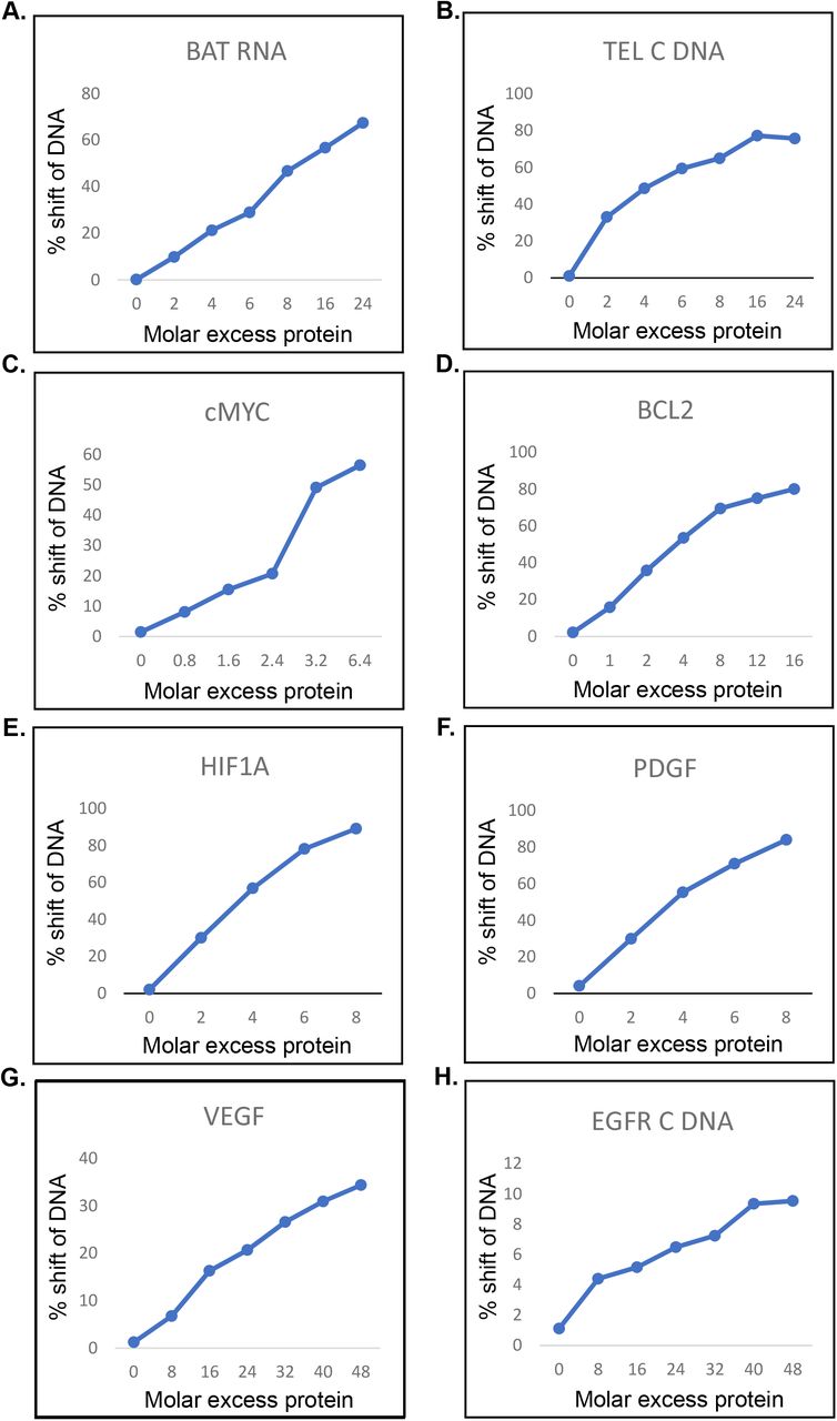
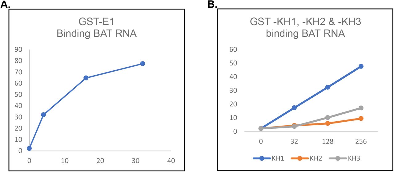
    Figure S1. Representative graphs showing quantification of hnRNP E1 binding to BAT RNA and poly-C–rich DNA tracts.

    (A) BAT RNA. (B) TEL DNA C. (C) c-MYC. (D) BCL2. (E) HIF1-a. (F) PDGF. (G) VEGF. (H) EGFR.

  • Figure S2.
    • Download figure
    • Open in new tab
    • Download powerpoint
    Figure S2. Representative graphs showing quantification of binding by hnRNP E1, KH1, KH2 and KH3 to BAT RNA.

    (A) Binding of hnRNP E1 to BAT RNA. (B) Binding of KH1, KH2, and KH3 to BAT RNA.

  • Figure S3.
    • Download figure
    • Open in new tab
    • Download powerpoint
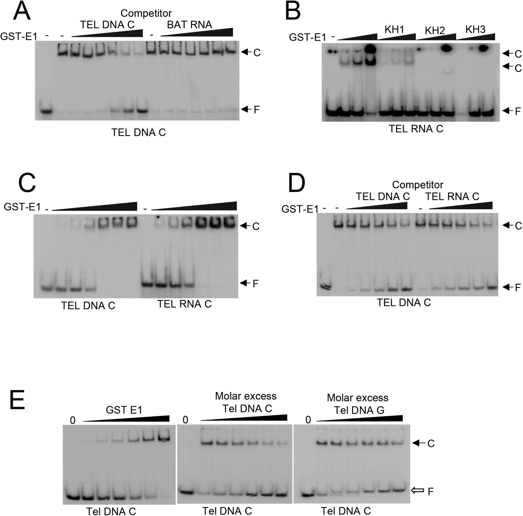
    Figure S3. Autoradiographs showing protein–nucleic acid interactions.

    (A) Competition of TEL DNA C and BAT RNA with TEL DNA C for binding to hnRNP E1. (B) Binding of hnRNP E1, KH1, KH2, and KH3 to TEL RNA C. (C) Electrophoretic mobility shift assay showing hnRNP E1 binding to TEL DNA C and TEL RNA C. (D) Competition of TEL DNA C and TEL RNA C with TEL DNA C for binding to hnRNP E1. (E) Binding of hnRNP E1 to TEL DNA C, and competition of TEL DNA C and TEL DNA G with TEL DNA C for binding to hnRNP E1.

  • Figure S4.
    • Download figure
    • Open in new tab
    • Download powerpoint
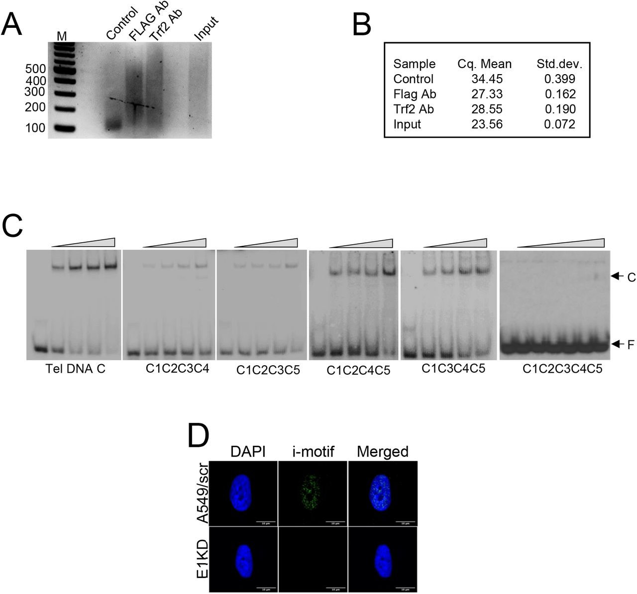
    Figure S4. Binding of hnRNP E1 to telomeric DNA.

    (A, B) Agarose gel showing fractionation of ChIP-PCR products generated by telomere primers (tel1 and tel2) from mouse cell line NMuMG. M, DNA molecular weight markers; Control: no antibody control, FLAG: FLAG antibody used to pull-down FLAG-tagged hnRNP E1, Trf2:Trf2 antibody used to pull-down Trf2. (C) Autoradiogram of electrophoretic mobility shift assay showing hnRNP E1 binding to mutants of TEL DNA C that contain mutations in four or five first “C”s of CCC repeats present in a 33 base deoxyoligonucleotide with five repeats of “CCCTAA.” (D) Immunofluorescence showing i-motifs in a single nucleus of A549 and E1KD cells. Fig 2J shows multiple nuclei.

  • Figure 3.
    • Download figure
    • Open in new tab
    • Download powerpoint
    Figure 3. Role of hnRNP E1 in genome integrity.

    (A, B) Immunofluorescence showing γ-H2AX activation in E1KD cells. (C) Western blot showing γ-H2AX activation in E1KD cells. (D) Immunofluorescence showing increase in and colocalization of γ-H2AX and G4 foci in E1KD cells. (E) ChIP-qPCR analysis showing γ-H2AX localization at c-MYC promoter of scrambled (Ascr) and hnRNP E1 knockdown (E1KD) cells.

  • Figure 4.
    • Download figure
    • Open in new tab
    • Download powerpoint
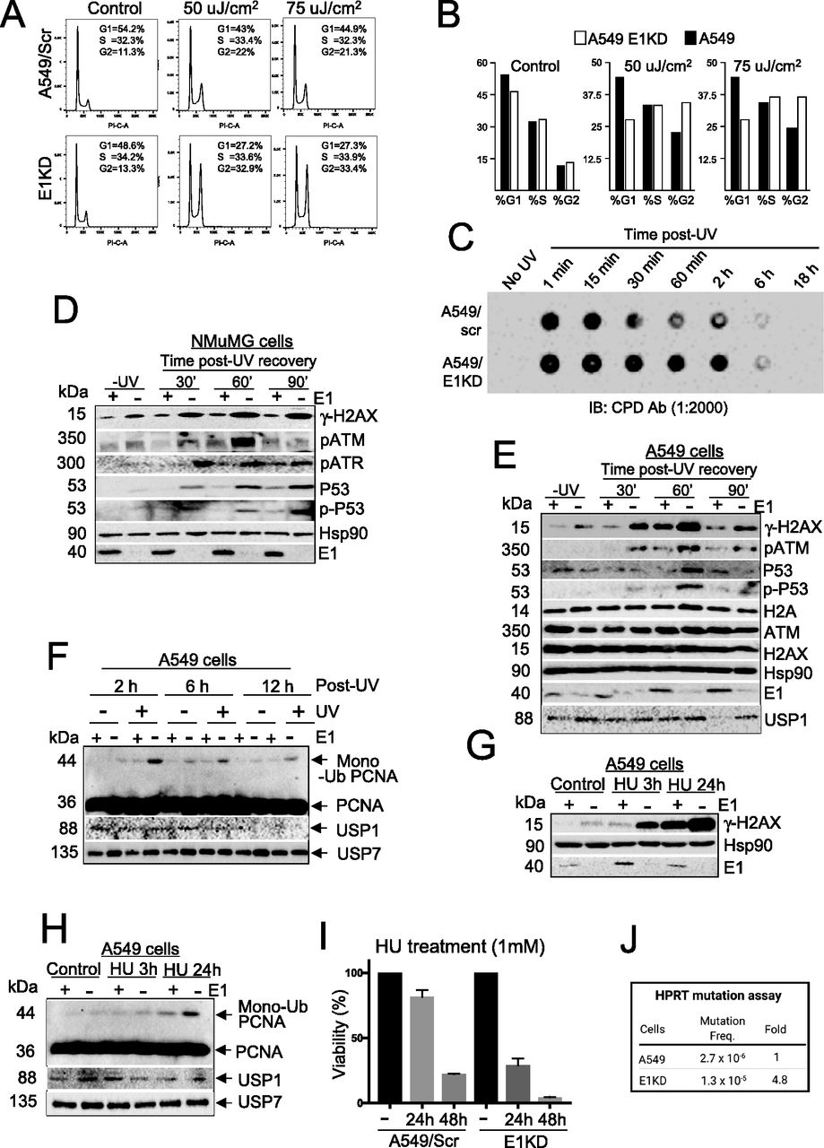
    Figure 4. E1KD phenotype acts synergistically with HU/UV and promotes mutation.

    (A, B) Cell cycle distribution of A549 control and E1KD cells exposed to UV. (C) Blot showing CPD incorporation in wild-type and E1KD cells. (D, E) Western blots showing effects of UV on checkpoint proteins in A549 control and E1KD (D) cells, and checkpoint proteins in NMuMG control and E1KD cells (E). (F) Western blot showing proliferating cell nuclear antigen monoubiquitination, Usp1, and Usp7 in A549 control and E1KD cells recovering from UV exposure (h, hours). (G, H) Western blots showing γ-H2AX activation (G) and proliferating cell nuclear antigen monoubiquitination along with Usp1 and Usp7 (H) in A549 control and E1KD cells treated with HU. (I) Histogram of clonogenic assay with A549 control and E1KD cells exposed to HU. (J) Mutation frequency in A549 control and E1KD cells.

  • Figure 5.
    • Download figure
    • Open in new tab
    • Download powerpoint
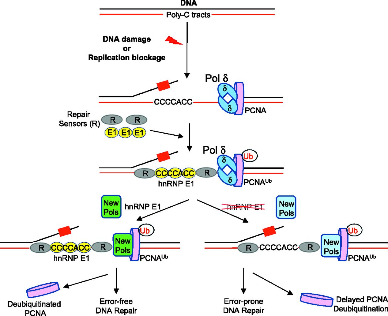
    Figure 5. Model showing the role of hnRNP E1 in genome integrity.

    Upon DNA damage, hnRNP E1 bound at polycytosine tracts activates damage signaling and repair with the loading and assistance of RPA (R). Proliferating cell nuclear antigen (PCNA) is monoubiquitinated, replaces DNA polymerase δ with TLS polymerases, which are often error-free, and finally PCNA is deubiquitinated. When DNA damage occurs in the absence of hnRNP E1, only RPA functions at polycytosine tracts albeit slowly, PCNA monoubiquitination is enhanced and lingers, error-prone TLS polymerases replace DNA polymerase δ, and signaling and repair are attenuated.

Supplementary Materials

  • Figures
  • Table S1 NMuMG sequences of 50–75 nucleotides with at least 50% “C,” which includes sequences with “C” clusters and potential hnRNP E1 binding sequences.

  • Table S2 List of (deoxyribo and oxyribo) oligonucleotides used in the experiments.

PreviousNext
Back to top
Download PDF
Article Alerts
Sign In to Email Alerts with your Email Address
Email Article

Thank you for your interest in spreading the word on Life Science Alliance.

NOTE: We only request your email address so that the person you are recommending the page to knows that you wanted them to see it, and that it is not junk mail. We do not capture any email address.

Enter multiple addresses on separate lines or separate them with commas.
Heterogeneous nuclear ribonucleoprotein E1 binds polycytosine DNA and monitors genome integrity
(Your Name) has sent you a message from Life Science Alliance
(Your Name) thought you would like to see the Life Science Alliance web site.
CAPTCHA
This question is for testing whether or not you are a human visitor and to prevent automated spam submissions.
Citation Tools
hnRNP E1 binds polycytosine DNA tracts
Bidyut K Mohanty, Joseph AQ Karam, Breege V Howley, Annamarie C Dalton, Simon Grelet, Toros Dincman, William S Streitfeld, Je-Hyun Yoon, Lata Balakrishnan, Walter J Chazin, David T Long, Philip H Howe
Life Science Alliance Jul 2021, 4 (9) e202000995; DOI: 10.26508/lsa.202000995

Citation Manager Formats

  • BibTeX
  • Bookends
  • EasyBib
  • EndNote (tagged)
  • EndNote 8 (xml)
  • Medlars
  • Mendeley
  • Papers
  • RefWorks Tagged
  • Ref Manager
  • RIS
  • Zotero
Share
hnRNP E1 binds polycytosine DNA tracts
Bidyut K Mohanty, Joseph AQ Karam, Breege V Howley, Annamarie C Dalton, Simon Grelet, Toros Dincman, William S Streitfeld, Je-Hyun Yoon, Lata Balakrishnan, Walter J Chazin, David T Long, Philip H Howe
Life Science Alliance Jul 2021, 4 (9) e202000995; DOI: 10.26508/lsa.202000995
del.icio.us logo Digg logo Reddit logo Twitter logo CiteULike logo Facebook logo Google logo Mendeley logo
  • Tweet Widget
  • Facebook Like
Issue Cover

In this Issue

Volume 4, No. 9
September 2021
  • Table of Contents
  • Cover (PDF)
  • About the Cover
  • Masthead (PDF)
Advertisement

Jump to section

  • Article
    • Abstract
    • Introduction
    • Results
    • Discussion
    • Materials and Methods
    • Acknowledgements
    • References
  • Figures & Data
  • Info
  • Metrics
  • Reviewer Comments
  • PDF

Subjects

  • Molecular Biology

Related Articles

  • No related articles found.

Cited By...

  • No citing articles found.
  • Google Scholar

More in this TOC Section

  • Neutrophils delay repair in Wallerian degeneration
  • Vacuoles drive nuclear and rDNA morphology
  • Cdh5-lineage–independent origin of dermal lymphatics
Show more Research Article

Similar Articles

EMBO Press LogoRockefeller University Press LogoCold Spring Harbor Logo

Content

  • Home
  • Newest Articles
  • Current Issue
  • Archive
  • Subject Collections

For Authors

  • Submit a Manuscript
  • Author Guidelines
  • License, copyright, Fee

Other Services

  • Alerts
  • Twitter
  • RSS Feeds

More Information

  • Editors & Staff
  • Reviewer Guidelines
  • Feedback
  • Licensing and Reuse
  • Privacy Policy

ISSN: 2575-1077
© 2022 Life Science Alliance LLC

Life Science Alliance is registered as a trademark in the U.S. Patent and Trade Mark Office and in the European Union Intellectual Property Office.