


Article Figures & Data
Figures
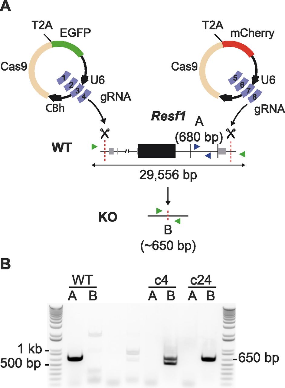
- Figure 1. Deletion of Resf1 in embryonic stem cells (ESCs).
(A) Scheme of the deletion strategy used at the Resf1 locus. The line diagram shows Resf1, with lines representing introns and non-transcribed regions, thick black boxes represent coding regions of exons and thin grey boxes represent UTRs. Resf1 was deleted by targeting Cas9 via sets of gRNAs complementary to sites (red dotted lines) lying upstream of the transcription start site and downstream of the polyadenylation signal of the Resf1 gene. (A, B) Deletion of Resf1 was assessed by PCR using primer pairs nested within intron IV (A, blue triangles) or flanking the targeted sites (B, green triangles). To delete Resf1, wild-type (WT) ESCs were transfected with four CBh-eSpCas9-T2a-EGFP and four CBh-eSpCas9-T2a-mCherry plasmids carrying eight distinct gRNAs. (B) Electrophoresis of PCRs from WT ESCs and putative Resf1−/− ESC clones (c4 and c24) using primers A or B.
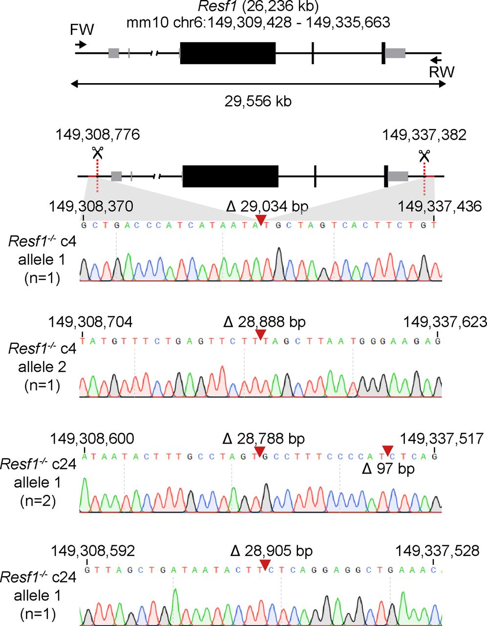
- Figure S1. Sequence analysis of putative Resf1−/− ES cell lines.
Cartoon of the Resf1 locus. Resf1 size and genome coordinates (mm10 genome assembly) are shown at the top. Thick black boxes represent coding exons, thin grey boxes represent non-coding exons, lines represent introns and non-transcribed regions. Forward (FW) and reverse (RV) primers used to analyse deletions of Resf1 are indicated as arrows. The distance between FW and RV primers is shown by the double arrow. Sites 5′ of Resf1 start codon and 3′ from Resf1 polyadenylation signal targeted by CRISPR/Cas9 are shown as red dotted lines. Deletion of Resf1 was validated in putative Resf1−/− clones 4 (c4) and 24 (c24) by PCR using FW and RV primers. Sequence tracks of the amplified region in individual alleles of Resf1−/− c4 and c24 cell lines. Complementary coordinates of the first and the last shown residue are shown. Red arrows indicate deletion events with indicated size of the deletion.
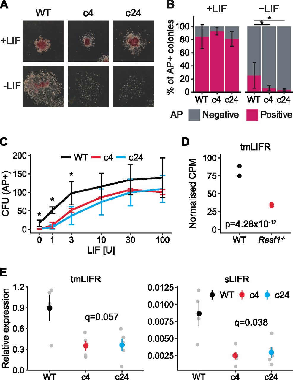
- Figure 2. Deletion of Resf1 reduces embryonic stem cell (ESC) self-renewal by decreasing expression of LIFR.
(A, B, C) Clonal ESC self-renewal assays. (A) Representative images of the colonies formed by the indicated ESCs in the presence or absence of leukemia inhibitory factor (LIF). Colonies were stained for AP 6 d after plating. (B) Proportion of AP+ colonies formed by WT, Resf1−/− c4, and Resf1−/− c24 ESCs in the presence or absence of LIF. Bars represent mean ± SD (n = 5; *q < 0.05; Wilcoxon rank-sum test). (C) Number of AP+ CFU generated by WT or Resf1−/− ESCs at different LIF concentrations (U/ml); mean ± SD (n = 4). (D) CPM for ENSMUST00000171588 transmembrane LIF receptor (tmLIFR) transcript in wild-type and Resf1−/− ESCs; n = 2 RNA-seq data from Fukuda et al (2018). Adjusted P-value calculated by DESeq is shown, (E) Quantification of tmLIFR and soluble LIFR (sLIFR) transcript levels in wild-type, Resf1−/− c4, and Resf1−/− c24 ESCs by quantitative PCR on reverse-transcribed RNA. Grey points show individual data points. Coloured point ranges represent mean ± SE; n = 4 for wild-type cells, n = 5 for Resf1−/− cells. FDR corrected P-values are shown (two-tailed t test).
- Figure S2. Self-renewal of Resf1−/− embryonic stem cells (ESCs) at low leukemia inhibitory factor (LIF) concentrations and capacity of NANOG and ESRRB to confer LIF-independent self-renewal in Resf1−/− ESCs.
(A) Photography of colonies formed by wild-type (WT) and Resf1−/− clonal cell lines (c4 and c24) after 6-d culture in serum medium supplemented with no (0) or 1 U/ml LIF. Colonies were stained for AP. (B) Images of colonies stained for AP of WT or Resf1−/− ESCs stably expressing dsRed, Flag-Nanog, or Flag-Esrrb transgenes cultured in the presence (+) or absence (−) of LIF (100 U/ml) for 8 d.
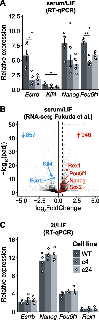
- Figure 3. Effect of Resf1 deletion on the expression of pluripotency markers in naïve pluripotent stem cells.
(A) mRNA levels of the indicated transcripts were determined in wild-type (WT) and Resf1−/− embryonic stem cells (c4 and c24) cultured in serum/leukemia inhibitory factor (LIF) medium (n = 5). (B) Volcano plot comparing transcriptomes of WT and Resf1−/− embryonic stem cells (Fukuda et al, 2018). Dashed lines represent significance thresholds. Number of significantly down-regulated (blue) and up-regulated (red) genes are shown. Selected pluripotency transcription factors are highlighted. (C) As (A) but cells were maintained in 2i/LIF medium. Bars and whiskers represent mean ± SEM (n = 4). Scatter plots represent individual data points. *q < 0.05 (two-tailed t test).
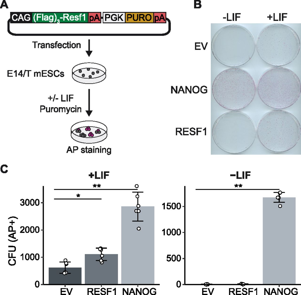
- Figure 4. Effect of episomal expression of Resf1 on embryonic stem cell (ESC) self-renewal.
(A) Strategy to assess the effect of episomal expression of RESF1 on ESC self-renewal. E14/T ESCs were transfected with the plasmid shown and cultured in the presence of puromycin in medium with or without leukemia inhibitory factor (LIF). (B) After 8 d, colonies were stained for AP. Empty vector (EV) and a plasmid encoding Nanog in place of Resf1 provided controls. (C) Quantification of colony numbers from (B). Bars represent mean ± SD (n = 6) and scatter plots individual data points; **q < 0.01, *q < 0.05 (Wilcoxon rank-sum test).
- Figure 5. Resf1 is dispensable for NANOG and ESRRB function to sustain leukemia inhibitory factor-independent self-renewal.
(A) Co-immunoprecipitation of Flag-RESF1 and HA-NANOG from nuclear extracts of embryonic stem cells (ESCs) episomally expressing HA-NANOG alone (−) or HA-NANOG plus Flag-RESF1 (+). (B) Schematic representation of wild-type (WT) or Resf1−/− ESCs with stably integrated transgenes in which the puromycin resistance gene (PURO) is linked in the same transcript to either dsRed, Flag-Nanog or Flag-Esrrb. Black boxes represent coding exons, grey boxes represent non-coding exons. (C) Immunoblot analysis of Flag expression after stable integration of dsRed, Flag-Nanog, or Flag-Esrrb expression cassettes in WT and Resf1−/− ESCs (c4, c24); anti-LAMIN was used as a loading control. Relative Flag signal over LAMIN control is shown below. (B, D) Quantification of alkaline positive (AP+) CFUs formed by cells described in (B) after 8-d culture in the presence or absence of leukemia inhibitory factor. Bars represent mean ± SD (n = 3).
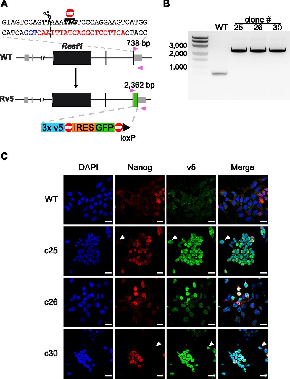
- Figure 6. Epitope tagging of the endogenous Resf1 gene.
(A) Cartoon representation of Resf1 in wild-type (WT) and Resf1-tagged (Rv5) cells. Lines represent introns and non-transcribed regions; thick boxes represent coding exons; thin grey boxes represent non-coding exons. Resf1 was tagged by inserting 3× v5-tag epitopes, stop codon, internal ribosome entry site, GFP, stop codon, and a loxP site in front of the Resf1 stop codon using CRISPR/Cas9. The complementary sequence of the gRNA (red) with the PAM sequence (blue) is shown. The expected cleavage site is indicated by a dotted line close to the Resf1 stop codon. Genotyping primers (pink triangles) and expected PCR product sizes are shown. (B) Genotyping of WT and Rv5 clones using primers flanking the insertion site. (C) Immunostaining of WT and Rv5 clones for RESF1-v5 and NANOG. White arrows indicate cells expressing v5 but no NANOG. White scale bars represent 25 μm.
- Figure S3. Resf1 expression in epiblast and primordial germ cells (PGCs).
(A) RT-qPCR analysis of Resf1 mRNA levels normalised to TBP mRNA in E14Tg2a embryonic stem cells cultured in 2i/leukemia inhibitory factor or serum/leukemia inhibitory factor medium, epiblast-like cells (EpiLCs), epiblast stem cells (EpiSCs), and day 4 PGC-like cells (PGCLCs D4). Individual data points are shown as well as the median expression levels (red bar). (B, C) Resf1 expression in single cell RNA sequencing dataset of mouse embryo at the indicated embryonic (E) stages. (B) Summary of Resf1 mRNA expression in epiblast and PGCs between embryonic stages E6.5 and E8.5. The proportion of single cells expressing Resf1 at individual stages is shown. (B, C) Boxplots showing Resf1 mRNA expression levels in PGCs and epiblast cells from (B). Box represents interquartile range; horizontal lines are medians and points are outliers.
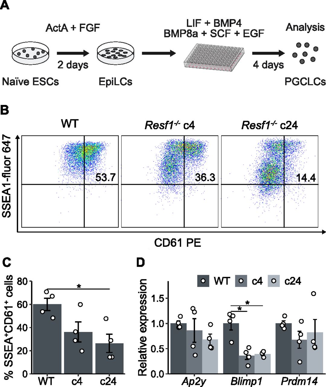
- Figure 7. Effect of Resf1 deletion on PGCLC differentiation.
(A) Scheme of differentiation of naïve embryonic stem cells into primordial germ cell-like cells (PGCLCs). Embryonic stem cells (ESCs) are treated with Activin A and Fgf2 for 2 d to form EpiLCs. EpiLCs are then aggregated in the presence of the indicated cytokines. (B) Representative scatter plots of SSEA1 and CD61 expression measured by flow cytometry after 4 d of PGCLC differentiation using the indicated cell lines. Numbers represent percentage of CD61+SSEA1+ population. (B, C) Quantification of CD61+SSEA1+ cell populations shown in (B) (n = 4). (D) Relative expression of the indicated primordial germ cell markers in WT or Resf1−/− cells after 4 d of PGCLC differentiation (n = 4). Bars represent mean ± SEM. Scatter plots represent individual data points. *q < 0.05 (two-tailed t test).
- Figure S4. Expression of EpiLC markers in Resf1−/− EpiLCs.
(A) Photographs of wild-type, Resf1−/− c4, and Resf1−/− c24 EpiLCs. White scale bars represent 100 μm. (B) mRNA levels of the indicated transcripts were measured in wild-type (WT) and Resf1−/− EpiLCs (c4 and c24) by RT-qPCR. Bars and whiskers represent mean ± SD (n = 3). Scatter plots represent individual data points.
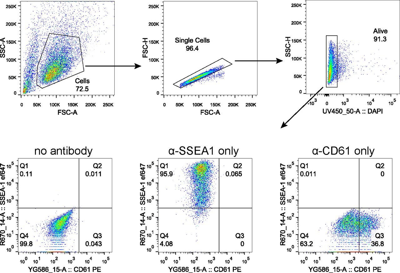
- Figure S5. Gating strategy for quantification of PGCLC population.
Representative scatter plots indicating gating strategy to quantify PGCLC population marked by co-expression of SSEA1 and CD61. Gating for live cells was performed using DAPI. Samples without any antibody staining and samples stained with a single antibody were used to gate SSEA1+CD61+ population as shown.


Supplementary Materials
Table S1. List of used primers and antibodies. [LSA-2021-01190_TableS1.xlsx]


In this Issue
Subjects
Related Articles
Cited By...
- No citing articles found.