Advertisement

Main menu

  • Home
  • Articles
    • Newest Articles
    • Current Issue
    • Methods & Resources
    • Archive
    • Subjects
  • Collections
  • Submit
    • Submit a Manuscript
    • Author Guidelines
    • License, Copyright, Fee
    • FAQ
    • Why Submit
  • About
    • About Us
    • Editors & Staff
    • Board Members
    • Licensing and Reuse
    • Reviewer Guidelines
    • Privacy Policy
    • Advertise
    • Contact Us
    • LSA LLC
  • Alerts
  • Other Publications
    • EMBO Press
    • The EMBO Journal
    • EMBO reports
    • EMBO Molecular Medicine
    • Molecular Systems Biology
    • Rockefeller University Press
    • Journal of Cell Biology
    • Journal of Experimental Medicine
    • Journal of General Physiology
    • Cold Spring Harbor Laboratory Press
    • Genes & Development
    • Genome Research

User menu

  • My alerts

Search

  • Advanced search
Life Science Alliance
  • Other Publications
    • EMBO Press
    • The EMBO Journal
    • EMBO reports
    • EMBO Molecular Medicine
    • Molecular Systems Biology
    • Rockefeller University Press
    • Journal of Cell Biology
    • Journal of Experimental Medicine
    • Journal of General Physiology
    • Cold Spring Harbor Laboratory Press
    • Genes & Development
    • Genome Research
  • My alerts
Life Science Alliance

Advanced Search

  • Home
  • Articles
    • Newest Articles
    • Current Issue
    • Methods & Resources
    • Archive
    • Subjects
  • Collections
  • Submit
    • Submit a Manuscript
    • Author Guidelines
    • License, Copyright, Fee
    • FAQ
    • Why Submit
  • About
    • About Us
    • Editors & Staff
    • Board Members
    • Licensing and Reuse
    • Reviewer Guidelines
    • Privacy Policy
    • Advertise
    • Contact Us
    • LSA LLC
  • Alerts
  • Follow lsa Template on Twitter
Research Article
Source Data
Transparent Process
Open Access

Dystrophin involvement in peripheral circadian SRF signalling

View ORCID ProfileCorinne A Betts  Correspondence email, Aarti Jagannath, Tirsa LE van Westering, Melissa Bowerman, View ORCID ProfileSubhashis Banerjee, Jinhong Meng, Maria Sofia Falzarano, View ORCID ProfileLara Cravo, Graham McClorey, Katharina E Meijboom, Amarjit Bhomra, View ORCID ProfileWooi Fang Lim, Carlo Rinaldi, View ORCID ProfileJohn R Counsell, Katarzyna Chwalenia, Elizabeth O’Donovan, Amer F Saleh, Michael J Gait, Jennifer E Morgan, Alessandra Ferlini, View ORCID ProfileRussell G Foster, Matthew JA Wood
Corinne A Betts
1Department of Paediatrics, University of Oxford, South Parks Road, Oxford, UK
Roles: Conceptualization, Data curation, Formal analysis, Funding acquisition, Investigation, Visualization, Methodology, Project administration, Writing—original draft, review, and editing
  • Find this author on Google Scholar
  • Find this author on PubMed
  • Search for this author on this site
  • ORCID record for Corinne A Betts
  • For correspondence: corinne.betts@paediatrics.ox.ac.uk
Aarti Jagannath
2Sleep and Circadian Neuroscience Institute (SCNi), Nuffield Department of Clinical Neurosciences, Oxford Molecular Pathology Institute, Dunn School of Pathology, University of Oxford, Oxford, UK
Roles: Conceptualization, Data curation, Formal analysis, Investigation, Methodology, Writing—review and editing
  • Find this author on Google Scholar
  • Find this author on PubMed
  • Search for this author on this site
Tirsa LE van Westering
1Department of Paediatrics, University of Oxford, South Parks Road, Oxford, UK
Roles: Data curation, Formal analysis, Investigation, Methodology, Writing—review and editing
  • Find this author on Google Scholar
  • Find this author on PubMed
  • Search for this author on this site
Melissa Bowerman
1Department of Paediatrics, University of Oxford, South Parks Road, Oxford, UK
3School of Medicine, Keele University, Staffordshire, Wolfson Centre for Inherited Neuromuscular Disease, The Robert Jones and Agnes Hunt Orthopaedic Hospital, Oswestry, UK
Roles: Formal analysis, Investigation, Writing—review and editing
  • Find this author on Google Scholar
  • Find this author on PubMed
  • Search for this author on this site
Subhashis Banerjee
1Department of Paediatrics, University of Oxford, South Parks Road, Oxford, UK
Roles: Formal analysis, Investigation, Writing—review and editing
  • Find this author on Google Scholar
  • Find this author on PubMed
  • Search for this author on this site
  • ORCID record for Subhashis Banerjee
Jinhong Meng
4Dubowitz Neuromuscular Centre, Molecular Neurosciences Section, Developmental Neuroscience Programme, University College London Great Ormond Street Institute of Child Health, London, UK
7National Institute for Health Research Great Ormond Street Hospital Biomedical Research Centre, London, UK
Roles: Investigation, Methodology
  • Find this author on Google Scholar
  • Find this author on PubMed
  • Search for this author on this site
Maria Sofia Falzarano
5Department of Medical Sciences, Unit of Medical Genetics, University of Ferrara, Ferrara, Italy
Roles: Resources, Investigation
  • Find this author on Google Scholar
  • Find this author on PubMed
  • Search for this author on this site
Lara Cravo
1Department of Paediatrics, University of Oxford, South Parks Road, Oxford, UK
Roles: Investigation, Methodology
  • Find this author on Google Scholar
  • Find this author on PubMed
  • Search for this author on this site
  • ORCID record for Lara Cravo
Graham McClorey
1Department of Paediatrics, University of Oxford, South Parks Road, Oxford, UK
Roles: Formal analysis, Writing—review and editing
  • Find this author on Google Scholar
  • Find this author on PubMed
  • Search for this author on this site
Katharina E Meijboom
1Department of Paediatrics, University of Oxford, South Parks Road, Oxford, UK
Roles: Formal analysis, Investigation
  • Find this author on Google Scholar
  • Find this author on PubMed
  • Search for this author on this site
Amarjit Bhomra
1Department of Paediatrics, University of Oxford, South Parks Road, Oxford, UK
Roles: Formal analysis, Investigation, Methodology
  • Find this author on Google Scholar
  • Find this author on PubMed
  • Search for this author on this site
Wooi Fang Lim
1Department of Paediatrics, University of Oxford, South Parks Road, Oxford, UK
Roles: Formal analysis, Investigation, Methodology
  • Find this author on Google Scholar
  • Find this author on PubMed
  • Search for this author on this site
  • ORCID record for Wooi Fang Lim
Carlo Rinaldi
1Department of Paediatrics, University of Oxford, South Parks Road, Oxford, UK
6Muscular Dystrophy UK Oxford Neuromuscular Centre, University of Oxford, Oxford, UK
Roles: Formal analysis
  • Find this author on Google Scholar
  • Find this author on PubMed
  • Search for this author on this site
John R Counsell
4Dubowitz Neuromuscular Centre, Molecular Neurosciences Section, Developmental Neuroscience Programme, University College London Great Ormond Street Institute of Child Health, London, UK
7National Institute for Health Research Great Ormond Street Hospital Biomedical Research Centre, London, UK
Roles: Investigation, Methodology
  • Find this author on Google Scholar
  • Find this author on PubMed
  • Search for this author on this site
  • ORCID record for John R Counsell
Katarzyna Chwalenia
1Department of Paediatrics, University of Oxford, South Parks Road, Oxford, UK
Roles: Formal analysis, Investigation, Methodology
  • Find this author on Google Scholar
  • Find this author on PubMed
  • Search for this author on this site
Elizabeth O’Donovan
8Medical Research Council, Laboratory of Molecular Biology, Francis Crick Avenue, Cambridge, UK
Roles: Resources
  • Find this author on Google Scholar
  • Find this author on PubMed
  • Search for this author on this site
Amer F Saleh
8Medical Research Council, Laboratory of Molecular Biology, Francis Crick Avenue, Cambridge, UK
9Functional and Mechanistic Safety, Clinical Pharmacology and Safety Sciences, R&D, AstraZeneca, Cambridge, UK
Roles: Resources
  • Find this author on Google Scholar
  • Find this author on PubMed
  • Search for this author on this site
Michael J Gait
8Medical Research Council, Laboratory of Molecular Biology, Francis Crick Avenue, Cambridge, UK
Roles: Resources, Supervision, Writing—review and editing
  • Find this author on Google Scholar
  • Find this author on PubMed
  • Search for this author on this site
Jennifer E Morgan
4Dubowitz Neuromuscular Centre, Molecular Neurosciences Section, Developmental Neuroscience Programme, University College London Great Ormond Street Institute of Child Health, London, UK
7National Institute for Health Research Great Ormond Street Hospital Biomedical Research Centre, London, UK
Roles: Formal analysis, Supervision, Writing—review and editing
  • Find this author on Google Scholar
  • Find this author on PubMed
  • Search for this author on this site
Alessandra Ferlini
5Department of Medical Sciences, Unit of Medical Genetics, University of Ferrara, Ferrara, Italy
Roles: Resources, Supervision, Writing—review and editing
  • Find this author on Google Scholar
  • Find this author on PubMed
  • Search for this author on this site
Russell G Foster
2Sleep and Circadian Neuroscience Institute (SCNi), Nuffield Department of Clinical Neurosciences, Oxford Molecular Pathology Institute, Dunn School of Pathology, University of Oxford, Oxford, UK
Roles: Data curation, Formal analysis, Supervision
  • Find this author on Google Scholar
  • Find this author on PubMed
  • Search for this author on this site
  • ORCID record for Russell G Foster
Matthew JA Wood
1Department of Paediatrics, University of Oxford, South Parks Road, Oxford, UK
6Muscular Dystrophy UK Oxford Neuromuscular Centre, University of Oxford, Oxford, UK
Roles: Supervision, Funding acquisition, Writing—review and editing
  • Find this author on Google Scholar
  • Find this author on PubMed
  • Search for this author on this site
Published 13 August 2021. DOI: 10.26508/lsa.202101014
  • Article
  • Figures & Data
  • Info
  • Metrics
  • Reviewer Comments
  • PDF
Loading

Article Figures & Data

Figures

  • Supplementary Materials
  • Figure 1.
    • Download figure
    • Open in new tab
    • Download powerpoint
    Figure 1. Disruption of RhoA-actin-serum response factor (SRF) pathway in dystrophic cells and patient muscle biopsies.

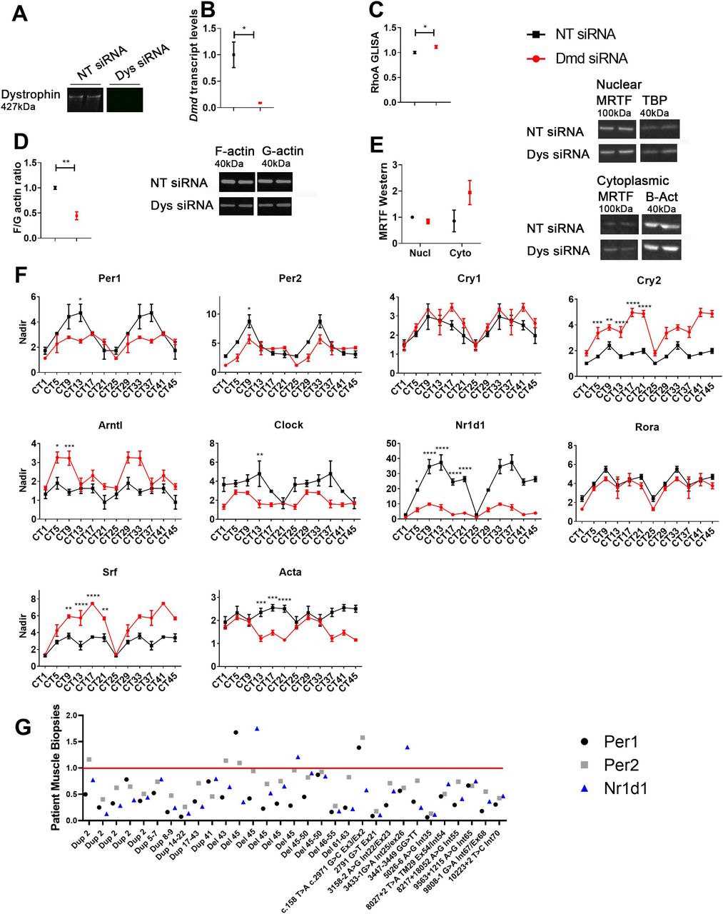
    To mimic dystrophic conditions, H2K 2B4 myotubes were transfected with siRNAs targeting the Dmd gene (Dmd siRNA, red), and a non-targeting (NT siRNA, black) siRNA was used for control. (A, B) Dystrophin was successfully down-regulated at (A) protein and (B) the transcript level. (C, D, E) In addition, the absence of dystrophin resulted in (C) altered RhoA activity, (D) reduced F/G-actin levels, and (E) greater cytoplasmic MRTF accumulation (loading controls: nuclear TBP, and cytoplasmic β-actin). For gene transcript studies, cells were placed is serum free media and collected over a 24-h time course (double plotted-48 h-to show oscillation pattern). (F) Circadian time (CT) gene expression data indicates diurnal oscillation patterns and down-regulation of pertinent core clock genes (Per1 and Per2) and down-stream targets for RhoA-actin-SRF (Nr1d1 and Acta1; housekeeping gene—Gapdh). (G) Muscle biopsies were obtained from an array of patients with different mutations or deletions in the dystrophin genes, and indicate down-regulation of RhoA-actin-SRF target genes in most cases (housekeeping genes—RPL13a). All samples were normalised to a pooled skeletal muscle sample from two healthy volunteers, as indicated by red line. For RhoA GLISA, F/G actin ratio and MRTF Westerns, data were normalised to NT control (n = 3; two-tailed t test). For RT-qPCR CT data, the nadir was determined as the minimum value across both treatments (Dmd and NT siRNAs) and CTs and applied to all samples; nadir normalised to 1 (n = 2–3 for each siRNA and time point; two-way ANOVA with Bonferroni post hoc test performed). Mean values reported with SEM; ***P < 0.001, **P < 0.01, *P < 0.05. For uncropped Western blot images and loading controls, see Source Data Fig 1.

    Source data are available for this figure.

    Source Data for Figure 1[LSA-2021-01014_SdataF1.tif]

  • Figure 2.
    • Download figure
    • Open in new tab
    • Download powerpoint
    Figure 2. Diurnal changes in RhoA-MRTF-serum response factor (SRF) cascade components in tibialis anterior of dystrophin-utrophin (dKO) mouse model.

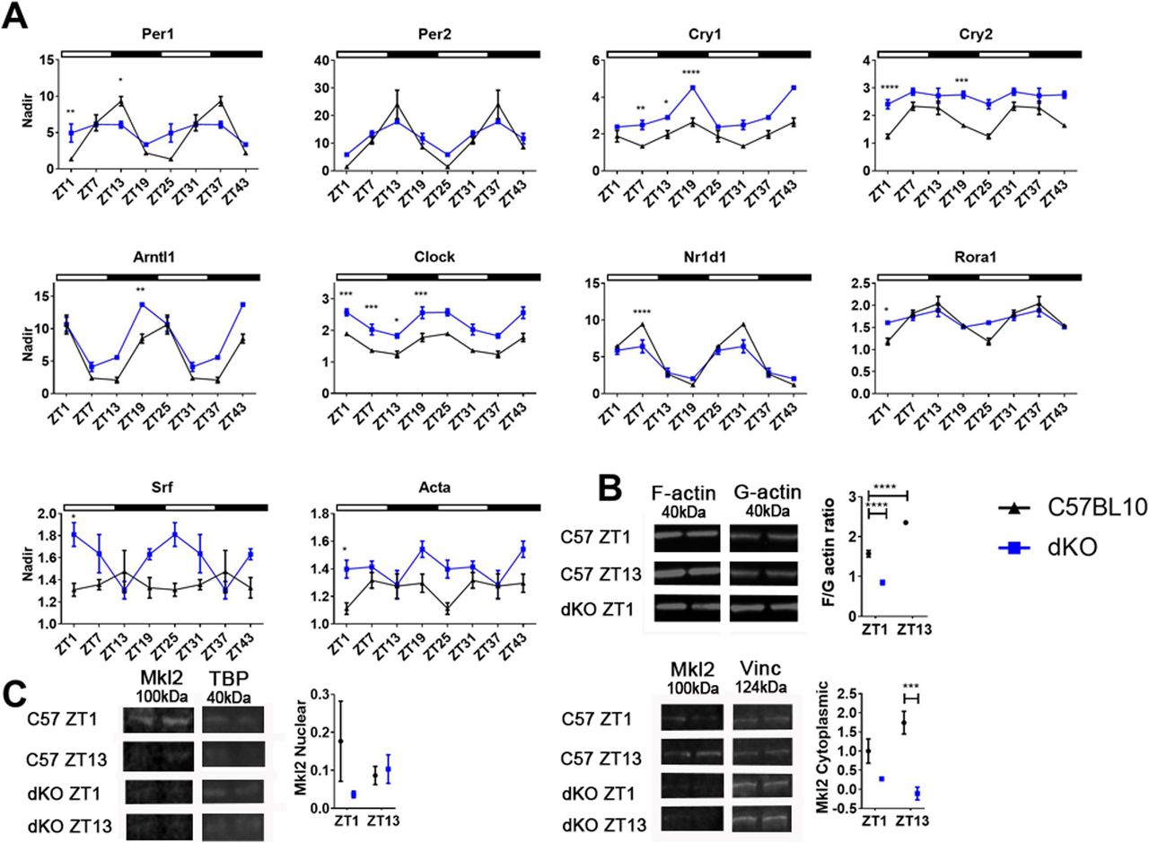
    (A) Tissues were collected over a 24-h time course and double plotted (48 h) to better illustrate the (A) oscillation pattern of core clock genes in tibialis anterior (TA) of 5-wk-old dKO animals which were significantly altered compared to C57BL10 (Zeitgeber- ZT). Down-stream targets for RhoA-actin-SRF pathway (Nr1d1, Rora1, and Acta) and indeed Srf were also altered (Gapdh used as housekeeping gene). (B, C) F/G-actin protein ratios in dKO mice were significantly down-regulated, (C) as were cytoplasmic MRTF fraction levels (loading control: nuclear TBP, and cytoplasmic vinculin). In addition, C57BL10 animals exhibit diurnal changes in F/G actin ratio and MRTF fraction levels. Light and dark periods represented by outlined (light) and solid bars (dark). For RT-qPCR ZT data, the nadir was determined as the minimum value across both genotypes and ZTs and applied to all samples; nadir normalised to 1 (n = 3–4 for each for each genotype and time-point). For F/G actin ratio and MRTF westerns, data were normalised to C57BL10 (n = 3–4). One or two-way ANOVA with Bonferroni post hoc test performed. Mean values reported with SEM; ***P < 0.001, **P < 0.01, *P < 0.05. For uncropped Western blot images and loading controls, see Source Data Fig 2.

    Source data are available for this figure.

    Source Data for Figure 2[LSA-2021-01014_SdataF2.tif]

  • Figure S1.
    • Download figure
    • Open in new tab
    • Download powerpoint
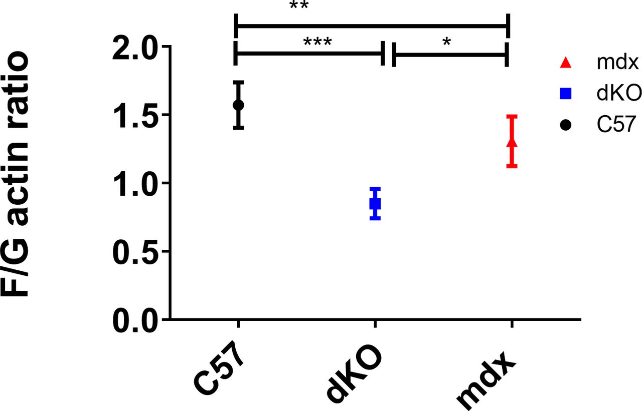
    Figure S1. F/G actin ratios in C57BL10, and mdx and dKO littermates.

    F/G-actin protein ratios from the tibialis anterior (TA) of 5-wk-old dKO were significantly lower than littermate mdx and C57BL10 animals (ZT1). N = 3–4 for each for each genotype; one-way ANOVA with Bonferroni post hoc test performed. Mean values reported with SEM; ***P < 0.001, **P < 0.01, *P < 0.05.

  • Figure 3.
    • Download figure
    • Open in new tab
    • Download powerpoint
    Figure 3. Perturbed circadian rest-activity and response to jet-lag in dystrophic mouse model.

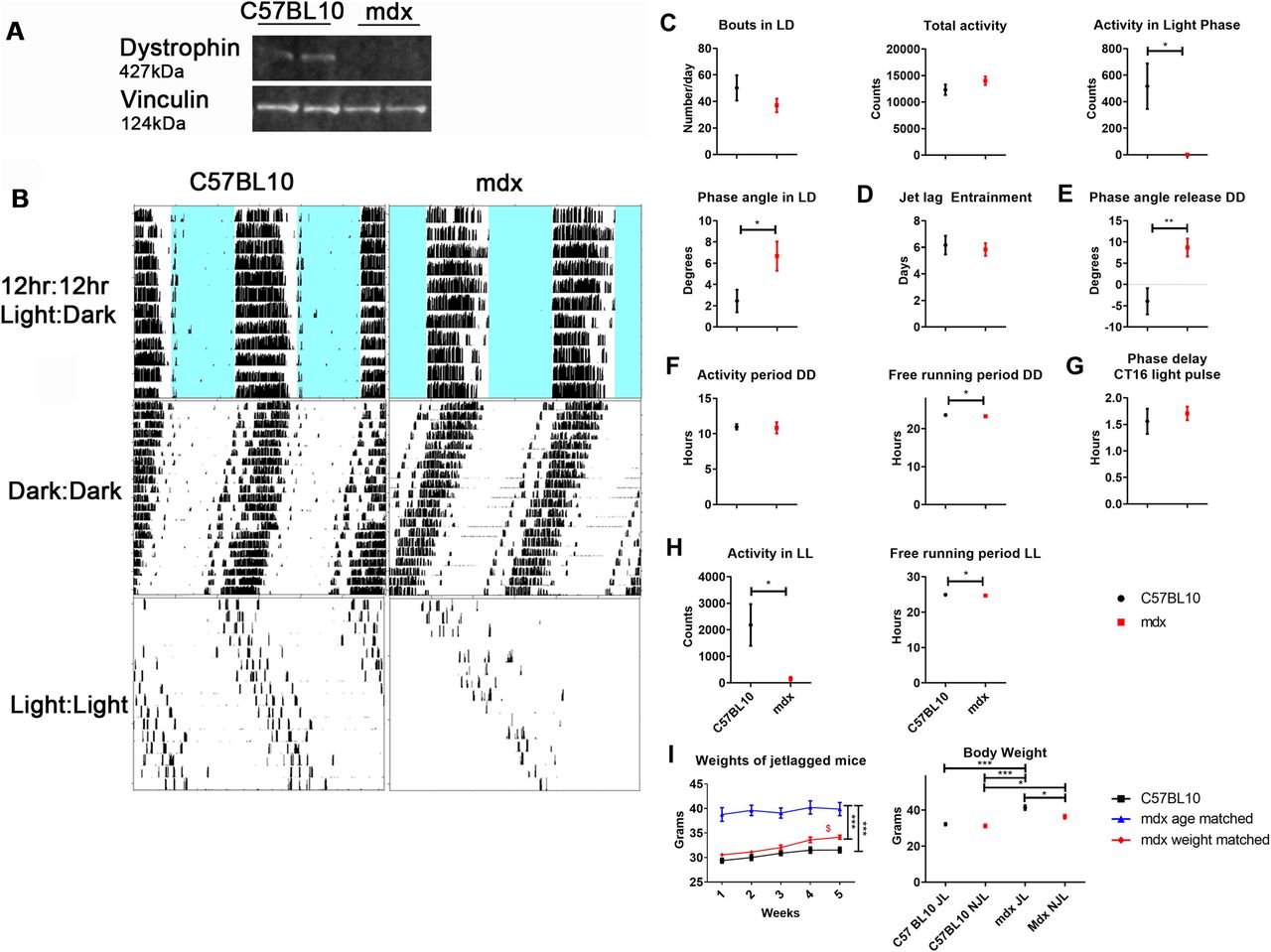
    (A) Dystrophin Western blot shows the absence of protein in the SCN of mdx animals (vinculin used as loading control). (B) Locomotor behaviour of mdx and C57BL10 mice were assessed and representative wheel-running activity blots (B) shown; 12 h:12 h light:dark cycle (LD), dark:dark (DD), and light:light (LL). (C, D, E, F, G, H) Data analysed and represented as graphs: (C) behaviour during LD, (D) after 6-h phase advancements, (E) phase angle on release into DD, (F) behaviour during DD, (G) phase delay after a light pulse 4-h post exercise commencement, (H) and behaviour during LL. (I) Weights of mice undergoing 5-wk jetlag protocol assessed (C57BL10, mdx age matched and mdx weight matched), as well as total body weight of jetlagged (JL) and non-jetlagged (NJL) mdx and C57BL10 cohorts. t test for comparisons between two groups (two-tailed). For weights recorded over the 5 wk of jetlag: black asterisk on right-one-way ANOVA comparing all three cohorts; dollar sign-one way ANOVA with Bonferroni post hoc between each cohort at each time point. For body weights with JL and NJL, one-way ANOVA with Bonferroni post hoc test performed (For wheel running activity n = 5–6, jetlag study n = 3–4; ***P < 0.001, **P < 0.01, *P < 0.05). For uncropped dystrophin Western blot and loading control, see Source Data Fig 3.

    Source data are available for this figure.

    Source Data for Figure 3[LSA-2021-01014_SdataF3.tif]

  • Figure 4.
    • Download figure
    • Open in new tab
    • Download powerpoint
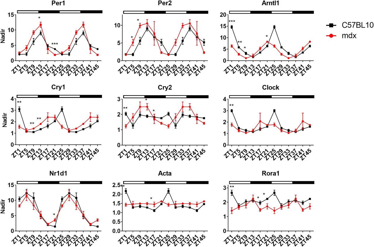
    Figure 4. Altered expression of clock genes in tibialis anterior of mdx mouse model.

    Tissues were collected over a 24-h time course and double plotted (48 h) to better illustrate the oscillation pattern of core clock genes in tibialis anterior (TA) of 20 wk old mdx animals which were significantly altered compared with C57BL10 (Zeitgeber- ZT). Down-stream targets for RhoA-actin-serum response factor pathway (Nr1d1, Acta, and Rora1) were also altered (Atp5b used as housekeeping gene). Light and dark periods represented by outlined (light) and solid bars (dark). The nadir was determined as the minimum value across both genotypes and ZTs and applied to all samples; nadir normalised to one (n = 3–4 for each for each genotype and time-point; two-way ANOVA with Bonferroni post hoc test performed). Mean values reported with SEM; ***P < 0.001, **P < 0.01, *P < 0.05.

  • Figure S2.
    • Download figure
    • Open in new tab
    • Download powerpoint
    Figure S2. Utrophin protein and transcript levels altered in dystrophic models.

    (A, B) Transcript levels (left), Western blot quantification (middle), and representative Western blots (right; total protein quantified for normalisation) for utrophin shown for (A) mdx and C57BL10 mice and (B) H2K 2B4 myotubes. (C) UTRN gene expression down-regulated in all but one Duchenne muscular dystrophy muscle biopsy sample. (D) Linear regression plotting PER1, PER2, and NR1D1 versus UTRN gene expression indicates significant correlation pattern with PER1 (R = 0.1687) and PER2 (R = 0.4542). Data normalised to NT siRNA or C57BL10; t test for comparisons between two groups (two-tailed; ***P < 0.001, **P < 0.01, *P < 0.05). N = 3–4. For uncropped Western blot images and loading controls, see Source Data Fig S2.

    Source data are available for this figure.

    Source Data for Figure S2[LSA-2021-01014_SdataFS2.tif]

  • Figure S3.
    • Download figure
    • Open in new tab
    • Download powerpoint
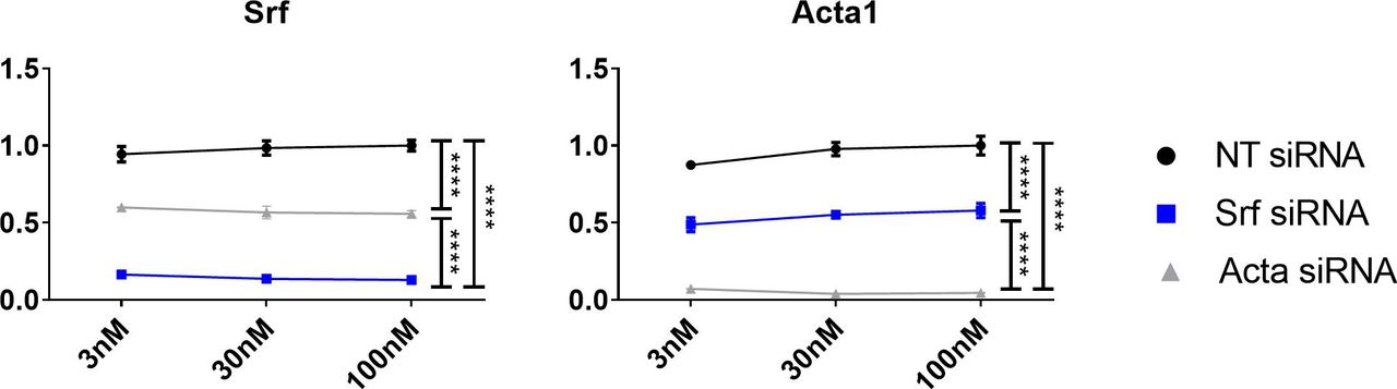
    Figure S3. In vitro dose response curves for Srf and Acta.

    H2K 2B4 myotubes were transfected with 3 nM, 30 nM and 100 nM of siRNA targeting the Srf, Acta and Dmd genes (Srf siRNA, blue; and Acta siRNA, grey), and a non-targeting (NT siRNA, black) siRNA was used for control. Gene expression data indicates significant and sustained knock-down of respective genes at low doses (housekeeping gene— Gapdh). Data normalised to NT siRNA; n = 3; two-way ANOVA with Bonferroni post hoc test performed. Mean values reported with SEM; ***P < 0.001, **P < 0.01, *P < 0.05.

  • Figure 5.
    • Download figure
    • Open in new tab
    • Download powerpoint
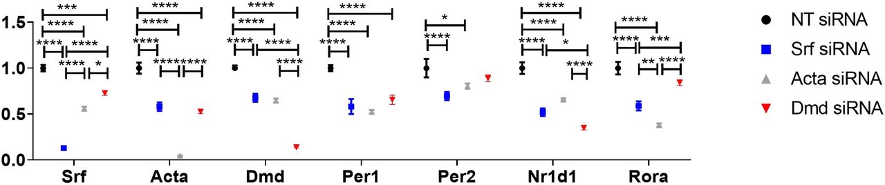
    Figure 5. Abrogation of alternative RhoA-actin-serum response factor (SRF) components leads to a reduction in the expression of SRF target genes.

    H2K 2B4 myotubes were transfected with 100 nM siRNAs targeting the Srf, Acta and Dmd genes (Srf siRNA, blue; Acta siRNA, grey; and Dmd siRNA, red), and a non-targeting (NT siRNA, black) siRNA was used for control. Gene expression data indicate successful knock-down of respective genes (Srf, Acta and Dmd) and down-regulation of pertinent core clock genes (Per1 and Per2) and other down-stream targets for RhoA-actin-SRF (Nr1d1 and Acta1; housekeeping gene—Gapdh). Data normalised to NT siRNA; n = 3; two-way ANOVA with Bonferroni post hoc test performed. Mean values reported with SEM; ***P < 0.001, **P < 0.01, *P < 0.05.

  • Figure 6.
    • Download figure
    • Open in new tab
    • Download powerpoint
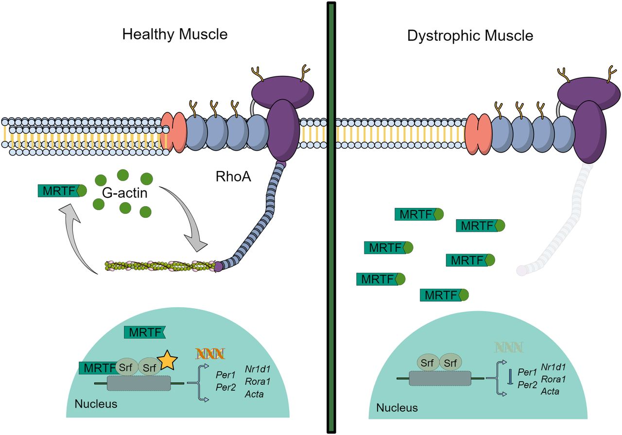
    Figure 6. Schematic illustrating RhoA-actin-serum response factor (SRF) signalling pathway.

    In healthy muscle, systemic cues activate RhoA in peripheral tissues which in turn regulates diurnal polymerisation (F-actin) and de-polymerisation (globular (G)-actin) of actin. During the de-polymerisation stage, G-actin preferentially binds to myocardin-related transcription factor (MRTF). However, when this shifts to the polymerisation phase, the G-actin pool diminishes and unbound MRTF translocates into the nucleus and influences expression of SRF. SRF activation regulates transcription of SRF target genes including the core clock genes—Per1 and Per2, as well as secondary loop clock genes—Rora1 and Nr1d1, and cytoskeletal genes such as Acta. When dystrophin is absent, F-actin is not stable and the cell shifts to the de-polymerised state. G-actin binds to MRTF and therefore SRF transcription is hampered.

Supplementary Materials

  • Figures
  • Table S1 SiRNA and PCR primer IDs or sequences.

PreviousNext
Back to top
Download PDF
Article Alerts
Sign In to Email Alerts with your Email Address
Email Article

Thank you for your interest in spreading the word on Life Science Alliance.

NOTE: We only request your email address so that the person you are recommending the page to knows that you wanted them to see it, and that it is not junk mail. We do not capture any email address.

Enter multiple addresses on separate lines or separate them with commas.
Dystrophin involvement in peripheral circadian SRF signalling
(Your Name) has sent you a message from Life Science Alliance
(Your Name) thought you would like to see the Life Science Alliance web site.
CAPTCHA
This question is for testing whether or not you are a human visitor and to prevent automated spam submissions.
Citation Tools
Dystrophin and circadian SRF signalling
Corinne A Betts, Aarti Jagannath, Tirsa LE van Westering, Melissa Bowerman, Subhashis Banerjee, Jinhong Meng, Maria Sofia Falzarano, Lara Cravo, Graham McClorey, Katharina E Meijboom, Amarjit Bhomra, Wooi Fang Lim, Carlo Rinaldi, John R Counsell, Katarzyna Chwalenia, Elizabeth O’Donovan, Amer F Saleh, Michael J Gait, Jennifer E Morgan, Alessandra Ferlini, Russell G Foster, Matthew JA Wood
Life Science Alliance Aug 2021, 4 (10) e202101014; DOI: 10.26508/lsa.202101014

Citation Manager Formats

  • BibTeX
  • Bookends
  • EasyBib
  • EndNote (tagged)
  • EndNote 8 (xml)
  • Medlars
  • Mendeley
  • Papers
  • RefWorks Tagged
  • Ref Manager
  • RIS
  • Zotero
Share
Dystrophin and circadian SRF signalling
Corinne A Betts, Aarti Jagannath, Tirsa LE van Westering, Melissa Bowerman, Subhashis Banerjee, Jinhong Meng, Maria Sofia Falzarano, Lara Cravo, Graham McClorey, Katharina E Meijboom, Amarjit Bhomra, Wooi Fang Lim, Carlo Rinaldi, John R Counsell, Katarzyna Chwalenia, Elizabeth O’Donovan, Amer F Saleh, Michael J Gait, Jennifer E Morgan, Alessandra Ferlini, Russell G Foster, Matthew JA Wood
Life Science Alliance Aug 2021, 4 (10) e202101014; DOI: 10.26508/lsa.202101014
del.icio.us logo Digg logo Reddit logo Twitter logo CiteULike logo Facebook logo Google logo Mendeley logo
  • Tweet Widget
  • Facebook Like
Issue Cover

In this Issue

Volume 4, No. 10
October 2021
  • Table of Contents
  • Cover (PDF)
  • About the Cover
  • Masthead (PDF)
Advertisement

Jump to section

  • Article
    • Abstract
    • Introduction
    • Results
    • Discussion
    • Materials and Methods
    • Acknowledgements
    • References
  • Figures & Data
  • Info
  • Metrics
  • Reviewer Comments
  • PDF

Subjects

  • Molecular Biology

Related Articles

  • No related articles found.

Cited By...

  • No citing articles found.
  • Google Scholar

More in this TOC Section

  • Neutrophils delay repair in Wallerian degeneration
  • Vacuoles drive nuclear and rDNA morphology
  • Cdh5-lineage–independent origin of dermal lymphatics
Show more Research Article

Similar Articles

EMBO Press LogoRockefeller University Press LogoCold Spring Harbor Logo

Content

  • Home
  • Newest Articles
  • Current Issue
  • Archive
  • Subject Collections

For Authors

  • Submit a Manuscript
  • Author Guidelines
  • License, copyright, Fee

Other Services

  • Alerts
  • Twitter
  • RSS Feeds

More Information

  • Editors & Staff
  • Reviewer Guidelines
  • Feedback
  • Licensing and Reuse
  • Privacy Policy

ISSN: 2575-1077
© 2022 Life Science Alliance LLC

Life Science Alliance is registered as a trademark in the U.S. Patent and Trade Mark Office and in the European Union Intellectual Property Office.