Advertisement

Main menu

  • Home
  • Articles
    • Newest Articles
    • Current Issue
    • Methods & Resources
    • Archive
    • Subjects
  • Collections
  • Submit
    • Submit a Manuscript
    • Author Guidelines
    • License, Copyright, Fee
    • FAQ
    • Why Submit
  • About
    • About Us
    • Editors & Staff
    • Board Members
    • Licensing and Reuse
    • Reviewer Guidelines
    • Privacy Policy
    • Advertise
    • Contact Us
    • LSA LLC
  • Alerts
  • Other Publications
    • EMBO Press
    • The EMBO Journal
    • EMBO reports
    • EMBO Molecular Medicine
    • Molecular Systems Biology
    • Rockefeller University Press
    • Journal of Cell Biology
    • Journal of Experimental Medicine
    • Journal of General Physiology
    • Cold Spring Harbor Laboratory Press
    • Genes & Development
    • Genome Research

User menu

  • My alerts

Search

  • Advanced search
Life Science Alliance
  • Other Publications
    • EMBO Press
    • The EMBO Journal
    • EMBO reports
    • EMBO Molecular Medicine
    • Molecular Systems Biology
    • Rockefeller University Press
    • Journal of Cell Biology
    • Journal of Experimental Medicine
    • Journal of General Physiology
    • Cold Spring Harbor Laboratory Press
    • Genes & Development
    • Genome Research
  • My alerts
Life Science Alliance

Advanced Search

  • Home
  • Articles
    • Newest Articles
    • Current Issue
    • Methods & Resources
    • Archive
    • Subjects
  • Collections
  • Submit
    • Submit a Manuscript
    • Author Guidelines
    • License, Copyright, Fee
    • FAQ
    • Why Submit
  • About
    • About Us
    • Editors & Staff
    • Board Members
    • Licensing and Reuse
    • Reviewer Guidelines
    • Privacy Policy
    • Advertise
    • Contact Us
    • LSA LLC
  • Alerts
  • Follow lsa Template on Twitter
Research Article
Source Data
Transparent Process
Open Access

Small molecule inhibitors and a kinase-dead expressing mouse model demonstrate that the kinase activity of Chk1 is essential for mouse embryos and cancer cells

View ORCID ProfileSomsundar V Muralidharan, View ORCID ProfileLisa M Nilsson, View ORCID ProfileMattias F Lindberg, View ORCID ProfileJonas A Nilsson  Correspondence email
Somsundar V Muralidharan
Department of Surgery, Sahlgrenska Cancer Center, Institute of Clinical Sciences at University of Gothenburg, Gothenburg, Sweden
  • Find this author on Google Scholar
  • Find this author on PubMed
  • Search for this author on this site
  • ORCID record for Somsundar V Muralidharan
Lisa M Nilsson
Department of Surgery, Sahlgrenska Cancer Center, Institute of Clinical Sciences at University of Gothenburg, Gothenburg, Sweden
  • Find this author on Google Scholar
  • Find this author on PubMed
  • Search for this author on this site
  • ORCID record for Lisa M Nilsson
Mattias F Lindberg
Department of Surgery, Sahlgrenska Cancer Center, Institute of Clinical Sciences at University of Gothenburg, Gothenburg, Sweden
  • Find this author on Google Scholar
  • Find this author on PubMed
  • Search for this author on this site
  • ORCID record for Mattias F Lindberg
Jonas A Nilsson
Department of Surgery, Sahlgrenska Cancer Center, Institute of Clinical Sciences at University of Gothenburg, Gothenburg, Sweden
  • Find this author on Google Scholar
  • Find this author on PubMed
  • Search for this author on this site
  • ORCID record for Jonas A Nilsson
  • For correspondence: jonas.a.nilsson@surgery.gu.se
Published 22 June 2020. DOI: 10.26508/lsa.202000671
  • Article
  • Figures & Data
  • Info
  • Metrics
  • Reviewer Comments
  • PDF
Loading

Article Figures & Data

Figures

  • Figure 1.
    • Download figure
    • Open in new tab
    • Download powerpoint
    Figure 1. Chk1 inhibitors reduce expression of Chk1 and induce DNA damage.

    (A) λ820 mouse lymphoma cells were treated with LY2603618 for 24 h, and cells were analyzed by immunoblotting for Chk1 and loading control β-actin. (B) λ820 mouse lymphoma cells were treated with CCT245737 and cells were analyzed by immunoblotting for apoptosis marker cleaved PARP, total or phosphorylated (p)Chk1 and DNA damage marker phosphorylated histone 2AX (γH2Ax). (C) λ820 mouse lymphoma cells were treated with LY2603618 or ATR inhibitor VE821 for 24 h, and cells were analyzed by immunoblotting for total Chk1, pChk1, phosphorylated (p)RPA2, and loading control β-actin.

    Source data are available for this figure.

    Source Data for Figure 1[LSA-2020-00671_SdataF1.zip]

  • Figure 2.
    • Download figure
    • Open in new tab
    • Download powerpoint
    Figure 2. Generation of a kinase-dead Chk1 mouse model.

    (A) Clustering analysis of Chk1 from yeast, human, mouse, and frog. Arrow indicated aspartate 130 (D130). (B) Targeting strategy to generate a kinase-dead Chek1 allele. Top part of the mouse Chek1 gene. Middle part of the Chek1 gene showing targeted allele in ES cells. Bottom part of the Chek1 gene showing the Chek1 gene after removal of neo cassette in an Flp-deleted mouse. (C) PCR genotyping of three offspring after mating between a heterozygous Chek1 knockin mouse (Chek1D130A/wt). (B) PCR primers (NDEL1 and 2) are shown in (B). (D) Results of genotyping of embryos or offspring from time pregnancies between Chek1D130A/wt females and Chek1D130A/wt males. (E) Results of genotyping of offspring from crosses of heterozygous Chek1 knockin males or females to C57BL/6 mice.

  • Figure S1.
    • Download figure
    • Open in new tab
    • Download powerpoint
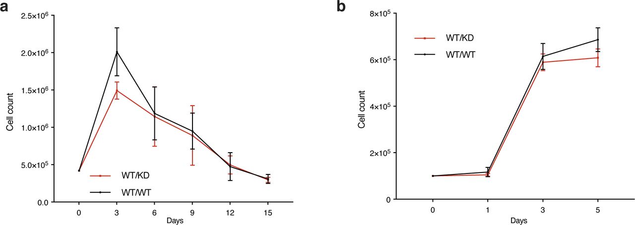
    Figure S1. Heterozygosity of a kinase-dead Chk1 expressing allele does not impact growth of MEFs.

    (A) Timed pregnancies between Chek1D130A/+ mice and C57BL/6 mice were used to make MEFs. Passage 3 MEFs were seeded at 0.5 million cells in a 5-cm plate and cultured for 3 d. Cells were counted, and 0.5 million cells were plated and cultured for 3 d again. The procedure was continued until the cells no longer were able to proliferate in 3 d. (B) Growth curves of MEFs grown for 5 d.

  • Figure 3.
    • Download figure
    • Open in new tab
    • Download powerpoint
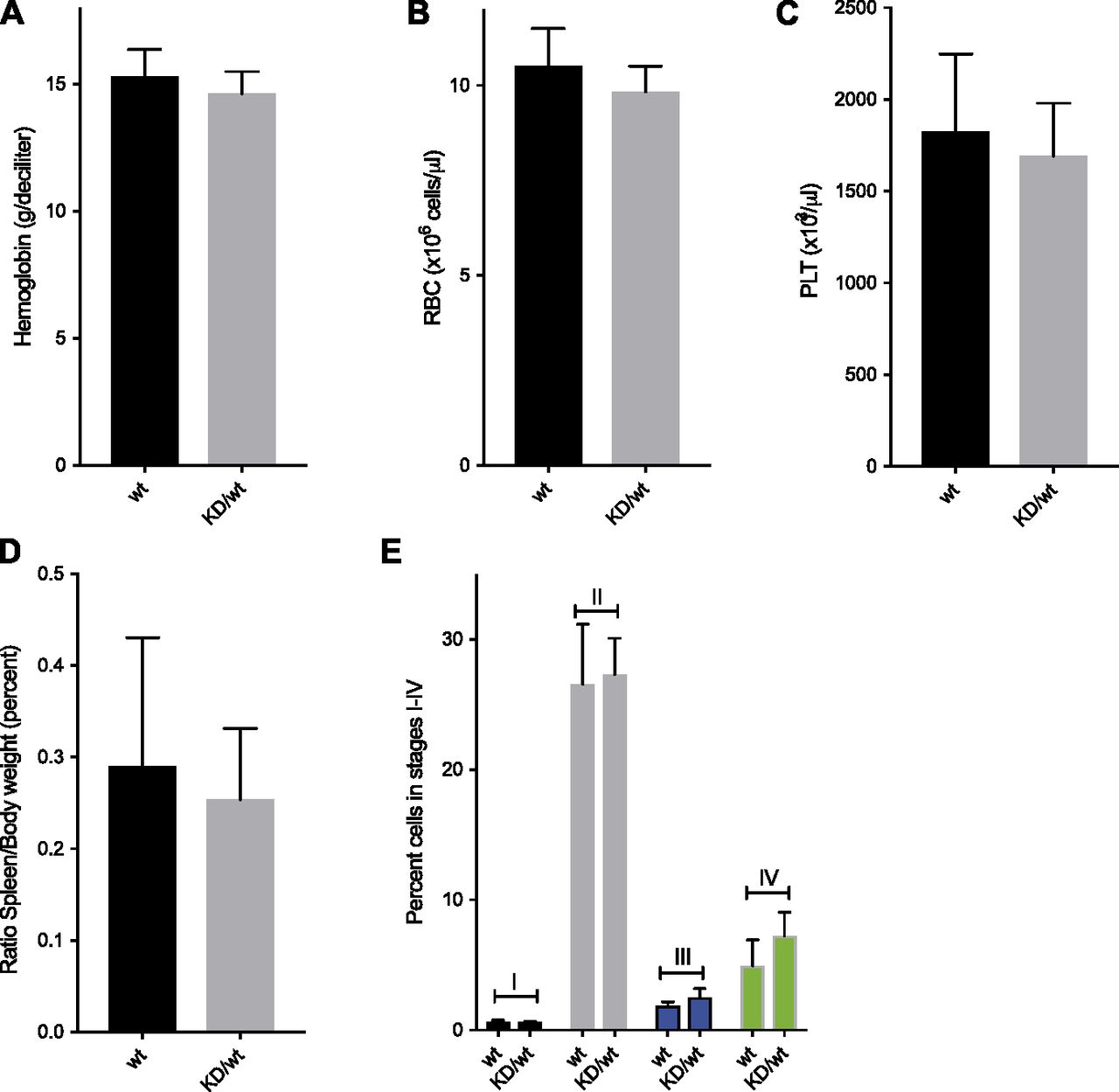
    Figure 3. Heterozygous Chek1 knockin mouse mice do not have a problem with erythropoiesis.

    (A, B, C, D) 21 wild-type (wt) and 15 heterozygous Chek1 knockin (KD/wt) 45–60-wk-old mice were analyzed for hemoglobin content (A), red blood cell count (B), platelet count, and spleen weight (D). (E) Six wt and three KD/wt mice were analyzed for different stages (I–IV) of erythroid development using Ter119/CD71 staining and flow cytometry. Representative flow cytometry plots are seen in Fig S2.

  • Figure S2.
    • Download figure
    • Open in new tab
    • Download powerpoint
    Figure S2. Bone marrows from wild-type and Chek1KD/w mice were analyzed for different stages (I–IV) of erythroid development using Ter119/CD71 staining and flow cytometry.

    Shown are representative flow cytometry plots and the data from multiple mice is seen in Fig 3E.

  • Figure 4.
    • Download figure
    • Open in new tab
    • Download powerpoint
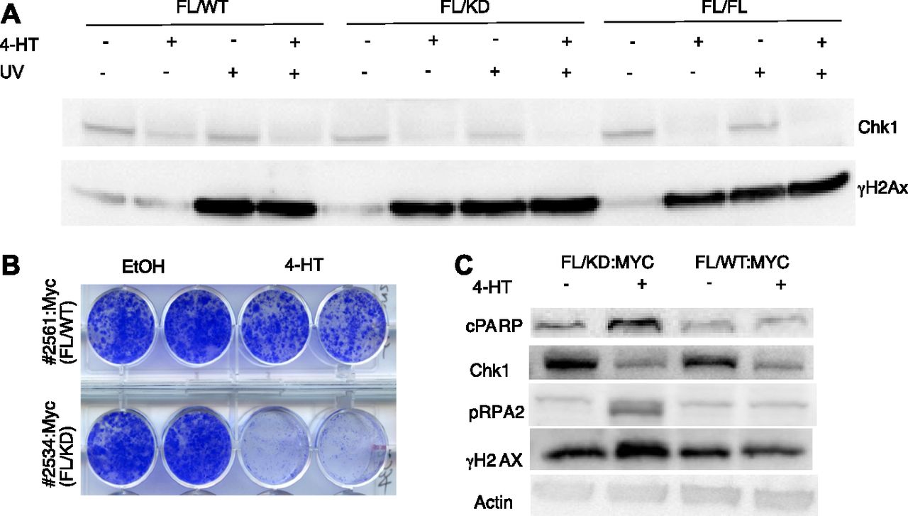
    Figure 4. Chk1 kinase activity is essential to suppress DNA damage signaling activation and for fibroblast viability.

    (A) Mouse fibroblasts from CreER mice carrying one floxed Chek1 allele and a wild-type (FL/WT), kinase-dead (FL/KD), or another floxed allele (FL/FL) were treated with vehicle or 4-hydroxytamoxifen (4HT) for 4 d and/or with UVB for 30 s (∼700 J/m2) and harvested 2 h after radiation or mock. Immunoblotting was performed for Chk1 and γH2Ax. (B) Mouse fibroblast expressing CreER, and a floxed and a kinase-dead or wild-type Chek1 allele were transduced with a Myc-expressing retrovirus. Cells were treated with vehicle or tamoxifen and long-term survival was determined by staining cell monolayers. (A, C) Cells manipulated the same as in (A) were analyzed by immunoblotting for indicated markers of apoptosis (PARP), replication stress (threonine 21 phosphorylated-RPA2), and DNA damage (γH2Ax).

  • Figure 5.
    • Download figure
    • Open in new tab
    • Download powerpoint
    Figure 5. Expression of only a kinase-dead allele of Chek1 suppresses lymphoma growth.

    (A) Kaplan–Meier survival analysis of λ-Myc; Cdkn2a+/− mice carrying the indicated Chek1 genotypes. (A, B, C, D) Lymphomas from mice generated from mice in (A) were transplanted into eight C57BL/6 mice per indicated genotype. When white blood counts reached between 20 and 50 × 103 cells/μl, four mice received vehicle or tamoxifen by i.p. injection. White blood count was followed until relapse or when palpable tumors required euthanasia of the animals.

  • Figure S3.
    • Download figure
    • Open in new tab
    • Download powerpoint
    Figure S3. λ-Myc lymphomas of indicated Chek1 genotypes were transplanted into C57BL/6 recipient mice.

    When white blood count was above normal, mice were treated with tamoxifen to induce Cre-mediated deletion of the floxed Chek1 allele. Tumors were harvested from the mice when becoming palpable and protein and DNA were extracted. (A) Western blot analysis of protein from indicated lymphomas. The blots were probed with antibodies against human Myc to confirm that the lymphoma were λ-Myc lymphomas, with anti-Chk1 to investigate if Chk1 had been deleted, with anti-γH2ax to analyze if DNA damage was induced and with anti–β-actin as a loading control. (B) PCR analysis with primers spanning one of the loxP sites in the genome of floxed mice. The larger band disappears if floxing occurs. (C) PCR analysis with primers detecting a DNA sequence that is created if Cre recombines in the genome of mice with floxed Chek1 alleles.

  • Figure 6.
    • Download figure
    • Open in new tab
    • Download powerpoint
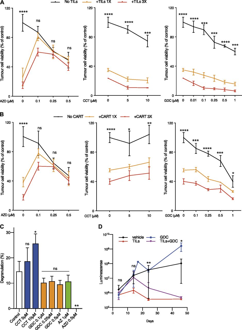
    Figure 6. Chk1 inhibition does not interfere with T-cell–mediated killing of melanoma cells.

    (A) 33F8-luciferase expressing melanoma cells were treated with indicated Chk1 inhibitors in the absence or presence of autologous TILs (1:1 or 3:1 TILs vs melanoma cells). Luciferin was added and viability was measured in a 96-well plate luminometer. (A, B) The same experimental setup as in (A) but using HER2-directed CAR-T cells instead of TILs. (C) 33F8 cells were mixed with autologous TILs in the presence of indicated inhibitors. Surface expression of CD107a as a marker of T-cell degranulation was determined by flow cytometry. (D) NOD/SCID/IL2 receptor gamma knockout mice transgenic for human interleukin-2 (hIL2-NOG mice) were transplanted with 33F8-luciferase melanoma cells. When tumors were detectable by IVIS imaging, TILs were injected and mice were dosed in indicated groups of animals (n = 4). Mice were followed with IVIS imaging until tumor size reached the ethics limit (10 mm on the shortest side). Shown are median IVIS signal and standard error of the mean.

PreviousNext
Back to top
Download PDF
Article Alerts
Sign In to Email Alerts with your Email Address
Email Article

Thank you for your interest in spreading the word on Life Science Alliance.

NOTE: We only request your email address so that the person you are recommending the page to knows that you wanted them to see it, and that it is not junk mail. We do not capture any email address.

Enter multiple addresses on separate lines or separate them with commas.
Small molecule inhibitors and a kinase-dead expressing mouse model demonstrate that the kinase activity of Chk1 is essential for mouse embryos and cancer cells
(Your Name) has sent you a message from Life Science Alliance
(Your Name) thought you would like to see the Life Science Alliance web site.
CAPTCHA
This question is for testing whether or not you are a human visitor and to prevent automated spam submissions.
Citation Tools
Chk1 kinase activity is needed for mouse embryos and tumors
Somsundar V Muralidharan, Lisa M Nilsson, Mattias F Lindberg, Jonas A Nilsson
Life Science Alliance Jun 2020, 3 (8) e202000671; DOI: 10.26508/lsa.202000671

Citation Manager Formats

  • BibTeX
  • Bookends
  • EasyBib
  • EndNote (tagged)
  • EndNote 8 (xml)
  • Medlars
  • Mendeley
  • Papers
  • RefWorks Tagged
  • Ref Manager
  • RIS
  • Zotero
Share
Chk1 kinase activity is needed for mouse embryos and tumors
Somsundar V Muralidharan, Lisa M Nilsson, Mattias F Lindberg, Jonas A Nilsson
Life Science Alliance Jun 2020, 3 (8) e202000671; DOI: 10.26508/lsa.202000671
del.icio.us logo Digg logo Reddit logo Twitter logo CiteULike logo Facebook logo Google logo Mendeley logo
  • Tweet Widget
  • Facebook Like
Issue Cover

In this Issue

Volume 3, No. 8
August 2020
  • Table of Contents
  • Cover (PDF)
  • About the Cover
  • Masthead (PDF)
Advertisement

Jump to section

  • Article
    • Abstract
    • Introduction
    • Results
    • Discussion
    • Materials and Methods
    • Acknowledgements
    • References
  • Figures & Data
  • Info
  • Metrics
  • Reviewer Comments
  • PDF

Subjects

  • Genetics, Gene Therapy & Genetic Disease
  • Cell Biology
  • Cancer

Related Articles

  • No related articles found.

Cited By...

  • No citing articles found.
  • Google Scholar

More in this TOC Section

  • Neutrophils delay repair in Wallerian degeneration
  • Vacuoles drive nuclear and rDNA morphology
  • Cdh5-lineage–independent origin of dermal lymphatics
Show more Research Article

Similar Articles

EMBO Press LogoRockefeller University Press LogoCold Spring Harbor Logo

Content

  • Home
  • Newest Articles
  • Current Issue
  • Archive
  • Subject Collections

For Authors

  • Submit a Manuscript
  • Author Guidelines
  • License, copyright, Fee

Other Services

  • Alerts
  • Twitter
  • RSS Feeds

More Information

  • Editors & Staff
  • Reviewer Guidelines
  • Feedback
  • Licensing and Reuse
  • Privacy Policy

ISSN: 2575-1077
© 2022 Life Science Alliance LLC

Life Science Alliance is registered as a trademark in the U.S. Patent and Trade Mark Office and in the European Union Intellectual Property Office.