Advertisement

Main menu

  • Home
  • Articles
    • Newest Articles
    • Current Issue
    • Methods & Resources
    • Archive
    • Subjects
  • Collections
  • Submit
    • Submit a Manuscript
    • Author Guidelines
    • License, Copyright, Fee
    • FAQ
    • Why submit
  • About
    • About Us
    • Editors & Staff
    • Board Members
    • Licensing and Reuse
    • Reviewer Guidelines
    • Privacy Policy
    • Advertise
    • Contact Us
    • LSA LLC
  • Alerts
  • Other Publications
    • EMBO Press
    • The EMBO Journal
    • EMBO reports
    • EMBO Molecular Medicine
    • Molecular Systems Biology
    • Rockefeller University Press
    • Journal of Cell Biology
    • Journal of Experimental Medicine
    • Journal of General Physiology
    • Cold Spring Harbor Laboratory Press
    • Genes & Development
    • Genome Research

User menu

  • My alerts

Search

  • Advanced search
Life Science Alliance
  • Other Publications
    • EMBO Press
    • The EMBO Journal
    • EMBO reports
    • EMBO Molecular Medicine
    • Molecular Systems Biology
    • Rockefeller University Press
    • Journal of Cell Biology
    • Journal of Experimental Medicine
    • Journal of General Physiology
    • Cold Spring Harbor Laboratory Press
    • Genes & Development
    • Genome Research
  • My alerts
Life Science Alliance

Advanced Search

  • Home
  • Articles
    • Newest Articles
    • Current Issue
    • Methods & Resources
    • Archive
    • Subjects
  • Collections
  • Submit
    • Submit a Manuscript
    • Author Guidelines
    • License, Copyright, Fee
    • FAQ
    • Why submit
  • About
    • About Us
    • Editors & Staff
    • Board Members
    • Licensing and Reuse
    • Reviewer Guidelines
    • Privacy Policy
    • Advertise
    • Contact Us
    • LSA LLC
  • Alerts
  • Follow lsa Template on Twitter
Research Article
Transparent Process
Open Access

Laminin is the ECM niche for trophoblast stem cells

View ORCID ProfileDaiji Kiyozumi, Itsuko Nakano, Ryoko Sato-Nishiuchi, Satoshi Tanaka, View ORCID ProfileKiyotoshi Sekiguchi  Correspondence email
Daiji Kiyozumi
1Laboratory of Extracellular Matrix Biochemistry, Institute for Protein Research, Osaka University, Osaka, Japan
  • Find this author on Google Scholar
  • Find this author on PubMed
  • Search for this author on this site
  • ORCID record for Daiji Kiyozumi
Itsuko Nakano
1Laboratory of Extracellular Matrix Biochemistry, Institute for Protein Research, Osaka University, Osaka, Japan
  • Find this author on Google Scholar
  • Find this author on PubMed
  • Search for this author on this site
Ryoko Sato-Nishiuchi
1Laboratory of Extracellular Matrix Biochemistry, Institute for Protein Research, Osaka University, Osaka, Japan
  • Find this author on Google Scholar
  • Find this author on PubMed
  • Search for this author on this site
Satoshi Tanaka
2Graduate School of Agricultural and Life Sciences, The University of Tokyo, Tokyo, Japan
  • Find this author on Google Scholar
  • Find this author on PubMed
  • Search for this author on this site
Kiyotoshi Sekiguchi
1Laboratory of Extracellular Matrix Biochemistry, Institute for Protein Research, Osaka University, Osaka, Japan
  • Find this author on Google Scholar
  • Find this author on PubMed
  • Search for this author on this site
  • ORCID record for Kiyotoshi Sekiguchi
  • For correspondence: sekiguch@protein.osaka-u.ac.jp
Published 14 January 2020. DOI: 10.26508/lsa.201900515
  • Article
  • Figures & Data
  • Info
  • Metrics
  • Reviewer Comments
  • PDF
Loading

Article Figures & Data

Figures

  • Supplementary Materials
  • Figure S1.
    • Download figure
    • Open in new tab
    • Download powerpoint
    Figure S1.

    Diagram illustrating the niche environment for TSCs. FGF4 and nodal from the epiblast act on TSCs as niche factors. The inset shows the region illustrated in the main figure. The diagram is based on that in the study by (Tanaka et al 1998).

  • Figure 1.
    • Download figure
    • Open in new tab
    • Download powerpoint
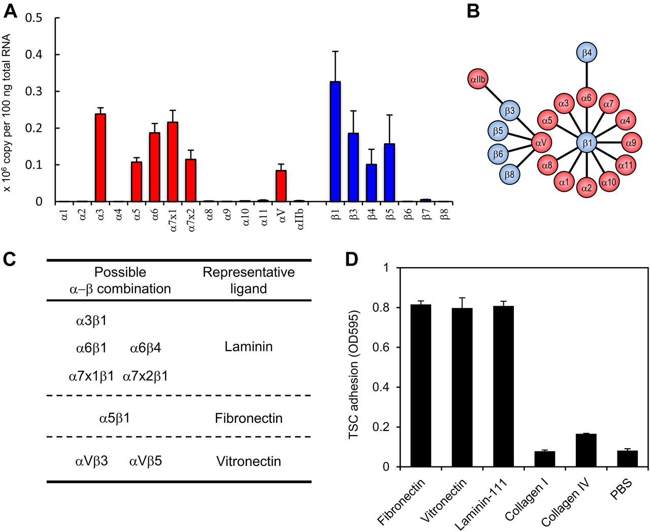
    Figure 1. Expression profile of integrin subunits in TSCs.

    (A) Transcript expression of integrin subunits in TSCs. Data represent means ± SD (n = 3). (B) Diagrammatic representation of a known integrin αβ heterodimer. The diagram is based on those in Hynes (2002) and Barczyk et al (2010). (C) Heterodimeric αβ integrin subtypes expressed in TSCs. (D) Adhesion of TSCs to defined substrates. Data represent means ± SD (n = 3).

  • Figure 2.
    • Download figure
    • Open in new tab
    • Download powerpoint
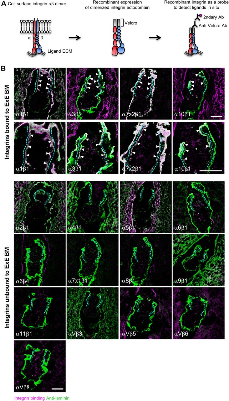
    Figure 2. Characterization of the ECM niche for TSCs in vivo.

    (A) Schematic views of in situ integrin ligand detection. Left, integrin αβ dimer on the cell surface; middle, αβ dimerized recombinant integrin ectodomain; right, in situ integrin ligand detection. (B) Comprehensive analyses of the ECM niche using E5.5 embryonic sections. Magenta, in situ binding of recombinant integrins; green, immunoreactivities for anti-laminin α1 (for integrins α3β1, α6β1, α6β4, α7x1β1, α7x2β1, α8β1, α10β1, α11β1, αVβ5, αVβ6, and αVβ8) and anti-laminin γ1 (for integrins α1β1, α2β1, α4β1, α5β1, α9β1, and αVβ3) mAbs; white, area double-positive for magenta and green signals; dotted lines, ExE basement membrane. The arrowheads indicate recombinant integrins bound to the ExE basement membrane. Bars, 50 μm.

  • Figure 3.
    • Download figure
    • Open in new tab
    • Download powerpoint
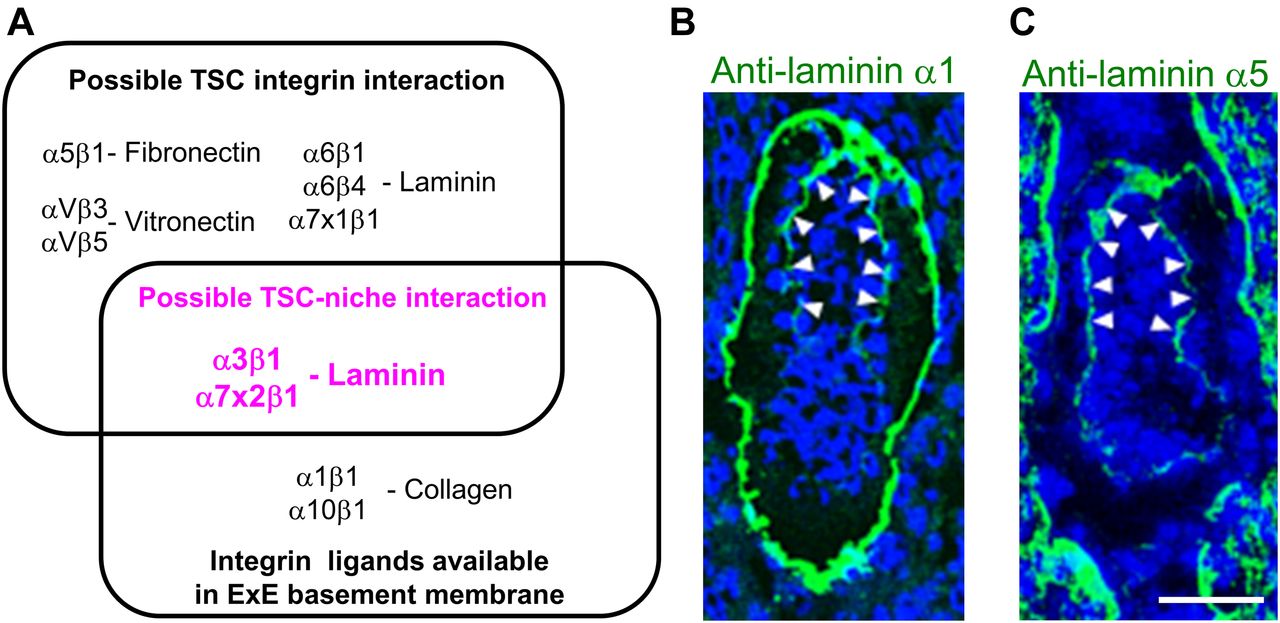
    Figure 3. The only integrin ligand available for TSCs is laminin.

    (A) Venn diagram representing possible TSC–integrin interactions, integrin ligands available in the ExE basement membrane, and possible TSC–niche interactions (magenta). (B, C) Localizations of the laminin α1 and α5 chains in E5.5 embryos. Wild-type E5.5 embryos were immunostained with anti-laminin α1 (B) and anti-laminin α5 (C) antibodies. Arrowheads, ExE basement membrane. Bar, 50 μm.
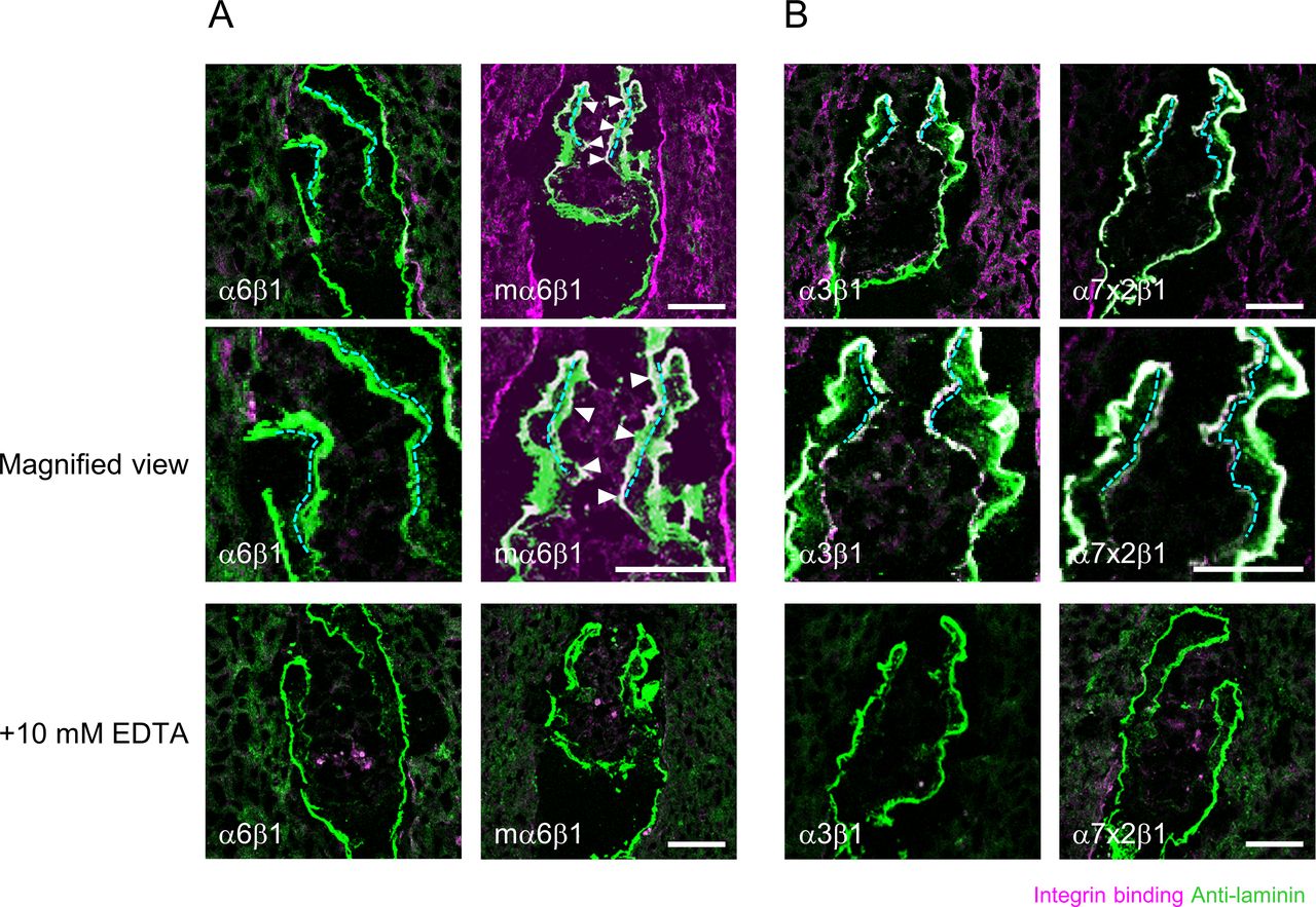
  • Figure S2.
    • Download figure
    • Open in new tab
    • Download powerpoint
    Figure S2.

    In situ integrin overlay assays with recombinant laminin-binding integrins. (A) E5.5 embryonic sections were subjected to in situ integrin overlay assays using recombinant human integrin a6b1 or mouse integrin α6β1 (mα6β1) in the absence or presence of 10 mM EDTA. Mouse, but not human, integrin α6β1 gave signals on ExE basement membranes. (B) E5.5 embryonic sections were subjected to in situ integrin overlay assays using recombinant integrin a3b1 and a7x2b1 in the absence or presence of 10 mM EDTA. Magenta, in situ binding of recombinant integrin; green, anti-laminin immunoreactivity; white, area double-positive for magenta and green signals; dotted lines, ExE basement membrane. The arrowheads indicate recombinant integrins binding to the ExE basement membrane. Bars, 50 μm.

  • Figure 4.
    • Download figure
    • Open in new tab
    • Download powerpoint
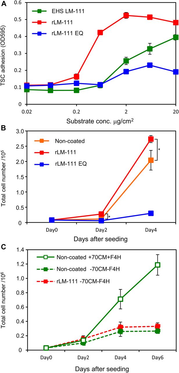
    Figure 4. TSCs proliferate on laminin-111.

    (A) TSC adhesion on various integrin ligands. Cell culture dishes were coated with increasing concentrations of EHS laminin-111 (EHS LM-111), recombinant laminin-111 (rLM-111), and recombinant laminin-111 EQ mutant (rLM-111 EQ). (B) TSC proliferation on non-coated, rLM-111–coated, and rLM-111 EQ–coated dishes. *P < 0.05, significant difference between non-coated and laminin-111–coated dishes by t test. (C) TSC proliferation on non-coated or rLM-111–coated dishes. 70CM+F4H was added (Non-coated +70CM+F4H) or depleted (Non-coated −70CM−F4H and rLM-111 −70CM−F4H). Data represent means ± SD (n = 3).

  • Figure S3.
    • Download figure
    • Open in new tab
    • Download powerpoint
    Figure S3.

    Inhibition of TSC adhesion by integrin-blocking Abs. TSCs were seeded onto culture dishes coated with 70CM or laminin-111 (LM111) in the presence of function-blocking antibodies against integrin αV, β1, or β3, or combinations of αV and β1, αV and β3, or β1 and β3. The results were normalized to the no Ab control. Data represent means ± SD (n = 4).

  • Figure 5.
    • Download figure
    • Open in new tab
    • Download powerpoint
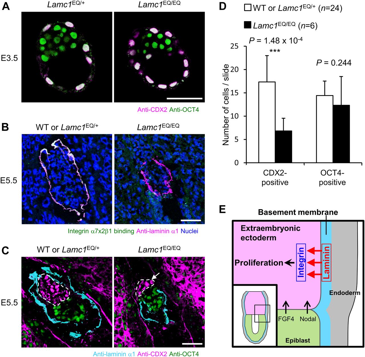
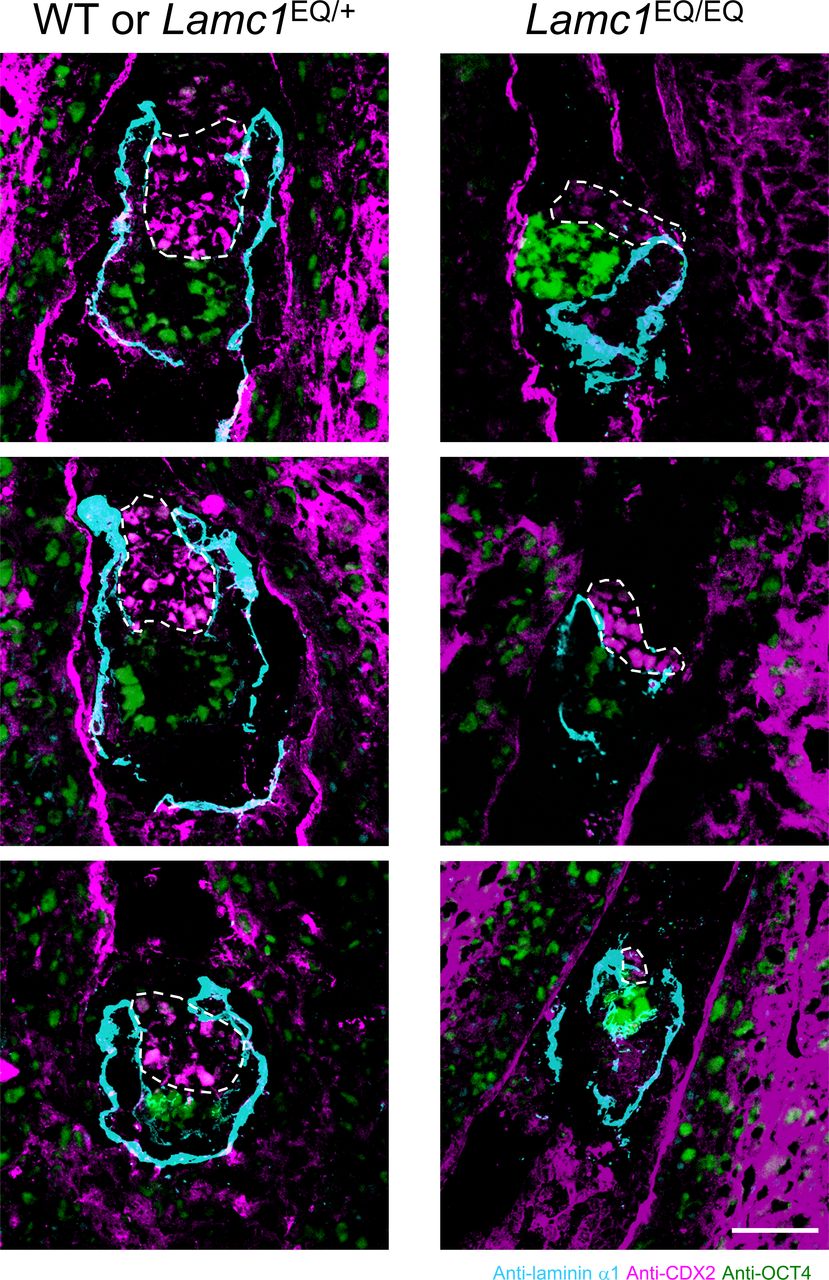
    Figure 5. Laminin–integrin interactions contribute to TSC expansion in vivo.

    (A) Expression of undifferentiated TSC marker CDX2 in trophectoderm cells in Lamc1EQ/+ and Lamc1EQ/EQ blastocysts. Bar, 50 μm. (B) Loss of integrin α7x2β1 binding to the basement membrane in E5.5 Lamc1EQ/EQ embryos. Magenta, anti-laminin α1 antibody binding; green, recombinant integrin α7x2β1 binding; white, area double-positive for magenta and green signals. Bar, 50 μm. (C) Morphologies of E5.5 control and Lamc1EQ/EQ embryos visualized by immunofluorescence. Cyan, laminin α1; magenta, CDX2; green, OCT4. ExE cells are enclosed by dotted lines. The arrow indicates CDX2-positive cells detached from the laminin-positive basement membrane. Bar, 50 μm. (D) Quantification of CDX2-positive and OCT4-positive cells in control WT and Lamc1EQ/+ and Lamc1EQ/EQ E5.5 egg cylinders. Data represent means ± SD (n = 24 and 6 for WT or Lamc1EQ/+ and Lamc1EQ/EQ, respectively). ***P < 0.001, significant difference by Welch’s t test. (E) Diagram illustrating the dependence of TSCs on laminin–integrin interactions in the mouse conceptus. In addition to FGF4 and nodal from the epiblast, laminin also acts on TSCs as an ECM niche through binding to integrin receptors. The inset shows the region illustrated in the main figure. The diagram is based on that in Tanaka et al (1998).

  • Figure S4.
    • Download figure
    • Open in new tab
    • Download powerpoint
    Figure S4.

    Morphologies of E5.5 control and Lamc1EQ/EQ embryos. Cyan, laminin α1; magenta, CDX2; green, OCT4. ExE cells are enclosed by dotted lines. Bar, 50 μm.

Supplementary Materials

  • Figures
  • Table S1 Primers used for quantitative RT-PCR.

PreviousNext
Back to top
Download PDF
Article Alerts
Sign In to Email Alerts with your Email Address
Email Article

Thank you for your interest in spreading the word on Life Science Alliance.

NOTE: We only request your email address so that the person you are recommending the page to knows that you wanted them to see it, and that it is not junk mail. We do not capture any email address.

Enter multiple addresses on separate lines or separate them with commas.
Laminin is the ECM niche for trophoblast stem cells
(Your Name) has sent you a message from Life Science Alliance
(Your Name) thought you would like to see the Life Science Alliance web site.
CAPTCHA
This question is for testing whether or not you are a human visitor and to prevent automated spam submissions.
Citation Tools
Laminin as a trophoblast stem cell niche
Daiji Kiyozumi, Itsuko Nakano, Ryoko Sato-Nishiuchi, Satoshi Tanaka, Kiyotoshi Sekiguchi
Life Science Alliance Jan 2020, 3 (2) e201900515; DOI: 10.26508/lsa.201900515

Citation Manager Formats

  • BibTeX
  • Bookends
  • EasyBib
  • EndNote (tagged)
  • EndNote 8 (xml)
  • Medlars
  • Mendeley
  • Papers
  • RefWorks Tagged
  • Ref Manager
  • RIS
  • Zotero
Share
Laminin as a trophoblast stem cell niche
Daiji Kiyozumi, Itsuko Nakano, Ryoko Sato-Nishiuchi, Satoshi Tanaka, Kiyotoshi Sekiguchi
Life Science Alliance Jan 2020, 3 (2) e201900515; DOI: 10.26508/lsa.201900515
Reddit logo Twitter logo Facebook logo Mendeley logo
  • Tweet Widget
  • Facebook Like
Issue Cover

In this Issue

Volume 3, No. 2
February 2020
  • Table of Contents
  • Cover (PDF)
  • About the Cover
  • Masthead (PDF)
Advertisement

Jump to section

  • Article
    • Abstract
    • Introduction
    • Results and Discussion
    • Materials and Methods
    • Acknowledgements
    • References
  • Figures & Data
  • Info
  • Metrics
  • Reviewer Comments
  • PDF

Subjects

  • Cell Biology

Related Articles

  • No related articles found.

Cited By...

  • Laminin N-terminus {alpha}31 expression during development is lethal and causes widespread tissue-specific defects in a transgenic mouse model
  • Human Nai&#x0308;ve Epiblast Cells Possess Unrestricted Lineage Potential
  • Google Scholar

More in this TOC Section

  • Iodine-induced cellular toxicity
  • Filopodia from somatic cells shape oocyte quality
  • Dual role of H4K20me1 in axis patterning in Hydra
Show more Research Article

Similar Articles

EMBO Press LogoRockefeller University Press LogoCold Spring Harbor Logo

Content

  • Home
  • Newest Articles
  • Current Issue
  • Archive
  • Subject Collections

For Authors

  • Submit a Manuscript
  • Author Guidelines
  • License, copyright, Fee

Other Services

  • Alerts
  • Twitter
  • RSS Feeds

More Information

  • Editors & Staff
  • Reviewer Guidelines
  • Feedback
  • Licensing and Reuse
  • Privacy Policy

ISSN: 2575-1077
© 2023 Life Science Alliance LLC

Life Science Alliance is registered as a trademark in the U.S. Patent and Trade Mark Office and in the European Union Intellectual Property Office.