


Article Figures & Data
Figures
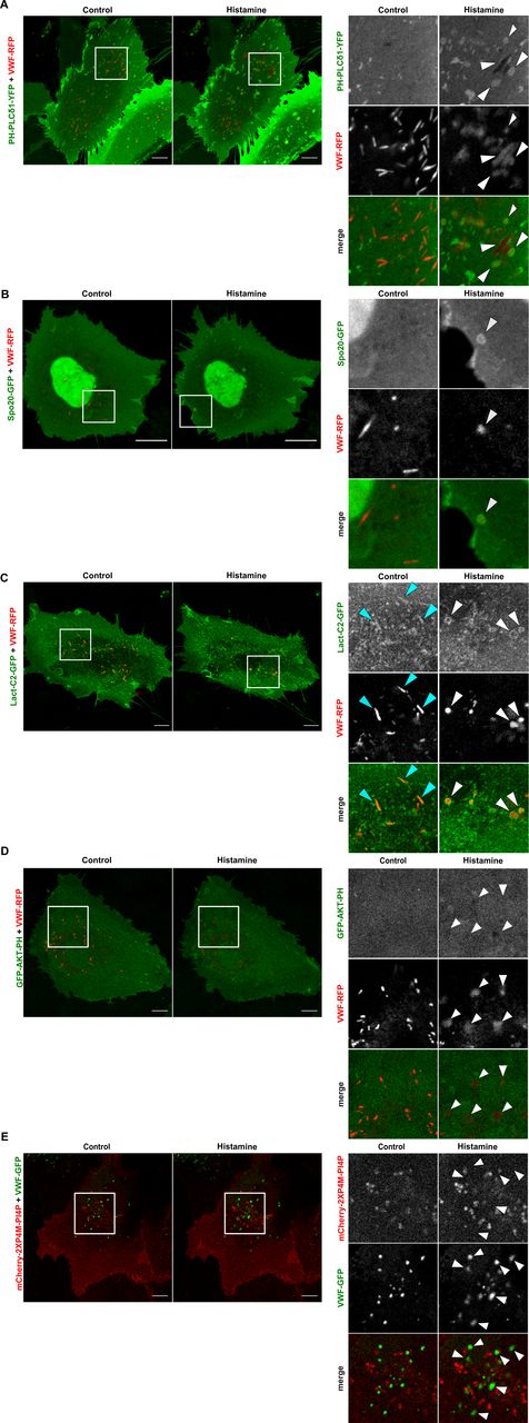
- Figure S1. PI(4,5)P2 and phosphatidic acid localize to Weibel–Palade body (WPB) fusion sites in histamine-stimulated HUVECs.
(A, B, C, D, E) HUVECs were transfected with either of PH-PLCδ1-YFP (A), Spo20-GFP (B), Lact-C2-GFP (C), GFP-AKT-PH (D), or mCherry-2XP4M-PI4P (E) and von-Willebrand factor-RFP or von-Willebrand factor-GFP as WPB marker and imaged by time-lapse confocal microscopy. Blue arrowheads denote a localization of phospholipid sensors on intracellular WPB at basal conditions and white arrowheads mark WPB-plasma membrane fusion sites after histamine stimulation. The ring-like structures positive for Lact-C2-GFP that appear at plasma membrane–WPB fusion sites in histamine-stimulated cells most likely represent a general membrane enrichment as they are not seen in the total internal reflection fluorescence imaging. Scale bar = 10 μm.
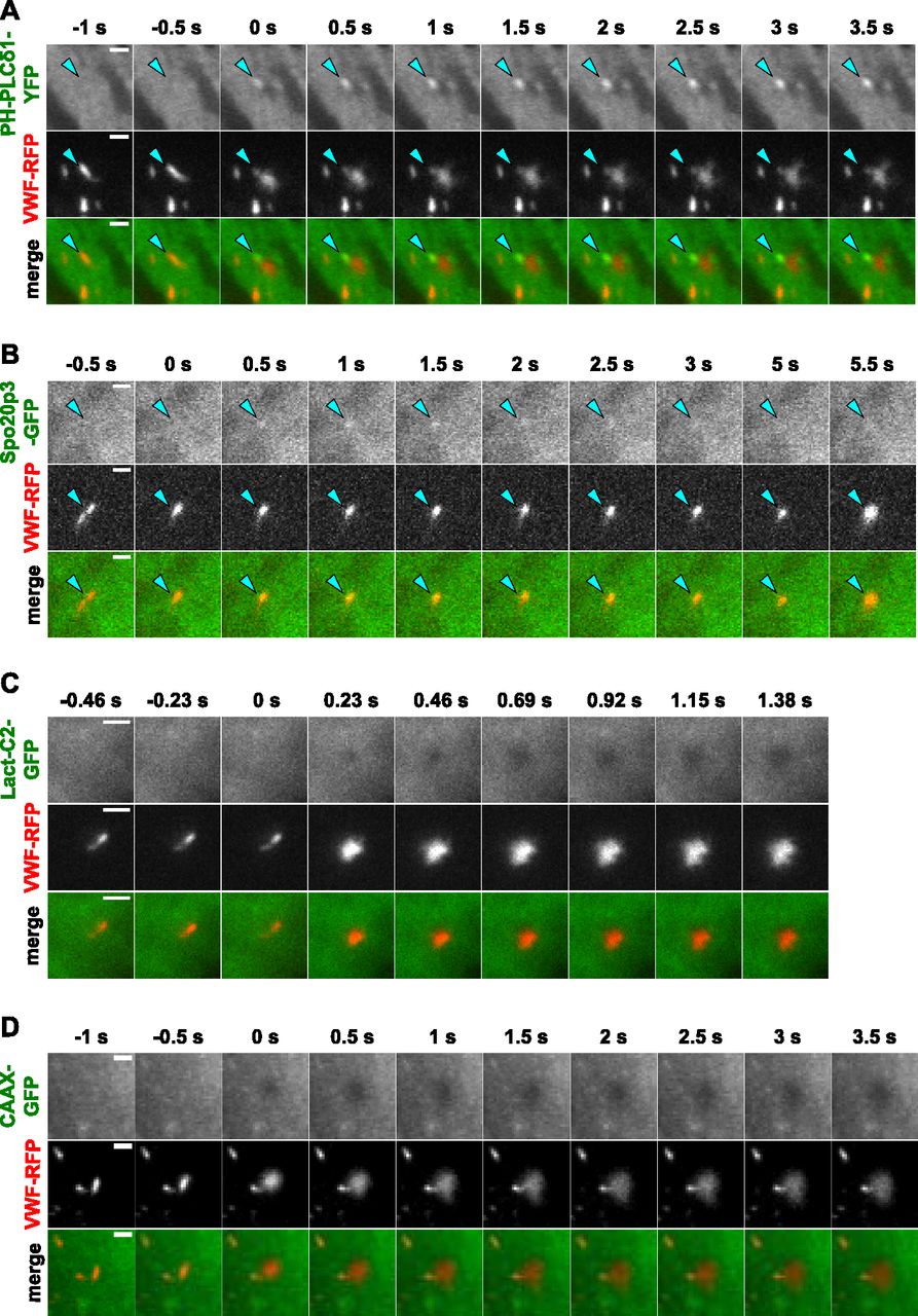
- Figure 1. PI(4,5)P2 and phosphatidic acid but not PS accumulate at Weibel–Palade body (WPB)–plasma membrane fusion sites.
(A, B, C, D) HUVECs expressing PH-PLCδ1-YFP (A), Spo20p3-GFP (B), Lact-C2-GFP (C), or CAAX-GFP (D) together with von-Willebrand factor-RFP as WPB marker were stimulated with 100 μM histamine and imaged by live-cell total internal reflection fluorescence microscopy. Still images show individual WPB undergoing exocytosis. Fusion occurred at t = 0 s. Note the transient enrichment of PH-PLCδ1-YFP and Spo20p3-GFP on the side of the actual fusion pore which appears darker in the total internal reflection fluorescence microscopy settings, most likely because the WPB membrane did not fully collapse into the plasma membrane during the time interval of the recordings. Scale bar = 2 μm.
- Figure 2. Neomycin treatment inhibits Ca2+-evoked von-Willebrand factor (VWF) secretion.
(A) HUVECs were treated with 2 mM neomycin for 20 min, incubated in agonist-free medium for 20 min, and then stimulated with 100 μM histamine for 20 min. VWF released into the cell culture supernatant was quantified by ELISA and secretion normalized to the total cellular VWF content (see the Materials and Methods section). n = 6, paired t test, ***P ≤ 0.001. Bars represent mean ± SEM. (B) HUVECs were treated as in (A), but stimulated with 10 μM ionomycin instead of histamine. n = 10, paired t test, ***P ≤ 0.001. Bars represent mean ± SEM. Note the marked reduction of evoked VWF secretion following neomycin treatment.
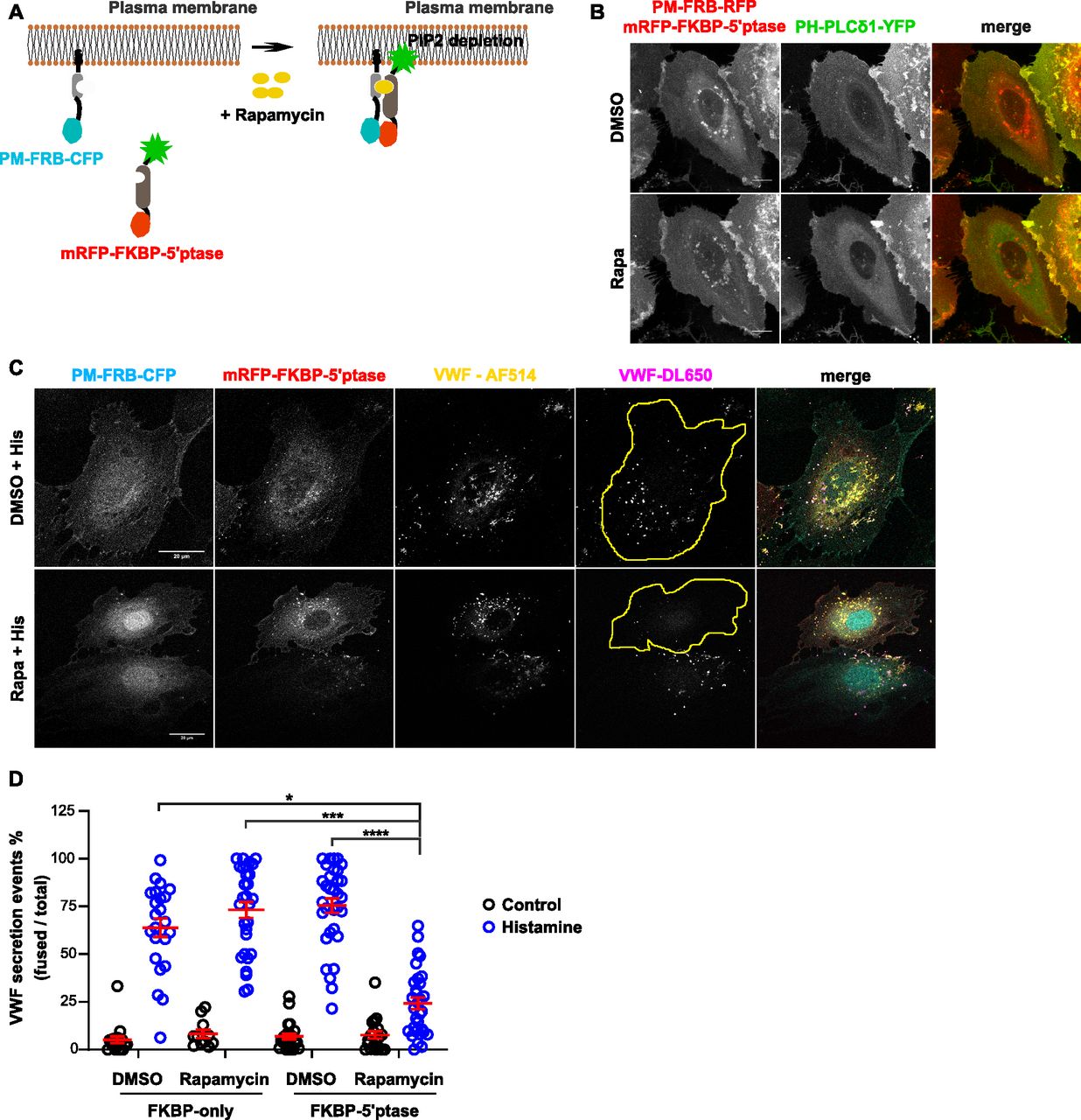
- Figure 3. Acute depletion of plasma membrane (PM) PI(4,5)P2 inhibits histamine-evoked Weibel–Palade body (WPB) exocytosis.
(A) Illustration depicting the rapamycin-inducible PI(4,5)P2 depletion system. A 5-ptase fused to mRFP-FKBP is expressed together with PM-targeted FRB-CFP. Rapamycin treatment induces the interaction of mRFP-FKBP-5-ptase with PM-FRB-CFP, causing a PM translocation of the 5-ptase and a resulting reduction of PM PI(4,5)P2. Adapted from Varnai et al (2006). (B) Representative maximum intensity projection images showing the PM localization of mRFP-FKBP-5-ptase and a PI(4,5)P2 depletion upon rapamycin treatment. HUVECs were transfected with PM-FRB-RFP, mRFP-FKBP-5-ptase, and the PI(4,5)P2 sensor PH-PLCδ1-YFP and live cell imaging was performed with time-lapse confocal microscopy, whereas rapamycin was added during acquisition. Translocation occurred between 3 and 7 min after rapamycin addition. In the flat HUVECs, this translocation is best seen in the rapamycin-induced increase of cytosolic PH-PLCδ1-YFP, which reflects itself in a more pronounced perinuclear fluorescence (thickest part of the cell containing most of the cytoplasm). Scale bar = 10 μM. (C, D) Representative confocal images and quantification of the effect of rapamycin-induced PM PI(4,5)P2 depletion on WPB exocytosis. HUVECs were transfected with PM-FRB-CFP and mRFP-FKPB-only or mRFP-FKBP-5-ptase. 24 h post-transfection cells were treated with rapamycin for 3 min, stimulated with histamine for 15 min, and subjected to the anti–von-Willebrand factor (VWF) antibody capture assay as described in the Materials and Methods section. VWF-AF514 labels total VWF, whereas VWF-DL650 only labels the secreted VWF and thus identifies sites of WPB exocytosis. Circumferences of cells positive for both PM-FRB-CFP and mRFP-FKBP-5-ptase are highlighted in yellow. (D) Shows a quantification of the anti-VWF antibody capture data from n = 3 independent experiments. Scatter plots represent mean ± SEM. Representative confocal images of the controls including mRFP-FKPB-only are shown in the Fig S2. Scale bar = 20 μM. Significance was tested with Kruskal–Wallis test, ***P < 0.0001.
- Figure S2. Acute depletion of plasma membrane (PM) PI(4,5)P2 interferes with transferrin internalization and histamine-evoked Weibel–Palade body (WPB) exocytosis.
(A, B) Transferrin uptake. HUVECs expressing PM-FRB-CFP and either mRFP-FKBP-5-ptase or mRFP-FKBP-only were subjected to control (DMSO) or rapamycin treatment and clathrin-mediated endocytosis was analyzed by uptake of AF488-labeled transferrin (Tf-AF488). (A) Confocal fluorescence images of the respective constructs and Tf-AF488 in individual cells (cellular circumference marked by a yellow line). (B) Total number of internalized Tf-AF488 puncta per cell quantified in n = 3 independent experiments. Significance tested with Kruskal–Wallis test (***P < 0.0001). Scatter plots represent mean ± SEM. (C) HUVECs transfected as in (A) were treated with rapamycin and/or histamine as indicated and subjected to the anti–von-Willebrand factor (VWF) antibody capture assay using AF514 and DL650 conjugated anti-VWF antibodies to label total and secreted VWF, respectively. Note that no effect on WPB number and distribution is observed after rapamycin treatment in resting cells and that histamine stimulation causes normal WPB exocytosis in the mRFP-FKBP–only expressing and rapamycin treated HUVECs. Scale bar = 20 μm.
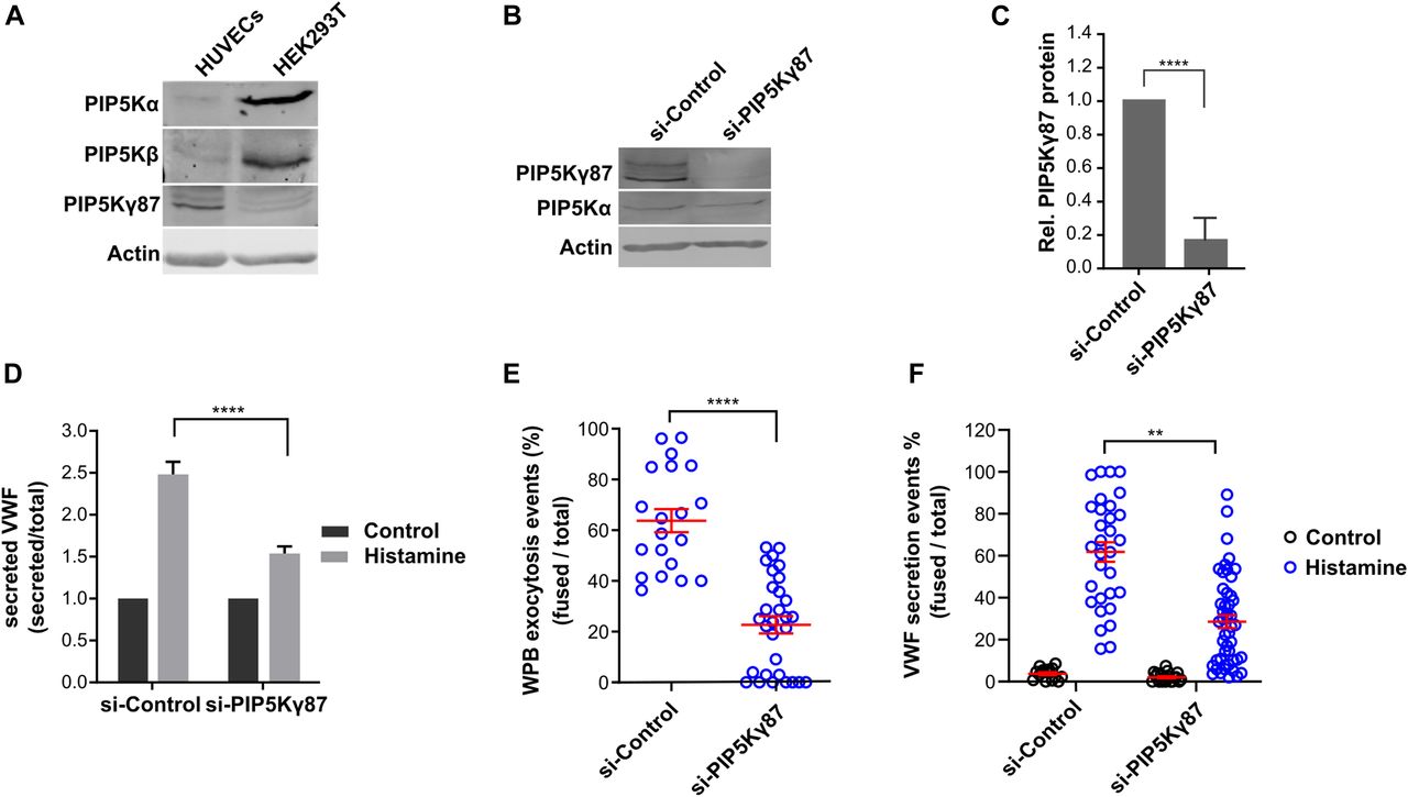
- Figure 4. PIP5Kγ87 knockdown decreases histamine-evoked Weibel–Palade body (WPB) exocytosis.
(A) Expression of PIP5Kα, PIP5Kβ, and PIP5Kγ in HUVECs and HEK293T. HUVECs and HEK293T cell lysates were subjected to Western blot analysis using isoform-specific antibodies. Actin served as loading control. (B, C) PIP5Kγ knockdown. HUVECs were transfected with either siRNA-control or siRNA-PIP5Kγ87, and the knockdown efficiency was analyzed by Western blot of total cell lysates and quantified. (D) Quantification of von-Willebrand factor (VWF) secretion in PIP5Kγ87 depleted HUVECs. HUVECs were transfected as in (B), stimulated with histamine for 15 min, and the amount of VWF released into the culture supernatant was measured by ELISA. Data were analyzed by two-way ANOVA Tukey test of n = 5 independent experiments, ****P < 0.0001. Bars represent mean ± SEM. (E) siRNA-control and siRNA-PIP5Kγ87 knockdown cells were transfected with VWF-RFP and cultured on μ-slide eight well glass bottom dishes. Live cell imaging was then performed using time-lapse confocal microscopy and histamine was added during acquisition. The percentage of VWF secretion events was calculated as the ratio of fusion events per total number of WPB present in the respective cell at basal condition, that is, before addition of histamine. n = 3 independent experiments, Mann Whitney test, ****P < 0.001. Scatter plots represent mean ± SEM. (F) HUVECs were transfected as in (B), and VWF secretion was measured using the anti-VWF antibody capture assay. The percentage of VWF secretion was calculated by dividing the total number of secretion events visualized by antibody capture by the total number of WPB present in the respective cell. n = 3 independent experiments, Kruskal–Wallis test, ****P < 0.001. Scatter plots represent mean ± SEM.
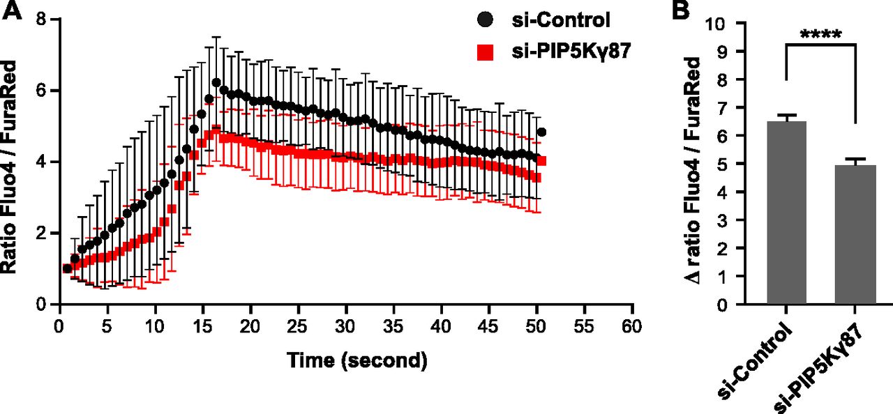
- Figure 5. PIP5Kγ87 knockdown partially inhibits intracellular calcium elevation in response to histamine stimulation.
PIP5Kγ87 control and knockdown cells were loaded with 2 μM of each, Fluo-4-AM and Fura Red-AM, and emission signals were recorded using live cell imaging by confocal microscopy. (A, B) The actual recordings for a representative cell are shown in (A), whereas (B) gives a quantification of the Ca2+ signals observed in n = 5 independent experiments with a total of 294 cells. Bars represent mean ± SEM. Significance was tested with Mann–Whitney test, ****P < 0.0001.
- Figure 6. The KRHH-PIP5Kγ87 mutant which is defective in phosphatidic acid binding is cytosolic and causes reduction of histamine-evoked Weibel–Palade body (WPB) exocytosis.
(A) Left, illustration highlighting the point mutations introduced in PIP5Kγ87. Right, localization of the different PIP5Kγ87 derivatives. HUVECs were transfected with either WT-PIP5Kγ87-GFP, KRHH-PIP5Kγ87-GFP, or KD-PIP5Kγ87-GFP and von-Willebrand factor (VWF)-RFP. Cells were fixed 15 h post-transfection, and the localization of the PIP5Kγ87 constructs was recorded by confocal microscopy. Scale bar = 10 μM. (B) HUVECs were transfected as in (A), and confocal live cell imaging was performed after addition of histamine. Still images show individual WPB undergoing exocytosis at t = 0 s. (C) Expression of KRHH-PIP5Kγ87-GFP interferes with histamine-evoked WPB exocytosis. Anti-VWF antibody capture assay of cells expressing WT-PIP5Kγ87-GFP, KRHH-PIP5Kγ87-GFP, or KD-PIP5Kγ87-GFP, respectively, in control or histamine-stimulated conditions. The number of fusion events and the total number of WPB were quantified using ImageJ and the percentage of WPB exocytosis events was calculated as the ratio of fusion events per total number of VWF-positive WPB. Data of n = 3 independent experiments were analyzed by Kruskal–Wallis test, **P < 0.01. Scatter plots represent mean ± SEM.
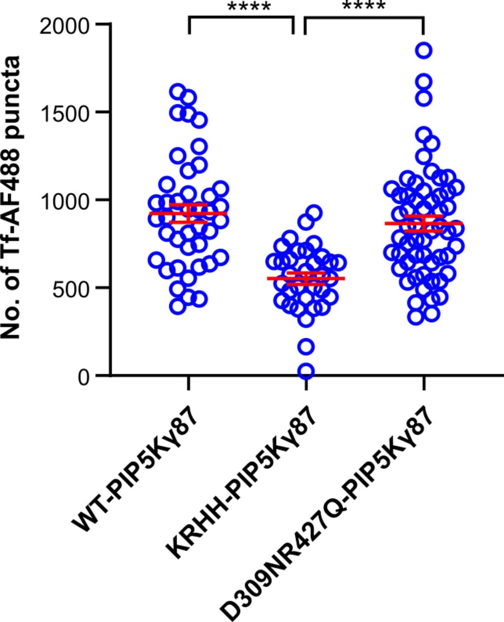
- Figure S3. Expression of KRHH-PIP5Kγ87-GFP affects transferrin endocytosis.
HUVECs expressing WT-PIP5Kγ87-GFP, KRHH-PIP5Kγ87-GFP, or KD-PIP5Kγ87-GFP, respectively, were subjected to the transferrin uptake assay using Tf-AF488. Tf-AF488 positive intracellular puncta were quantified using ImageJ in n = 3 independent experiments. Data were analyzed by Kruskal–Wallis test, ****P < 0.0001. Scatter plots represent mean ± SEM.
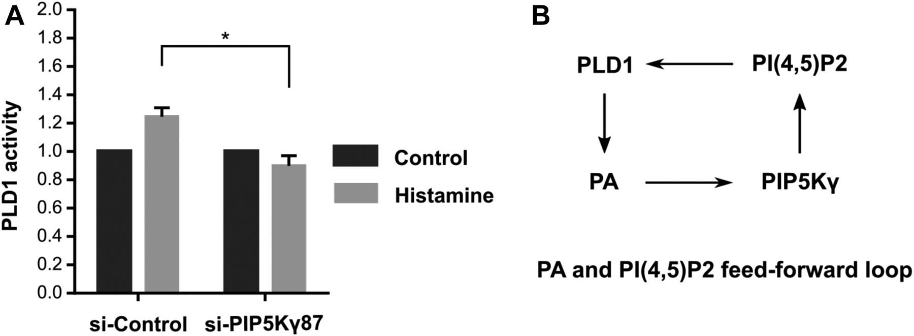
- Figure 7. PIP5Kγ87 depletion inhibits histamine-evoked PLD1 activation.
(A) HUVECs transfected with either siRNA-control or siRNA-PIP5Kγ87 were mock-treated (control) or treated with histamine (100 mM) for 20 min. PLD1 activity was then measured as described in the Materials and Methods section. Data from n = 3 independent experiments, statistical analysis used two-way ANOVA, *P < 0.05. Bars represent mean ± SEM. (B) Feed-forward model in which PI(4,5)P2 and phosphatidic acid regulate each other through recruitment/activation of the respective enzymes.


