


Article Figures & Data
Figures
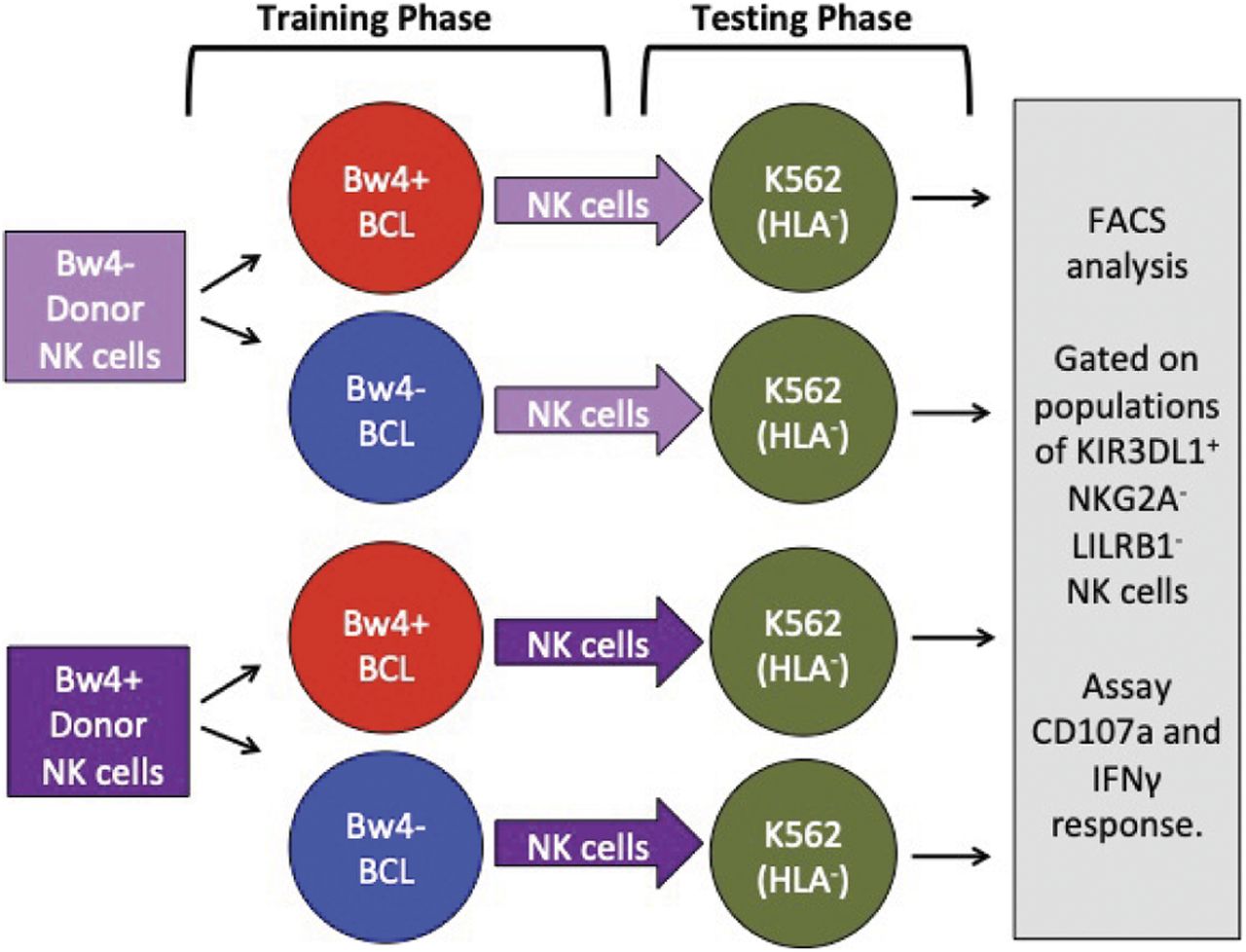
- Figure 1. General approach to in vitro NK cell education.
In the training phase (left), NK cells isolated from either Bw4+ or Bw4− donors are co-cultured with training cells that express either Bw4+HLA-B (educating) or Bw4−HLA-B (control). Testing lines are pretreated with etoposide VP-16. In the testing phase (right), K562 cells, which lack HLA class I, are added. The missing-self response of KIR3DL1+ NK cells to the HLA− targets is then measured by IFNγ and/or degranulation assays using flow cytometry.
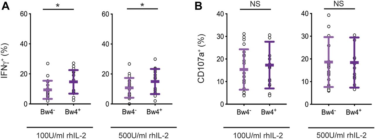
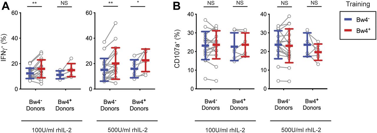
- Figure 2. After 5 d of in vitro cell culture, NK cell education is detectable by IFNγ production but not degranulation.
NK cells isolated from PBMCs were cultured in medium with rhIL-2 at the concentrations indicated. After 5 d, the K562 cells were added at a 10:1 E:T ratio, in medium with 500 U/ml rhIL-2 and anti-CD107a. 6 h later, the NK cells were stained and analyzed by flow cytometry. Shown are the mean ± SD of three experiments combined totaling 21 Bw4− donors and 18 Bw4+ donors. (A) Frequency of IFNγ+ cells in the viable KIR3DL1+NKG2A−LILRB1− NK cell gate. Shown are the results of a t test. *P < 0.05. (B) Frequency of CD107a+ cells in the viable KIR3DL1+NKG2A−LILRB1− NK cell gate. Shown are the results of a t test. NS = not significant.
Source data are available for this figure.
Source Data for Figure 2[LSA-2019-00434_SdataF2.xlsx]
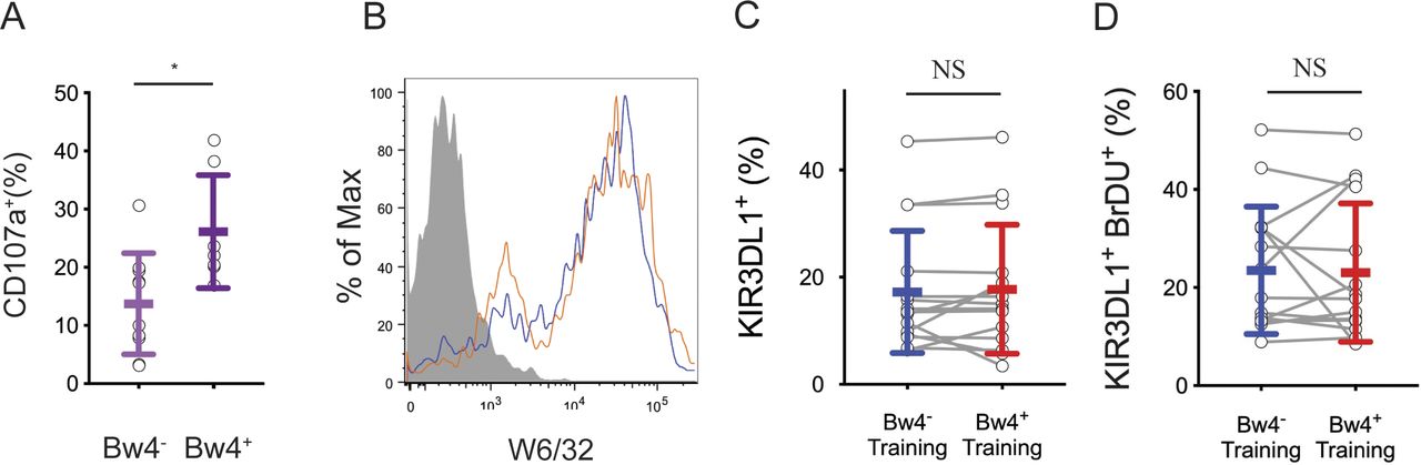
- Figure S1. In vivo NK cell education after 1 d of cell culture, HLA expression on training lines, and an analysis of NK cells after the training phase.
(A) NK cells from 10 Bw4− donors to 7 Bw4+ donors were isolated from PBMCs and cultured in media with 500 U/ml rhIL-2. 1 d later, K562 cells were added at a 10:1 E:T ratio in media with anti-CD107a. 6 h later, the NK cells were analyzed by flow cytometry. Shown is the frequency of CD107a+ cells in the KIR3DL1+KIR2D−NKG2A− gate. Shown is the result of an unpaired t test. *P < 0.05. (B) Flow cytometry histogram of cell lines Bw4+BCL (blue) and Bw4−BCL (orange) stained with anti-HLA class I antibody clone W6/32. HLA− K562 cells (gray filled) shown for reference. (C) NK cells were isolated from the PBMCs of 16 Bw4− donors and co-cultured with either Bw4+BCL cells or Bw4−BCL cells, each pretreated with etoposide. Training occurred in medium with 500 U/ml rhIL-2 and bromodeoxyuridine (BrDU) to track cell division. After 5 d, the NK cells were stained with a panel of surface antibodies and assayed by flow cytometry. Shown is the percentage of living KIR3DL1+ NK cells. The results of a paired t test is shown. (C, D) NK cells treated as in (C), but the percentage of living KIR3DL1+ BrDU+ NK cells is shown. The results of a paired t test is shown. NS = not significant.
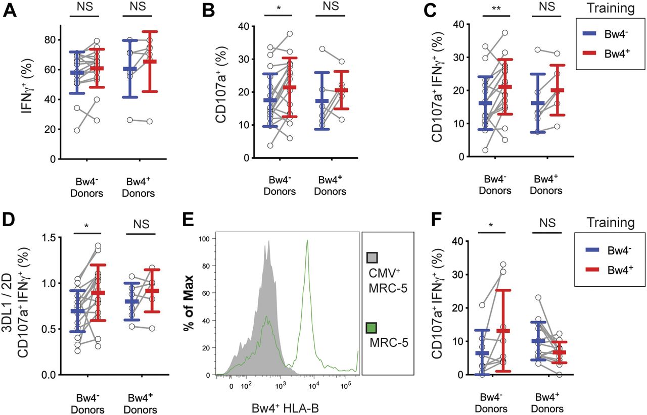
- Figure 3. In vitro training of KIR3DL1+ cells with Bw4+BCL improves the missing-self response by IFNγ production but not degranulation.
NK cells were isolated from the PBMCs of 17 Bw4− donors and six Bw4+ donors. NK cells were then co-cultured with either Bw4+BCL or Bw4−BCL at an 8:3 E:T ratio in medium with rhIL-2 at the concentrations indicated. After 5 d, K562 cells were added at a 10:1 E:T ratio, in medium with 500 U/ml rhIL-2 and anti-CD107a. 6 h later, NK cells were stained and analyzed by flow cytometry. All panels are representative of at least three replicate experiments. Mean ± SD are shown. (A) Frequency of IFNγ+ cells in the viable KIR3DL1+NKG2A−LILRB1− NK cell gate. Shown are the results of a Sidak’s multiple comparison test from paired two-way ANOVAs. **P < 0.01, *P < 0.05. (B) Frequency of CD107a+ cells in the viable KIR3DL1+NKG2A−LILRB1− NK cell gate. Shown are the results of a Sidak’s multiple comparison test from a paired two-way ANOVA. NS = not significant.
Source data are available for this figure.
Source Data for Figure 3[LSA-2019-00434_SdataF3.xlsx]
- Figure 4. In vitro training with Bw4+BCL and rhIL-12 maximizes the missing-self response of KIR3DL1+ NK cells.
For (A, B, C, D), NK cells were isolated from the PBMCs of 17 Bw4− donors and six Bw4+ donors. NK cells were then co-cultured with either Bw4+BCL or Bw4−BCL at an 8:3 E:T ratio in medium with 500 U/ml rhIL-2 and 50 ng/ml rhIL-12. After 5 d, K562 cells were added at a 10:1 E:T ratio, in medium with 500 U/ml rhIL-2 and anti-CD107a. 6 h later, NK cells were stained and analyzed by flow cytometry. (A, B, C, D) are representative of at least three replicate experiments. Mean ± SD are shown. (A) Frequency of IFNγ+ cells in the viable KIR3DL1+NKG2A−LILRB1− NK cell gate. Shown are the results of a Sidak’s multiple comparison test from a paired two-way ANOVA. (B) Frequency of CD107a+ cells in the viable KIR3DL1+NKG2A−LILRB1− NK cell gate. Shown are the results of a Sidak’s multiple comparison test from a paired two-way ANOVA. *P < 0.05. (C) Frequency of IFNγ+CD107a+ cells in the viable KIR3DL1+NKG2A−LILRB1− NK cell gate. Shown are the results of a Sidak’s multiple comparison test from a paired two-way ANOVA. **P < 0.01. (D) Frequency of IFNγ+CD107a+ cells in the viable KIR3DL1+NKG2A−LILRB1− NK cell gate divided by the frequency of IFNγ+CD107a+ cells in the viable KIR3DL1−KIR2D+NKG2A−LILRB1− NK cell gate. Shown are the results of a Sidak’s multiple comparison test from a paired two-way ANOVA. *P < 0.05. (E) MRC-5 cells were either co-cultured alone or with human CMV for 4 d. MRC-5 cells were then isolated and stained with anti-Bw4+HLA-B antibody and analyzed by flow cytometry. Viable MRC-5 cells are shown. (F) NK cells isolated from PBMCs were co-cultured with either Bw4+BCL or Bw4−BCL at an 8:3 E:T ratio. After 5 d, CMV-infected MRC-5 cells were added at a 10:1 E:T ratio. 6 h later, the NK cells were stained and analyzed by flow cytometry. Shown are the combined results of two experiments, representing three replicate experiments. The frequency of IFNγ+CD107a+ cells in the viable KIR3DL1+NKG2A−LILRB1− NK cell gate are shown. Shown are the results of a Sidak’s multiple comparison test from a paired two-way ANOVA. NS = not significant. *P < 0.05.
Source data are available for this figure.
Source Data for Figure 4[LSA-2019-00434_SdataF4.xlsx]
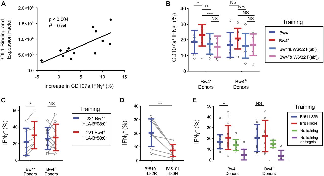
- Figure 5. The increased missing-self response of NK cells trained with Bw4+BCL is dependent on the binding of KIR3DL1 to HLA.
(A) Y-axis: AB&EF was assigned to each of 13 donors based on published KIR3DL1-HLA-B binding data (Boudreau et al, 2016), the HLA-B genotypes of Bw4+BCL, and the surface KIR3DL1 expression for each donor’s NK cells as measured by mean fluorescence intensity (MFI). X-axis: Difference between Bw4+BCL and Bw4−BCL training in the frequency of IFNγ+CD107a+ cells responding to missing-self, as presented in Fig 4D. Shown is the linear regression with the significance of non-zero slope and the goodness of fit calculation. These data are representative of at least three replicate experiments. (B) Bw4+BCL and Bw4−BCL were treated with F(ab′)2, fragments of the anti-HLA antibody W6/32. NK cells were isolated from PBMCs and co-cultured with either treated or untreated Bw4+BCL or Bw4−BCL. Cells were co-cultured at an 8:3 E:T ratio in medium with 500 U/ml rhIL-2 and 50 ng/ml rhIL-12. After 5 d, K562 cells were added at a 10:1 E:T ratio. 6 h later, the NK cells were stained and analyzed by flow cytometry. Shown is the frequency of IFNγ+CD107a+ cells in the viable KIR3DL1+NKG2A−LILRB1− NK cell gate. Shown are the combined results of two experiments comprising 11 Bw4− donors and 7 Bw4+ donors. These results represent at least three replicate experiments. Shown are mean ± SD and the results of a Sidak’s multiple comparison test from a paired two-way ANOVA. *P < 0.05, **P < 0.01, ***P < 0.001. (C) NK cells were isolated from PBMCs and co-cultured with 721.221 cells transfected to express either HLA-B*58:01 (Bw4+) or B*08:01 (Bw4−). The cells were co-cultured at an 8:3 E:T ratio in medium with 500 U/ml rhIL-2. After 3 d, the K562 cells were added at a 10:1 E:T ratio. 6 h later, the NK cells were stained and analyzed by flow cytometry. The frequency of IFNγ+ cells in the KIR3DL1+ gate is shown. Shown are combined data from two experiments comprising six Bw4− donors and nine Bw4+ donors. These data represent at least three replicate experiments. Shown are mean ± SD and the results of a Sidak’s multiple comparison tests from a paired two-way ANOVA. *P < 0.05. (D) 721.221 cells were transfected to express HLA-B*51:01 mutated to either abolish the Bw4 epitope (L82R, Bw4−), or leave it intact (I80N, Bw4+). The NK cells were isolated from PBMCs and co-cultured with either Bw4+I80N or Bw4−L82R in medium with 500 U/ml rhIL-2. 6 h later, the NK cells were stained with antibodies and analyzed by flow cytometry. The frequency of IFNγ+ cells in the KIR3DL1+ gate is shown. Shown are mean ± SD and the results of a t test. **P < 0.01. (E) NK cells were isolated from PBMC and co-cultured with 721.221 cells transfected to express HLA-B*51:01 with either the I80N (Bw4+) or L82R (Bw4−) mutation. Cells were co-cultured at an 8:3 E:T ratio in medium with 500 U/ml rhIL-2. Additional isolated NK cells were cultured alone in medium with 500 U/ml rhIL-2 (No training). After 3 d, K562 cells were added to co-cultures at a 10:1 E:T ratio, except for a portion of the NK cells cultured alone, which received only medium (No training or targets). 6 h later, NK cells were stained and analyzed by flow cytometry. The frequency of IFNγ+ cells in the KIR3DL1+NKG2A− gate is shown. Shown are the combined data comprising 20 Bw4− donors and 8 Bw4+ donors from two experiments. These data represent at least three replicate experiments. Shown are mean ± SD and the results of a Sidak’s multiple comparison tests from a paired two-way ANOVA. NS = not significant. *P < 0.05.
Source data are available for this figure.
Source Data for Figure 5[LSA-2019-00434_SdataF5.xlsx]
- Figure S2. A verification of F(ab′)2 efficacy, Bw4 expression on beads and mutated cell lines, and the results of NK cell training without etoposide treatment.
(A) Bw4+BCL cells, 221-B*58:01 cells, or K562 cells were each stained with W6/32 F(ab′)2 or left unstained for 30 min at either 37°C or 4°C. The cells were washed three times and then stained with fluorescently conjugated W6/32 antibody (anti-HLA class I) and assayed by flow cytometry. (B) Flow cytometry histograms of mutant HLA-B*51 cell lines. B*51-L82R cells and B*51-I80N were either stained with anti-HLA W6/32 (left panel) or anti-Bw4 epitope antibody (right panel), or left unstained, and assayed by flow cytometry. Dotted line = stained B*51-I80N. Red line = stained B*51-L82R. Green line = unstained B*51-I80N. Orange line = unstained B*51-L82R. (C) NK cells from nine Bw4− donors to five Bw4+ donors were co-cultured with either B*51-L82R or B*51-I80N, neither of which were treated with etoposide. After 3 d, the K562 cells were added to each co-culture at a 10:1 E:T ratio. After 6 h, the IFNγ response of KIR3DL1+ NK cells was assayed by flow cytometry. (D) NK cells were isolated from the PBMCs of 17 Bw4− donors and six Bw4+ donors. NK cells were then co-cultured with either Bw4+BCL or Bw4−BCL at an 8:3 E:T ratio in media with 500 U/ml rhIL-2. After 5 d, the NK cells were stained and analyzed by flow cytometry. Shown is the granzyme B expression as measured by MFI of cells in the living KIR3DL1+NKG2A−LILRB1− NK cell gate. Shown are mean ± SD and the results of a Sidak’s multiple comparison test from a paired two-way ANOVA. (E) Anti-CD2-DNAM1 beads were co-cultured for 24 h with NK cells from a Bw4+ donor or a Bw4− donor. Beads were then magnetically isolated from the co-cultures, stained with anti-Bw4 antibody, and assayed by flow cytometry. NS = not significant.
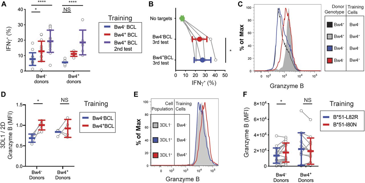
- Figure 6. Training NK cells from Bw4− donors with Bw4+BCL improves their missing-self response to multiple rounds of HLA− cells, and increases their expression of granzyme B.
(A) NK cells were isolated from the PBMCs of 13 Bw4− donors and six Bw4+ donors. Two replicate groups of NK cells were then co-cultured with Bw4+BCL and one group with Bw4−BCL, at an 8:3 E:T ratio in medium with 500 U/ml rhIL-2. After 5 d, K562 cells were added at a 10:1 E:T ratio (first testing). 6 h later, NK cells from co-cultures with Bw4−BCL and one of the Bw4+BCL co-cultures were stained and analyzed by flow cytometry. The other Bw4+BCL co-culture was rested for 24 h, after which more K562 cells were added at a 10:1 E:T ratio (second testing). 6 h later, NK cells were stained and analyzed by flow cytometry. The frequency of IFNγ+ cells in the viable KIR3DL1+NKG2A−LILRB1− NK cell gate is shown. These data represent at least three replicate experiments. Shown are mean ± SD and the results of a Sidak’s multiple comparison tests from a paired two-way ANOVA. *P < 0.05, ****P < 0.0001. (B) NK cells were isolated from the PBMCs of five Bw4− donors and co-cultured with either Bw4+BCL or Bw4−BCL at an 8:3 E:T ratio in medium with 500 U/ml rhIL-2 (day 1). On days 5, 7, and 9, K562 cells were added at a 10:1 E:T ratio in medium with 500 U/ml rhIL-2. 6 h after adding K562 cells on day 9, NK cells were stained and analyzed by flow cytometry. The frequency of IFNγ+ cells in the viable KIR3DL1+NKG2A−LILRB1− NK cell gate is shown. Shown are mean ± SD and the results of a paired t test. *P < 0.05. (C, D) NK cells were isolated from the PBMCs of four Bw4− donors and four Bw4+ donors. NK cells were then co-cultured with either Bw4+BCL or Bw4−BCL at an 8:3 E:T ratio in medium with 500 U/ml rhIL-2. After 5 d, K562 cells were added at a 10:1 E:T ratio, in medium with 500 U/ml rhIL-2. Two days later, the NK cells were stained and analyzed by flow cytometry. (C) Shown is a histogram of granzyme B expression of cells in the viable KIR3DL1+NKG2A−LILRB1− NK cell gate, obtained from concatenated flow cytometry data files for all eight donors. (D) Shown is the ratio between the granzyme B MFI for cells in the KIR3DL1+NKG2A−LILRB1− NK cell gate and the cells in the KIR2D+KIR3DL1−NKG2A−LILRB1− NK cell gate. Shown are mean ± SD and the results of a Sidak’s multiple comparison tests from a paired two-way ANOVA. *P < 0.05. (E, F) NK cells were isolated from PBMCs and co-cultured with 721.221 cells transfected to express HLA-B*51:01 with either the I80N (Bw4+) or L82R (Bw4−) mutation. The cells were co-cultured at an 8:3 E:T ratio in medium with 500 U/ml rhIL-2. After 5 d, K562 cells were added at a 10:1 E:T ratio. After 2 more days, NK cells were stained and analyzed by flow cytometry. (E) Shown is a histogram of granzyme B expression for viable cells in either the KIR3DL1+NKG2A−LILRB1− NK cell gate or the KIR3DL1−NKG2A−LILRB1− NK cells gate. These data were obtained from concatenated flow cytometry data files from seven Bw4− donors to five Bw4+ donors. (F) Shown is the granzyme B expression as measured by MFI of cells in the viable KIR3DL1+NKG2A−LILRB1− NK cell gate. These data were obtained from 18 Bw4− donors to 13 Bw4+ donors from three replicate experiments. Shown are mean ± SD and the results of a Sidak’s multiple comparison tests from a paired two-way ANOVA. NS = not significant. *P < 0.05.
Source data are available for this figure.
Source Data for Figure 6[LSA-2019-00434_SdataF6.xlsx]
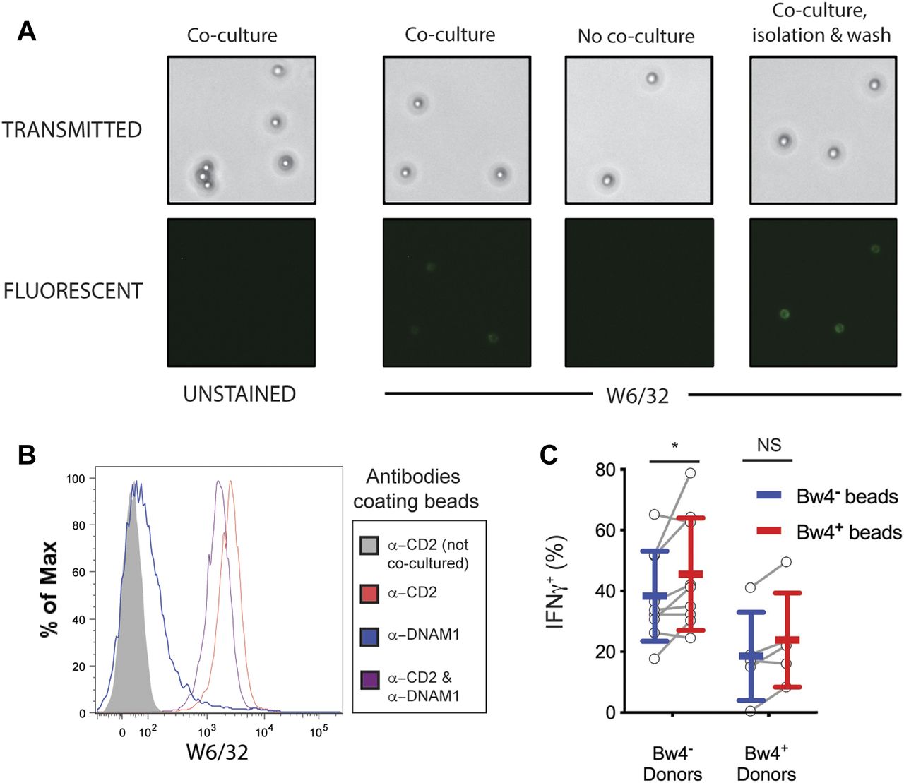
- Figure 7. Synthetic beads displaying Bw4+HLA-B are sufficient to educate KIR3DL1+ NK cells in vitro.
(A) Biotinylated antibodies against CD2 and DNAM1 were bound to streptavidin-coated paramagnetic beads and co-cultured with NK cells isolated from PBMC for 2 d in medium with 500 U/ml rhIL-2. The beads were separated with a magnet, stained with anti-HLA class I, and analyzed by light and fluorescent microscopy. (B) NK cells were co-cultured for 2 d with paramagnetic beads coated with either anti-DNAM1, anti-CD2, or both anti-DNAM1 and anti-CD2 antibodies. Co-cultures occurred in medium with 500 U/ml rhIL-2. Beads were isolated with a magnet, stained with anti-HLA class I and Alexa Fluor 700–labeled streptavidin, and analyzed by flow cytometry. Shown is the HLA signal of particles in the streptavidin+ gate, which excludes all cells. Representative of at least three replicate experiments. (C) Two sets of reverse-trogocytosed beads were constructed by co-culturing beads coated with anti-DNAM1 and anti-CD2 antibodies with NK cells from either Bw4+ or Bw4− donors. This produced beads coated in NK cell-derived membranes displaying Bw4+ or Bw4− HLA-B. Beads were then isolated with a magnet, washed, and co-cultured with NK cells isolated from either Bw4+ or Bw4− donors. Co-cultures occurred in medium with 500 U/ml rhIL-2. After 7 d, NK cells were separated from the beads with a magnet and combined with K562 cells at a 10:1 E:T ratio. 6 h later, the NK cells were stained with antibodies and analyzed by flow cytometry. Shown is combined data comprising nine Bw4− donors and five Bw4+ donors from three replicate experiments. Shown are mean ± SD and the results of a Sidak’s multiple comparison tests from a paired two-way ANOVA. NS = not significant. *P < 0.05.
Source data are available for this figure.
Source Data for Figure 7[LSA-2019-00434_SdataF7.xlsx]


Tables
- Table 1.
Results of in vitro training with Bw4 on the missing-self response of KIR3DL1+ NK cells.
Donor Allotype Bw4− % Change (IFNγ) % Change (CD107a) Bw4+ % Change (IFNγ) % Change (CD107a) Training Cell Allotype Bw4− Bw4+ Bw4− Bw4+ Type of Response to K562 IFNγ+ (%) CD107a+ (%) IFNγ+ (%) CD107a+ (%) IFNγ+ (%) CD107a+ (%) IFNγ+ (%) CD107a+ (%) Training conditions Low IL2 10.5 22.4 14.5 22.9 38.1** 2.2 6.9 20.7 11.6 19.1 68.1** −7.7 High IL2 14.8 19.8 19.7 18.7 33.1** −5.6 11.9 15.9 22.0 14.8 84.8* −6.9 NK cells were co-cultured with either Bw4+BCL or Bw4−BCL in medium with either 100 U/ml rhIL-2 or 500 U/ml rhIL-2. After 5 d, K562 cells were added at a 10:1 E:T ratio, in medium with 500 U/ml rhIL-2 and anti-CD107a. 6 h later, the NK cells were stained and analyzed by flow cytometry. Combined data from three experiments, totaling 25 Bw4− donors and 20 Bw4+ donors, are shown. The frequency of either CD107a+ or IFNγ+ NK cells in the viable KIR3DL1+NKG2A−LILRB1− NK cell gate is shown. Percent change was calculated as: ([Bw4+ training − Bw4− training]/Bw4− training). Bold type indicates a significant statistical result as assessed by Sidak’s multiple comparison test from paired two-way ANOVAs. Asterisks denote the degree of significance: **P < 0.01, *P < 0.05.


In this Issue
Related Articles
Cited By...
- No citing articles found.