Advertisement

Main menu

  • Home
  • Articles
    • Newest Articles
    • Current Issue
    • Methods & Resources
    • Archive
    • Subjects
  • Collections
  • Submit
    • Submit a Manuscript
    • Author Guidelines
    • License, Copyright, Fee
    • FAQ
    • Why Submit
  • About
    • About Us
    • Editors & Staff
    • Board Members
    • Licensing and Reuse
    • Reviewer Guidelines
    • Privacy Policy
    • Advertise
    • Contact Us
    • LSA LLC
  • Alerts
  • Other Publications
    • EMBO Press
    • The EMBO Journal
    • EMBO reports
    • EMBO Molecular Medicine
    • Molecular Systems Biology
    • Rockefeller University Press
    • Journal of Cell Biology
    • Journal of Experimental Medicine
    • Journal of General Physiology
    • Cold Spring Harbor Laboratory Press
    • Genes & Development
    • Genome Research

User menu

  • My alerts

Search

  • Advanced search
Life Science Alliance
  • Other Publications
    • EMBO Press
    • The EMBO Journal
    • EMBO reports
    • EMBO Molecular Medicine
    • Molecular Systems Biology
    • Rockefeller University Press
    • Journal of Cell Biology
    • Journal of Experimental Medicine
    • Journal of General Physiology
    • Cold Spring Harbor Laboratory Press
    • Genes & Development
    • Genome Research
  • My alerts
Life Science Alliance

Advanced Search

  • Home
  • Articles
    • Newest Articles
    • Current Issue
    • Methods & Resources
    • Archive
    • Subjects
  • Collections
  • Submit
    • Submit a Manuscript
    • Author Guidelines
    • License, Copyright, Fee
    • FAQ
    • Why Submit
  • About
    • About Us
    • Editors & Staff
    • Board Members
    • Licensing and Reuse
    • Reviewer Guidelines
    • Privacy Policy
    • Advertise
    • Contact Us
    • LSA LLC
  • Alerts
  • Follow lsa Template on Twitter
Research Article
Transparent Process
Open Access

ATP hydrolysis by KaiC promotes its KaiA binding in the cyanobacterial circadian clock system

Yasuhiro Yunoki, Kentaro Ishii, Maho Yagi-Utsumi, Reiko Murakami, Susumu Uchiyama, View ORCID ProfileHirokazu Yagi  Correspondence email, View ORCID ProfileKoichi Kato  Correspondence email
Yasuhiro Yunoki
1Graduate School of Pharmaceutical Sciences, Nagoya City University, Nagoya, Japan
3Institute for Molecular Science, National Institutes of Natural Sciences, Okazaki, Japan
  • Find this author on Google Scholar
  • Find this author on PubMed
  • Search for this author on this site
Kentaro Ishii
1Graduate School of Pharmaceutical Sciences, Nagoya City University, Nagoya, Japan
2Exploratory Research Center on Life and Living Systems (ExCELLS), National Institutes of Natural Sciences, Okazaki, Japan
3Institute for Molecular Science, National Institutes of Natural Sciences, Okazaki, Japan
  • Find this author on Google Scholar
  • Find this author on PubMed
  • Search for this author on this site
Maho Yagi-Utsumi
1Graduate School of Pharmaceutical Sciences, Nagoya City University, Nagoya, Japan
2Exploratory Research Center on Life and Living Systems (ExCELLS), National Institutes of Natural Sciences, Okazaki, Japan
3Institute for Molecular Science, National Institutes of Natural Sciences, Okazaki, Japan
  • Find this author on Google Scholar
  • Find this author on PubMed
  • Search for this author on this site
Reiko Murakami
1Graduate School of Pharmaceutical Sciences, Nagoya City University, Nagoya, Japan
  • Find this author on Google Scholar
  • Find this author on PubMed
  • Search for this author on this site
Susumu Uchiyama
2Exploratory Research Center on Life and Living Systems (ExCELLS), National Institutes of Natural Sciences, Okazaki, Japan
4Department of Biotechnology, Graduate School of Engineering, Osaka University, Osaka, Japan
  • Find this author on Google Scholar
  • Find this author on PubMed
  • Search for this author on this site
Hirokazu Yagi
1Graduate School of Pharmaceutical Sciences, Nagoya City University, Nagoya, Japan
  • Find this author on Google Scholar
  • Find this author on PubMed
  • Search for this author on this site
  • ORCID record for Hirokazu Yagi
  • For correspondence: hyagi@phar.nagoya-cu.ac.jp
Koichi Kato
1Graduate School of Pharmaceutical Sciences, Nagoya City University, Nagoya, Japan
2Exploratory Research Center on Life and Living Systems (ExCELLS), National Institutes of Natural Sciences, Okazaki, Japan
3Institute for Molecular Science, National Institutes of Natural Sciences, Okazaki, Japan
  • Find this author on Google Scholar
  • Find this author on PubMed
  • Search for this author on this site
  • ORCID record for Koichi Kato
  • For correspondence: kkato@excells.orion.ac.jp
Published 3 June 2019. DOI: 10.26508/lsa.201900368
  • Article
  • Figures & Data
  • Info
  • Metrics
  • Reviewer Comments
  • PDF
Loading

Article Figures & Data

Figures

  • Tables
  • Figure 1.
    • Download figure
    • Open in new tab
    • Download powerpoint
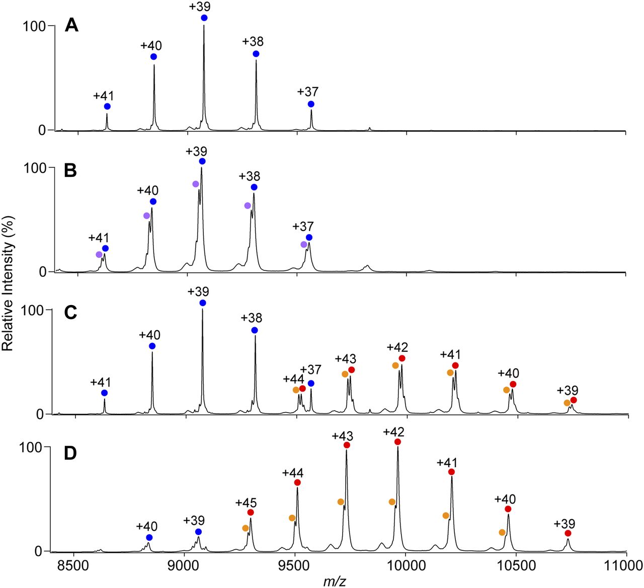
    Figure 1. KaiA–KaiC interaction depends on ATP hydrolysis.

    (A–D) Native mass spectra of (A, B) KaiCAA and (C, D) 6:3 mixtures of KaiCAA and KaiA in the presence of (A, C) 1 mM AMPPNP or (B, D) 1 mM ATP. After 5 h of incubation at 37°C with ATP or AMPPNP, the KaiC solutions with or without KaiA were immediately analyzed by nanoflow electrospray ionization MS. The blue and purple circles show the ion series of the KaiCAA homohexamer, whereas the orange and red circles show the 2:6 KaiA–KaiCAA hetero-octamer complexes. See Tables 1 and 2 for assignment details.

  • Figure S1.
    • Download figure
    • Open in new tab
    • Download powerpoint
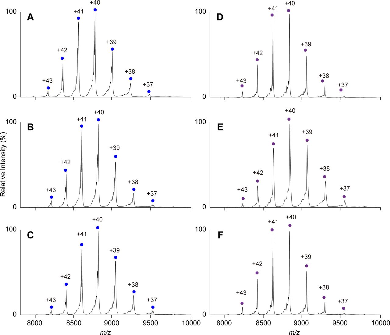
    Figure S1. Native MS characterization of KaiA–KaiCDD interaction with ATP or AMPPNP.

    (A–D) Native mass spectra of (A, B) KaiCDD and (C, D) 6:3 mixtures of KaiCDD and KaiA in the presence of (A, C) 1 mM AMPPNP or (B, D) 1 mM ATP. After 5 h of incubation at 37°C with the nucleotides, the KaiC solutions with or without KaiA were immediately analyzed by nanoflow electrospray ionization MS. The blue and purple circles show the ion series of the KaiCDD homohexamer, whereas the red circles show the 2:6 KaiA–KaiCAA hetero-octamer complexes. See Tables 1 and 2 for assignment details.

  • Figure S2.
    • Download figure
    • Open in new tab
    • Download powerpoint
    Figure S2. Native MS characterization of KaiCAA nucleotide state depending on external ATP/ADP condition.

    (A–F) Native mass spectra of KaiCAA mediated by (A–C) ATP and (D–F) AMPPNP. The KaiCAA hexamers incubated for 5 h at 37°C under (A, D) 100:0, (B, E) 75:25, and (C, F) 50:50 ATP/ADP conditions were immediately analyzed by nanoflow electrospray ionization MS. The blue and purple circles show the ion series of the KaiCAA hexamers containing seven ATP/five ADP molecules and 12 AMPPNP molecules, respectively.

  • Figure S3.
    • Download figure
    • Open in new tab
    • Download powerpoint
    Figure S3. Native MS characterization of nucleotide states of the CI and CII domains on KaiCAA.

    (A–C) Native mass spectra of KaiCAA mediated by ATP after trypsin digestion. After 5 h of incubation at 37°C in the presence of 1 mM ATP, KaiCAA was buffer-exchanged into 150 mM aqueous ammonium acetate and digested by 0.02 mg/ml trypsin for (A) 0 min, (B), 30 min, and (C) 60 min. The reaction mixture was directly analyzed by nanoflow electrospray ionization MS. The blue circles show the ion series of the KaiCAA homohexamer containing seven ATP and five ADP molecules, whereas the black circles show the hexameric CI domain (M1–S253) containing six ATP molecules. The CII domain was hardly detected as hexamer under the condition used here.

  • Figure S4.
    • Download figure
    • Open in new tab
    • Download powerpoint
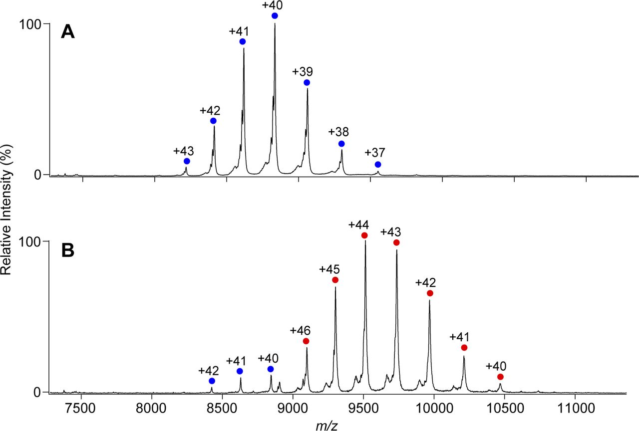
    Figure S4. Native MS characterization of the KaiA–KaiCAA/E77Q interaction.

    (A–D) Native mass spectra of (A) KaiCAA/E77Q and (B) a 6:3 mixtures of KaiCAA/E77Q and KaiA. After 5 h of incubation at 37°C in the presence of 1 mM ATP, the KaiCAA/E77Q solutions with or without KaiA were immediately analyzed by nanoflow electrospray ionization MS. The blue circles show the ion series of the KaiCAA/E77Q homohexamer, whereas the red circles show the 2:6 KaiA–KaiCAA/E77Q hetero-octamer complexes. See Table 2 for assignment details.

  • Figure S5.
    • Download figure
    • Open in new tab
    • Download powerpoint
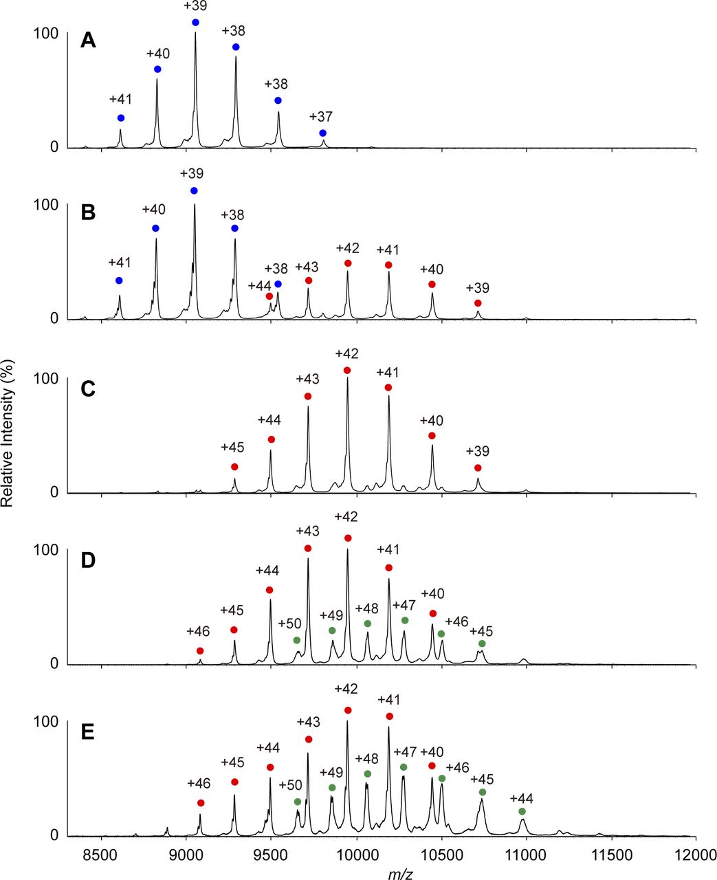
    Figure S5. Native MS analysis of KaiCAA-KaiA complex formation.

    (A–E) Native mass spectra of (A) KaiCAA and (B) 6:1, (C) 6:3, (D) 6:6, and, (E) 6:9 mixtures of KaiCAA and KaiA in the presence of 1 mM ATP. After 5 h of incubation at 37°C in the presence of 1 mM ATP, the KaiC solutions with or without KaiA were immediately analyzed by nanoflow electrospray ionization MS. The blue circles show the ion series of the KaiCAA homohexamer, whereas the red and green circles show the 2:6 and 4:6 KaiA–KaiCAA complexes, respectively.

  • Figure S6.
    • Download figure
    • Open in new tab
    • Download powerpoint
    Figure S6. 1H-15N HSQC spectra of KaiCAA and its C-terminally truncated mutant.

    (A–D) 1H-15N HSQC spectra of (A, B) KaiCAA and (C, D) the mutated KaiCAA lacking the C-terminal segment 487–518 in the presence of (A, C) 1 mM AMPPNP and (B, D) 1 mM ATP.

  • Figure 2.
    • Download figure
    • Open in new tab
    • Download powerpoint
    Figure 2. ATP hydrolysis–dependent conformational change of the C-terminal KaiA-binding region of KaiC.

    (A–C) 1H-15N HSQC spectrum of KaiCAA in the presence of (A) AMPPNP, (B) ATP, and (C) KaiA and ATP. NMR experiments were set up to take a total time of 3 h using the KaiC hexamer incubated with AMPPNP or ATP for 9 h. Assignments of the peaks from the C-terminal region are given in each spectrum. (D) Plot of relative peak intensity for KaiCAA resonances in the presence of AMPPNP versus ATP. (E) Plot of relative peak intensity for KaiCAA resonances in the presence versus absence of KaiA under the ATP condition. In (D) and (E), the residues that yielded no observable peaks under the AMPPNP condition are highlighted in red, whereas the asterisks indicate the proline residues and residues whose chemical shift perturbation data could not be obtained because of severe peak overlapping. (F) Crystal structure of two KaiC protomers in cartoon and surface representation, respectively, in the KaiC homohexameric ring mediated by AMPPNP (PDB ID code: 4O0M). In the crystal structure,the C-terminal region comprises a U-shaped A-loop (Glu487-Ile497) (orange) and a solvent-exposed C-tail (S498-S518), in which only the Ser498-Glu504 part (green) was modeled. The three residues (i.e., Gly488, Ile489, and Ile497) located in the A-loop, whose HSQC peaks were unobserved under the AMPPNP condition, are colored blue. The A-loop and AMPPNP molecule (red) are mediated by a loop comprising residues 415–430 (termed 422-loop, magenta).

  • Figure 3.
    • Download figure
    • Open in new tab
    • Download powerpoint
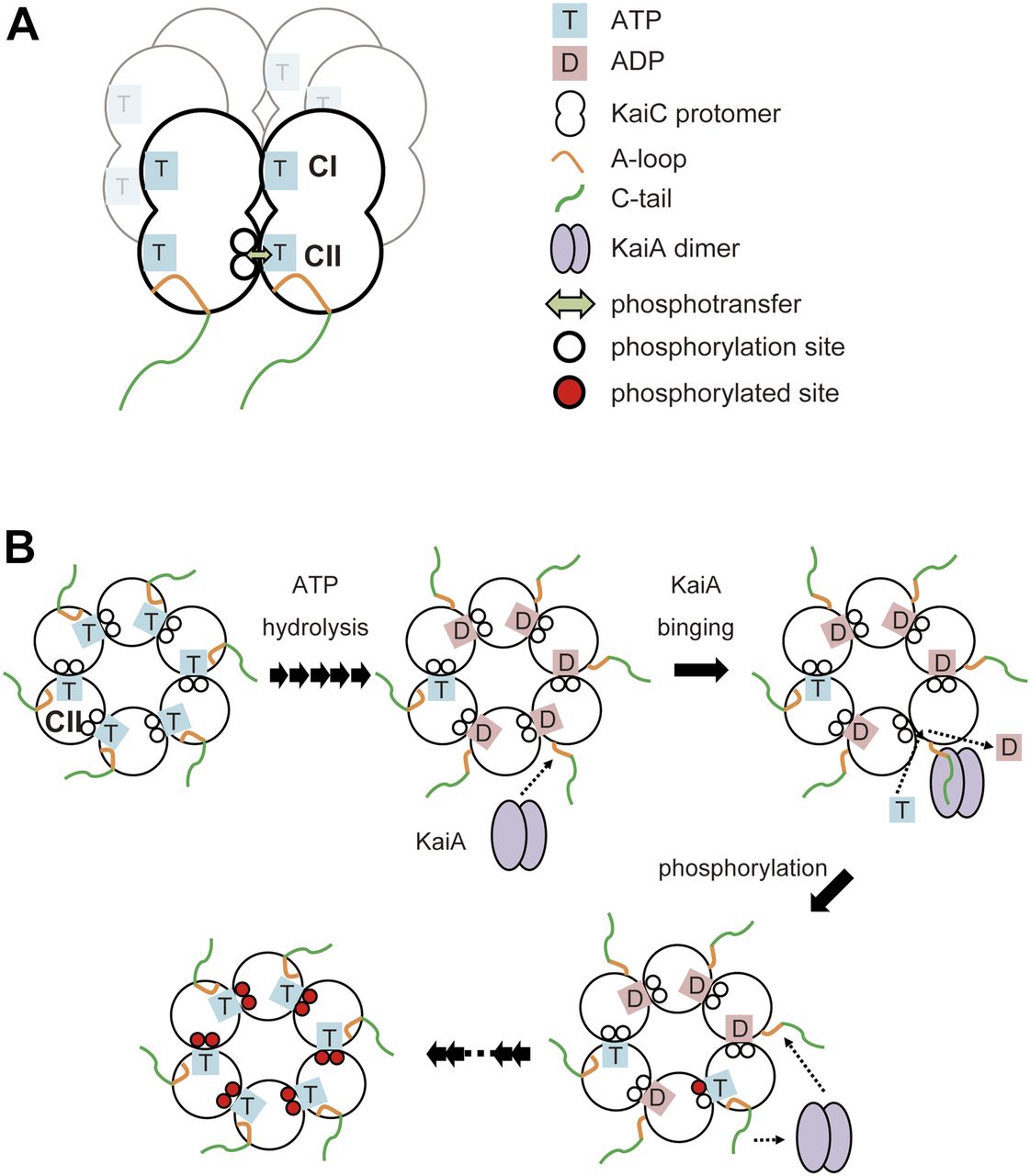
    Figure 3. The “fishing a line” mechanism coupling ATP hydrolysis and KaiA-mediated up-regulation of autophosphorylation in the KaiC hexamer.

    (A) While both CI and CII domains harbor nucleotide-binding sites and ATPase-active sites at the subunit interfaces, the autokinase activity is exerted only in the CII domain. This is because the autophosphorylation sites (i.e., Ser431 and Thr432) are spatially proximal to the ATP molecule accommodated in the CII domain of the neighboring protomer. (B) In the CII AAA+ ring hexamer, ATP hydrolysis releases the A-loop, which thereby becomes reactive with KaiA. KaiA binding to the C-terminal segments of KaiC facilitates ADP release and ATP incorporation. The rapid ATP/ADP turnover leads to the up-regulation of autophosphorylation of KaiC.

Tables

  • Figures
    • View popup
    Table 1.

    Summary of native MS characterization of KaiC and KaiA–KaiC complex formed in the presence of AMPPNP.

    Figure numberIon seriesAssignmentTheoretical mass (D)Experimental mass (D)Δm (D)aRelative quantity (%)
    Protein complexAMPPNP numberMg2+ number
    Fig 1ABlueKaiCAA61212353,850353,857 ± 10−7—
    Fig 1CBlueKaiCAA61212353,850353,855 ± 9−5—
    Fig 1CRedKaiCAA6/KaiA21112418,838418,896 ± 27−5853b
    Fig 1COrangeKaiCAA6/KaiA21012418,332418,412 ± 28−8047b
    Fig S2ABlueKaiCDD61212354,396354,375 ± 1221—
    Fig S2CBlueKaiCDD61212354,396354,447 ± 16−51—
    Fig S2CRedKaiCDD6/KaiA21212419,890420,076 ± 47−186—
    • ↵a Δm is the mass difference between the experimental mass and the theoretical mass.

    • ↵b Relative quantity of two ion series are shown.

    • View popup
    Table 2.

    Summary of native MS characterization of KaiC and KaiA–KaiC complex formed in the presence of ATP.

    Figure numberIon seriesAssignmentTheoretical mass (D)Experimental mass (D)Δm (D)aRelative quantity (%)
    Protein complexATP numberADP numberMg2+ number
    Fig 1BBlueKaiCAA67512353,462353,476 ± 18−1456b
    Fig 1BPurpleKaiCAA67312352,608352,593 ± 161544b
    Fig 1DBlueKaiCAA67512353,462353,461 ± 141—
    Fig 1DRedKaiCAA6/KaiA26512418,449418,445 ± 20467b
    Fig 1DOrangeKaiCAA6/KaiA201112417,969417,963 ± 20633b
    KaiCAA6/KaiA25512417,942417,963 ± 20−21
    Fig S1BBlueKaiCDD611012353,901353,902 ± 11−1—
    KaiCDD66612353,928353,902 ± 1126
    Fig S1DBlueKaiCDD66612353,928353,924 ± 9474b
    KaiCDD611012353,901353,924 ± 9−23
    Fig S1DPurpleKaiCDD66512353,501353,477 ± 92426b
    KaiCDD601212353,448353,477 ± 9−29
    Fig S1DRedKaiCDD6/KaiA221012419,102419,102 ± 320—
    KaiCDD6/KaiA27412419,075419,102 ± 32−27
    Fig S4ABlueKaiCAA/E77Q67512353,396353,383 ± 1313—
    Fig S4BBlueKaiCAA/E77Q612012353,796353,766 ± 2930—
    RedKaiCAA/E77Q6/KaiA27412418,533418,559 ± 59−26—
    • ↵a Δm is the mass difference between the experimental mass and the theoretical mass.

    • ↵b Relative quantity of two ion series are shown.

PreviousNext
Back to top
Download PDF
Article Alerts
Sign In to Email Alerts with your Email Address
Email Article

Thank you for your interest in spreading the word on Life Science Alliance.

NOTE: We only request your email address so that the person you are recommending the page to knows that you wanted them to see it, and that it is not junk mail. We do not capture any email address.

Enter multiple addresses on separate lines or separate them with commas.
ATP hydrolysis by KaiC promotes its KaiA binding in the cyanobacterial circadian clock system
(Your Name) has sent you a message from Life Science Alliance
(Your Name) thought you would like to see the Life Science Alliance web site.
CAPTCHA
This question is for testing whether or not you are a human visitor and to prevent automated spam submissions.
Citation Tools
ATP-dependent KaiA–KaiC interaction
Yasuhiro Yunoki, Kentaro Ishii, Maho Yagi-Utsumi, Reiko Murakami, Susumu Uchiyama, Hirokazu Yagi, Koichi Kato
Life Science Alliance Jun 2019, 2 (3) e201900368; DOI: 10.26508/lsa.201900368

Citation Manager Formats

  • BibTeX
  • Bookends
  • EasyBib
  • EndNote (tagged)
  • EndNote 8 (xml)
  • Medlars
  • Mendeley
  • Papers
  • RefWorks Tagged
  • Ref Manager
  • RIS
  • Zotero
Share
ATP-dependent KaiA–KaiC interaction
Yasuhiro Yunoki, Kentaro Ishii, Maho Yagi-Utsumi, Reiko Murakami, Susumu Uchiyama, Hirokazu Yagi, Koichi Kato
Life Science Alliance Jun 2019, 2 (3) e201900368; DOI: 10.26508/lsa.201900368
del.icio.us logo Digg logo Reddit logo Twitter logo CiteULike logo Facebook logo Google logo Mendeley logo
  • Tweet Widget
  • Facebook Like
Issue Cover

In this Issue

Volume 2, No. 3
June 2019
  • Table of Contents
  • Cover (PDF)
  • About the Cover
  • Masthead (PDF)
Advertisement

Jump to section

  • Article
    • Abstract
    • Introduction
    • Results and Discussion
    • Materials and Methods
    • Acknowledgements
    • References
  • Figures & Data
  • Info
  • Metrics
  • Reviewer Comments
  • PDF

Subjects

  • Biophysics
  • Molecular Biology
  • Structural Biology

Related Articles

  • No related articles found.

Cited By...

  • Bayesian Modeling Reveals Ultrasensitivity Underlying Metabolic Compensation in the Cyanobacterial Circadian Clock
  • Google Scholar

More in this TOC Section

  • NLRP3 controls ATM activation
  • RIF1 stabilizes replication domains
  • Chromatin context-dependent interferon response
Show more Research Article

Similar Articles

EMBO Press LogoRockefeller University Press LogoCold Spring Harbor Logo

Content

  • Home
  • Newest Articles
  • Current Issue
  • Archive
  • Subject Collections

For Authors

  • Submit a Manuscript
  • Author Guidelines
  • License, copyright, Fee

Other Services

  • Alerts
  • Twitter
  • RSS Feeds

More Information

  • Editors & Staff
  • Reviewer Guidelines
  • Feedback
  • Licensing and Reuse
  • Privacy Policy

ISSN: 2575-1077
© 2023 Life Science Alliance LLC

Life Science Alliance is registered as a trademark in the U.S. Patent and Trade Mark Office and in the European Union Intellectual Property Office.