Differential Susceptibility of Male Versus Female Laboratory Mice to Anaplasma phagocytophilum Infection

Abstract
:1. Introduction
2. Materials and Methods
2.1. Cultivation of Uninfected and A. phagocytophilum Infected Cell Lines
2.2. Literature Search
2.3. Infection of C57/Bl6J Mice
2.4. Evaluation of A. phagocytophilum Infection
2.5. Statistical Analysis
3. Results
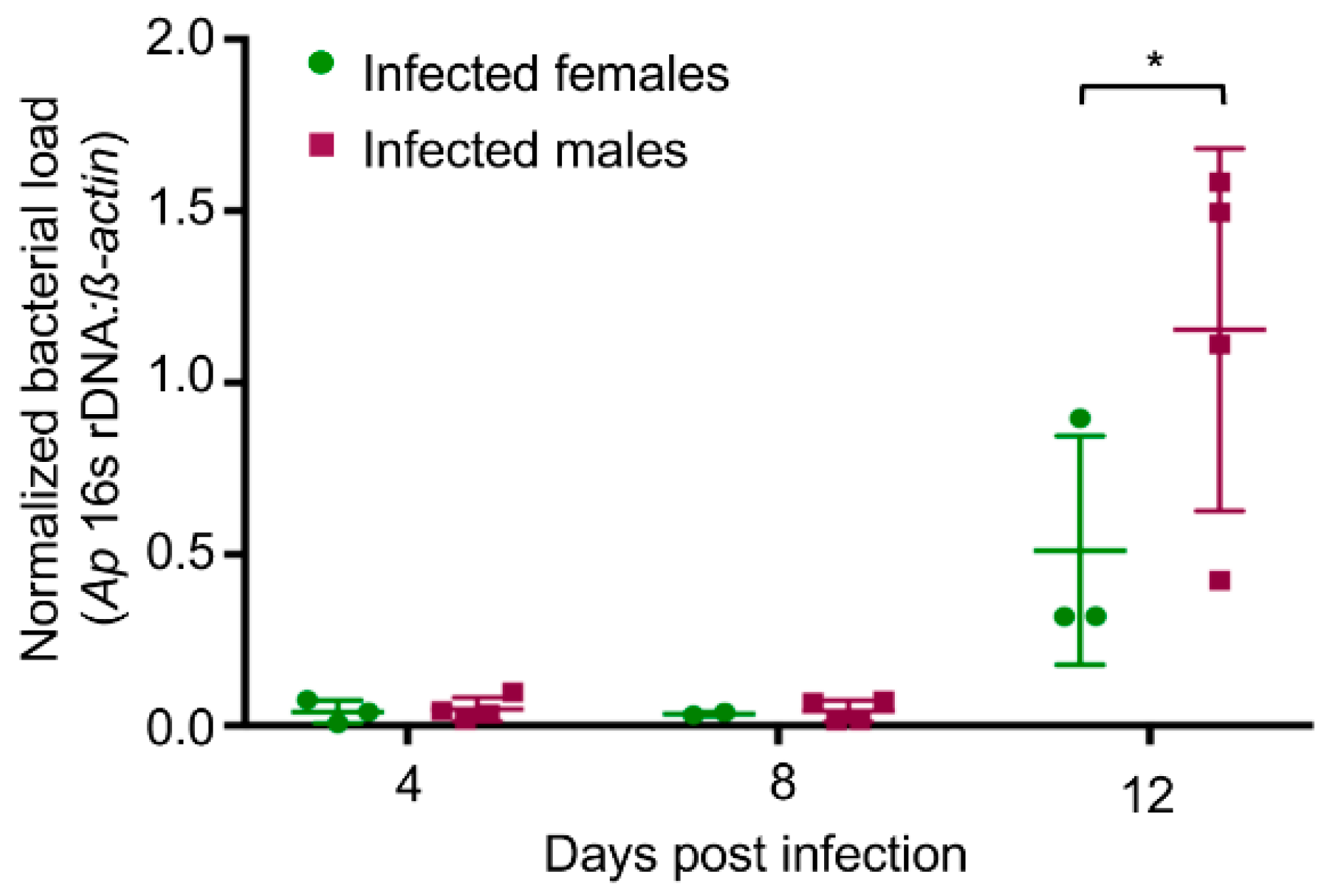
3.1. A. phagocytophilum Infected Male Mice Have Higher Peripheral Blood Bacterial DNA Levels than Infected Female Mice
3.2. A. phagocytophilum Infected Male Mice Exhibit Higher Percentages of Neutrophils Harboring Morulae and Splenomegaly Compared to Infected Female Mice
4. Discussion
Author Contributions
Funding
Conflicts of Interest
References
- Potluri, T.; Engle, K.; Fink, A.L.; Vom Steeg, L.G.; Klein, S.L. Sex reporting in preclinical microbiological and immunological research. MBio 2017, 8, e01868-17. [Google Scholar] [CrossRef] [PubMed]
- Holmes, C.B.; Hausler, H.; Nunn, P. A review of sex differences in the epidemiology of tuberculosis. Int. J. Tuberc. Lung Dis. 1998, 2, 96–104. [Google Scholar] [PubMed]
- Jimenez-Corona, M.E.; Garcia-Garcia, L.; DeRiemer, K.; Ferreyra-Reyes, L.; Bobadilla-del-Valle, M.; Cano-Arellano, B.; Canizales-Quintero, S.; Martínez-Gamboa, A.; Small, P.M.; Sifuentes-Osornio, J.; et al. Gender differentials of pulmonary tuberculosis transmission and reactivation in an endemic area. Thorax 2006, 61, 348–353. [Google Scholar] [CrossRef] [PubMed] [Green Version]
- Greig, J.E.; Carnie, J.A.; Tallis, G.F.; Ryan, N.J.; Tan, A.G.; Gordon, I.R.; Zwolak, B.; Leydon, J.A.; Guest, C.S.; Hart, W.G. An outbreak of Legionnaires’ disease at the Melbourne Aquarium, April 2000: Investigation and case-control studies. Med. J. Aust. 2004, 180, 566–572. [Google Scholar] [PubMed]
- Textoris, J.; Ban, L.H.; Capo, C.; Raoult, D.; Leone, M.; Mege, J.L. Sex-related differences in gene expression following Coxiella burnetii infection in mice: Potential role of circadian rhythm. PLoS ONE 2010, 5, e12190. [Google Scholar] [CrossRef] [PubMed]
- Berman, S.J.; Kundin, W.D. Scrub typhus in South Vietnam: A study of 87 cases. Ann. Intern. Med. 1973, 79, 26–30. [Google Scholar] [CrossRef] [PubMed]
- Chattopadhyay, S.; Richards, A.L. Scrub typhus vaccines: Past history and recent developments. Hum. Vaccin. 2007, 3, 73–80. [Google Scholar] [CrossRef] [PubMed]
- Deller, J.J., Jr.; Russell, P.K. An analysis of fevers of unknown origin in American soldiers in Vietnam. Ann. Intern. Med. 1967, 66, 1129–1143. [Google Scholar] [CrossRef] [PubMed]
- Gormley, T.S. A diagnosis of scrub typhus. Navy Med. 1996, 87, 20–22. [Google Scholar]
- Kelly, D.J.; Fuerst, P.A.; Ching, W.M.; Richards, A.L. Scrub typhus: The geographic distribution of phenotypic and genotypic variants of Orientia tsutsugamushi. Clin. Infect. Dis. 2009, 48 (Suppl. 3), S203–S230. [Google Scholar] [CrossRef] [PubMed]
- Kelly, D.J.; Richards, A.L.; Temenak, J.; Strickman, D.; Dasch, G.A. The past and present threat of rickettsial diseases to military medicine and international public health. Clin. Infect. Dis. 2002, 34 (Suppl. 4), S145–S169. [Google Scholar] [CrossRef] [PubMed]
- Sahni, S.K.; Narra, H.P.; Sahni, A.; Walker, D.H. Recent molecular insights into rickettsial pathogenesis and immunity. Future Microbiol. 2013, 8, 1265–1288. [Google Scholar] [CrossRef] [PubMed] [Green Version]
- Valbuena, G.; Walker, D.H. Approaches to vaccines against Orientia tsutsugamushi. Front. Cell. Infect. Microbiol. 2012, 2, 170. [Google Scholar] [CrossRef] [PubMed]
- Bakken, J.S.; Dumler, J.S. Human granulocytic anaplasmosis. Infect. Dis. Clin. N. Am. 2015, 29, 341–355. [Google Scholar] [CrossRef] [PubMed]
- Chen, S.M.; Dumler, J.S.; Bakken, J.S.; Walker, D.H. Identification of a granulocytotropic Ehrlichia species as the etiologic agent of human disease. J. Clin. Microbiol. 1994, 32, 589–595. [Google Scholar] [PubMed]
- Number of Reported Anaplasmosis Cases by Month of Onset, 2000–2016. Available online: https://www.cdc.gov/anaplasmosis/stats/index.html (accessed on 27 June 2018).
- Johan, S.B.; Paul, G.; Mark Van, E.; Denise, Z.B.; Oscar, L.S.; Sandy, M.; Krueth, J.; Tilden, R.L.; Asanovich, K.; Walls, J.; et al. Seroprevalence of human granulocytic ehrlichiosis among permanent residents of northwestern Wisconsin. Clin. Infect. Dis. 1998, 27, 1491–1496. [Google Scholar]
- Aguero-Rosenfeld, M.E.; Donnarumma, L.; Zentmaier, L.; Jacob, J.; Frey, M.; Noto, R.; Carbonaro, C.A.; Wormser, G.P. Seroprevalence of antibodies that react with Anaplasma phagocytophila, the agent of human granulocytic ehrlichiosis, in different populations in Westchester County, New York. J. Clin. Microbiol. 2002, 40, 2612–2615. [Google Scholar] [CrossRef] [PubMed]
- Bakken, J.S.; Dumler, J.S. Clinical diagnosis and treatment of human granulocytotropic anaplasmosis. Ann. N. Y. Acad. Sci. 2006, 1078, 236–247. [Google Scholar] [CrossRef] [PubMed]
- Hodzic, E.; Fish, D.; Maretzki, C.M.; De Silva, A.M.; Feng, S.; Barthold, S.W. Acquisition and transmission of the agent of human granulocytic ehrlichiosis by Ixodes scapularis ticks. J. Clin. Microbiol. 1998, 36, 3574–3578. [Google Scholar] [PubMed]
- Hodzic, E.; Ijdo, J.W.I.; Feng, S.; Katavolos, P.; Sun, W.; Maretzki, C.H.; Fish, D.; Fikrig, E.; Telford, S.R., III; Barthold, W.S. Granulocytic ehrlichiosis in the laboratory mouse. J. Infect. Dis. 1998, 177, 737–745. [Google Scholar] [CrossRef] [PubMed]
- Hodzic, E.; Feng, S.; Fish, D.; Leutenegger, C.M.; Freet, K.J.; Barthold, S.W. Infection of mice with the agent of human granulocytic ehrlichiosis after different routes of inoculation. J. Infect. Dis. 2001, 183, 1781–1786. [Google Scholar] [CrossRef] [PubMed]
- Sun, W.; Ijdo, J.W.; Telford, S.R., III; Hodzic, E.; Zhang, Y.; Barthold, S.W.; Fikrig, E. Immunization against the agent of human granulocytic ehrlichiosis in a murine model. J. Clin. Investig. 1997, 100, 3014–3018. [Google Scholar] [CrossRef] [PubMed]
- Borjesson, D.L.; Barthold, S.W. The mouse as a model for investigation of human granulocytic ehrlichiosis: Current knowledge and future directions. Comp. Med. 2002, 52, 403–413. [Google Scholar] [PubMed]
- Wang, X.; Shaw, D.K.; Hammond, H.L.; Sutterwala, F.S.; Rayamajhi, M.; Shirey, K.A.; Perkins, D.J.; Bonventre, J.V.; Velayutham, T.S.; Evans, S.M.; et al. The prostaglandin E2-EP3 receptor axis regulates Anaplasma phagocytophilum-mediated NLRC4 inflammasome activation. PLoS Pathog. 2016, 12, e1005803. [Google Scholar] [CrossRef] [PubMed]
- Carlyon, J.A.; Akkoyunlu, M.; Xia, L.; Yago, T.; Wang, T.; Cummings, R.D.; McEver, R.P.; Fikrig, E. Murine neutrophils require alpha1,3-fucosylation but not PSGL-1 for productive infection with Anaplasma phagocytophilum. Blood 2003, 102, 3387–3395. [Google Scholar] [CrossRef] [PubMed]
- Truchan, H.K.; VieBrock, L.; Cockburn, C.L.; Ojogun, N.; Griffin, B.P.; Wijesinghe, D.S.; Chalfant, C.E.; Carlyon, J.A. Anaplasma phagocytophilum Rab10-dependent parasitism of the trans-Golgi network is critical for completion of the infection cycle. Cell. Microbiol. 2016, 18, 260–281. [Google Scholar] [CrossRef] [PubMed]
- Troese, M.J.; Carlyon, J.A. Anaplasma phagocytophilum dense-cored organisms mediate cellular adherence through recognition of human P-selectin glycoprotein ligand 1. Infect. Immun. 2009, 77, 4018–4027. [Google Scholar] [CrossRef] [PubMed]
- Ojogun, N.; Kahlon, A.; Ragland, S.A.; Troese, M.J.; Mastronunzio, J.E.; Walker, N.J.; VieBrock, L.; Thomas, R.J.; Borjesson, D.L.; Fikrig, E.; et al. Anaplasma phagocytophilum outer membrane protein A interacts with sialylated glycoproteins to promote infection of mammalian host cells. Infect. Immun. 2012, 80, 3748–3760. [Google Scholar] [CrossRef] [PubMed]
- Livak, K.J.; Schmittgen, T.D. Analysis of relative gene expression data using real-time quantitative PCR and the 2(-Delta Delta C(T)) Method. Methods 2001, 25, 402–408. [Google Scholar] [CrossRef] [PubMed]
- Hodzic, E.; Borjesson, D.L.; Feng, S.; Barthold, S.W. Acquisition dynamics of Borrelia burgdorferi and the agent of human granulocytic ehrlichiosis at the host-vector interface. Vector Borne Zoonotic Dis. 2001, 1, 149–158. [Google Scholar] [CrossRef] [PubMed]
- Levin, M.L.; Ross, D.E. Acquisition of different isolates of Anaplasma phagocytophilum by Ixodes scapularis from a model animal. Vector Borne Zoonotic Dis. 2004, 4, 53–59. [Google Scholar] [CrossRef] [PubMed]
- Lin, Q.; Zhang, C.; Rikihisa, Y. Analysis of involvement of the RecF pathway in p44 recombination in Anaplasma phagocytophilum and in Escherichia coli by using a plasmid carrying the p44 expression and p44 donor loci. Infect. Immun. 2006, 74, 2052–2062. [Google Scholar] [CrossRef] [PubMed]
- Chen, G.; Severo, M.S.; Sakhon, O.S.; Choy, A.; Herron, M.J.; Felsheim, R.F.; Wiryawanc, H.; Liaoc, J.; Johnsd, J.L.; Munderlohb, U.G.; et al. Anaplasma phagocytophilum dihydrolipoamide dehydrogenase 1 affects host-derived immunopathology during microbial colonization. Infect. Immun. 2012, 80, 3194–3205. [Google Scholar] [CrossRef] [PubMed]
- Zhan, L.; Cao, W.-C.; Jiang, J.-F.; Zhang, X.-A.; Liu, Y.-X.; Wu, X.-M.; Zhang, W.-Y.; Zhang, P.-H.; Bian, C.-L.; Dumler, J.S.; et al. Anaplasma phagocytophilum from rodents and sheep, China. Emerg. Infect. Dis. 2010, 16, 764–768. [Google Scholar] [CrossRef] [PubMed]
- Choi, K.S.; Scorpio, D.G.; Dumler, J.S. Anaplasma phagocytophilum ligation to toll-like receptor (TLR) 2, but not to TLR4, activates macrophages for nuclear factor-kappa B nuclear translocation. J. Infect. Dis. 2004, 189, 1921–1925. [Google Scholar] [CrossRef] [PubMed]
- Wang, X.; Cheng, Z.; Zhang, C.; Kikuchi, T.; Rikihisa, Y. Anaplasma phagocytophilum p44 mRNA expression is differentially regulated in mammalian and tick host cells: Involvement of the DNA binding protein ApxR. J. Bacteriol. 2007, 189, 8651–8659. [Google Scholar] [CrossRef] [PubMed]
- Scorpio, D.G.; Choi, K.-S.; Dumler, J.S. Anaplasma phagocytophilum-related defects in CD8, NKT, and NK lymphocyte cytotoxicity. Front. Immunol. 2018, 9, 710. [Google Scholar] [CrossRef] [PubMed]
- Pedra, J.H.; Sutterwala, F.S.; Sukumaran, B.; Ogura, Y.; Qian, F.; Montgomery, R.R.; Flavell, R.A.; Fikrig, E. ASC/PYCARD and caspase-1 regulate the IL-18/IFN-gamma axis during Anaplasma phagocytophilum infection. J. Immunol. 2007, 179, 4783–4791. [Google Scholar] [CrossRef] [PubMed]
- Niu, H.; Xiong, Q.; Yamamoto, A.; Hayashi-Nishino, M.; Rikihisa, Y. Autophagosomes induced by a bacterial Beclin 1 binding protein facilitate obligatory intracellular infection. Proc. Natl. Acad. Sci. USA 2012, 109, 20800–20807. [Google Scholar] [CrossRef] [PubMed] [Green Version]
- Pedra, J.H.F.; Mattner, J.; Tao, J.; Kerfoot, S.M.; Davis, R.J.; Flavell, R.A.; Askenase, P.W.; Yin, Z.; Fikrig, E. c-Jun NH2-terminal kinase 2 inhibits gamma interferon production during Anaplasma phagocytophilum infection. Infect. Immun. 2008, 76, 308–316. [Google Scholar] [CrossRef] [PubMed]
- Kolbert, C.P.; Bruinsma, E.S.; Abdulkarim, A.S.; Hofmeister, E.K.; Tompkins, R.B.; Telford, S.R., III; Mitchell, P.D.; Adams-Stich, J.; Persing, D.H. Characterization of an immunoreactive protein from the agent of human granulocytic ehrlichiosis. J. Clin. Microbiol. 1997, 35, 1172–1178. [Google Scholar] [PubMed]
- Ravyn, M.D.; Lamb, L.J.; Jemmerson, R.; Goodman, J.L.; Johnson, R.C. Characterization of monoclonal antibodies to an immunodominant protein of the etiologic agent of human granulocytic ehrlichiosis. Am. J. Trop. Med. Hyg. 1999, 61, 171–176. [Google Scholar] [CrossRef] [PubMed]
- Kim, H.Y.; Rikihisa, Y. Characterization of monoclonal antibodies to the 44-kilodalton major outer membrane protein of the human granulocytic ehrlichiosis agent. J. Clin. Microbiol. 1998, 36, 3278–3284. [Google Scholar] [PubMed]
- Holden, K.; Hodzic, E.; Feng, S.; Freet, K.J.; Lefebvre, R.B.; Barthold, S.W. Coinfection with Anaplasma phagocytophilum alters Borrelia burgdorferi population distribution in C3H/HeN mice. Infect. Immun. 2005, 73, 3440–3444. [Google Scholar] [CrossRef] [PubMed]
- Thomas, V.; Anguita, J.; Barthold, S.W.; Fikrig, E. Coinfection with Borrelia burgdorferi and the agent of human granulocytic ehrlichiosis alters murine immune responses, pathogen burden, and severity of Lyme arthritis. Infect. Immun. 2001, 69, 3359–3371. [Google Scholar] [CrossRef] [PubMed]
- Banerjee, R.; Anguita, J.; Roos, D.; Fikrig, E. Cutting edge: Infection by the agent of human granulocytic ehrlichiosis prevents the respiratory burst by down-regulating gp91phox. J. Immunol. 2000, 164, 3946–3949. [Google Scholar] [CrossRef] [PubMed]
- Scorpio, D.G.; Akkoyunlu, M.; Fikrig, E.; Dumler, J.S. CXCR2 blockade influences Anaplasma phagocytophilum propagation but not histopathology in the mouse model of human granulocytic anaplasmosis. Clin. Diagn. Lab. Immunol. 2004, 11, 963–968. [Google Scholar] [CrossRef] [PubMed]
- Bunnell, J.E.; Trigiani, E.R.; Srinivas, S.R.; Dumler, J.S. Development and distribution of pathologic lesions are related to immune status and tissue deposition of human granulocytic ehrlichiosis agent-infected cells in a murine model system. J. Infect. Dis. 1999, 180, 546–550. [Google Scholar] [CrossRef] [PubMed]
- Teglas, M.B.; Foley, J. Differences in the transmissibility of two Anaplasma phagocytophilum strains by the North American tick vector species, Ixodes pacificus and Ixodes scapularis (Acari: Ixodidae). Exp. Appl. Acarol. 2006, 38, 47–58. [Google Scholar] [CrossRef] [PubMed]
- Ijdo, J.W.; Wu, C.; Telford Iii, S.R.; Fikrig, E. Differential expression of the p44 gene family in the agent of human granulocytic ehrlichiosis. Infect. Immun. 2002, 70, 5295–5298. [Google Scholar] [CrossRef] [PubMed]
- Choi, K.-S.; Webb, T.; Oelke, M.; Scorpio, D.G.; Dumler, J.S. Differential innate immune cell activation and proinflammatory response in Anaplasma phagocytophilum infection. Infect. Immun. 2007, 75, 3124–3130. [Google Scholar] [CrossRef] [PubMed]
- Johns, J.L.; Borjesson, D.L. Downregulation of CXCL12 signaling and altered hematopoietic stem and progenitor cell trafficking in a murine model of acute Anaplasma phagocytophilum infection. Innate Immun. 2012, 18, 418–428. [Google Scholar] [CrossRef] [PubMed]
- Ijdo, J.W.; Zhang, Y.; Hodzic, E.; Magnarelli, L.A.; Wilson, M.L.; Telford, S.R.; Barthold, S.W.; Fikrig, E. The early humoral response in human granulocytic ehrlichiosis. J. Infect. Dis. 1997, 176, 687–692. [Google Scholar] [CrossRef] [PubMed]
- Carlyon, J.A.; Ryan, D.; Archer, K.; Fikrig, E. Effects of Anaplasma phagocytophilum on host cell ferritin mrna and protein levels. Infect. Immun. 2005, 73, 7629–7636. [Google Scholar] [CrossRef] [PubMed]
- Birkner, K.; Steiner, B.; Rinkler, C.; Kern, Y.; Aichele, P.; Bogdan, C.; von Loewenich, F.D. The elimination of Anaplasma phagocytophilum requires CD4+ T cells, but is independent of Th1 cytokines and a wide spectrum of effector mechanisms. Eur. J. Immunol. 2008, 38, 3395–3410. [Google Scholar] [CrossRef] [PubMed]
- Lin, Q.; Rikihisa, Y. Establishment of cloned Anaplasma phagocytophilum and analysis of p44 gene conversion within an infected horse and infected SCID mice. Infect. Immun. 2005, 73, 5106–5114. [Google Scholar] [CrossRef] [PubMed]
- Blas-Machado, U.; de la Fuente, J.; Blouin, E.F.; Almazan, C.; Kocan, K.M.; Mysore, J.V. Experimental infection of C3H/HeJ mice with the NY18 isolate of Anaplasma phagocytophilum. Vet. Pathol. 2007, 44, 64–73. [Google Scholar] [CrossRef] [PubMed]
- Akkoyunlu, M.; Malawista, S.E.; Anguita, J.; Fikrig, E. Exploitation of interleukin-8-induced neutrophil chemotaxis by the agent of human granulocytic ehrlichiosis. Infect. Immun. 2001, 69, 5577–5588. [Google Scholar] [CrossRef] [PubMed]
- Banerjee, R.; Anguita, J.; Fikrig, E. Granulocytic ehrlichiosis in mice deficient in phagocyte oxidase or inducible nitric oxide synthase. Infect. Immun. 2000, 68, 4361–4362. [Google Scholar] [CrossRef] [PubMed]
- Xiong, Q.; Wang, X.; Rikihisa, Y. High-cholesterol diet facilitates Anaplasma phagocytophilum infection and up-regulates macrophage inflammatory protein-2 and CXCR2 expression in apolipoprotein e-deficient mice. J. Infect. Dis. 2007, 195, 1497–1503. [Google Scholar] [CrossRef] [PubMed]
- Pedra, J.H.; Tao, J.; Sutterwala, F.S.; Sukumaran, B.; Berliner, N.; Bockenstedt, L.K.; Flavell, R.A.; Yin, Z.; Fikrig, E. IL-12/23p40-dependent clearance of Anaplasma phagocytophilum in the murine model of human anaplasmosis. FEMS Immunol. Med. Microbiol. 2007, 50, 401–410. [Google Scholar] [CrossRef] [PubMed]
- Bunnell, J.E.; Magnarelli, L.A.; Dumler, J.S. Infection of laboratory mice with the human granulocytic ehrlichiosis agent does not induce antibodies to diagnostically significant Borrelia burgdorferi antigens. J. Clin. Microbiol. 1999, 37, 2077–2079. [Google Scholar] [PubMed]
- Johns, J.L.; MacNamara, K.C.; Walker, N.J.; Winslow, G.M.; Borjesson, D.L. Infection with Anaplasma phagocytophilum induces multilineage alterations in hematopoietic progenitor cells and peripheral blood cells. Infect. Immun. 2009, 77, 4070–4080. [Google Scholar] [CrossRef] [PubMed]
- Johns, J.L.; Discipulo, M.L.; Koehne, A.L.; Moorhead, K.A.; Nagamine, C.M. Influence of genetic background on hematologic and histopathologic alterations during acute granulocytic anaplasmosis in 129/SvEv and C57BL/6J mice lacking type I and type II interferon signaling. Comp. Med. 2017, 67, 127–137. [Google Scholar] [PubMed]
- Scorpio, D.G.; von Loewenich, F.D.; Göbel, H.; Bogdan, C.; Dumler, J.S. Innate immune response to Anaplasma phagocytophilum contributes to hepatic injury. Clin. Vaccin. Immunol. 2006, 13, 806–809. [Google Scholar] [CrossRef] [PubMed]
- Herron, M.J.; Ericson, M.E.; Kurtti, T.J.; Munderloh, U.G. The interactions of Anaplasma phagocytophilum, endothelial cells, and human neutrophils. Ann. N. Y. Acad. Sci. 2005, 1063, 374–382. [Google Scholar] [CrossRef] [PubMed]
- Wang, T.; Akkoyunlu, M.; Banerjee, R.; Fikrig, E. Interferon-γ deficiency reveals that 129Sv mice are inherently more susceptible to Anaplasma phagocytophilum than C57BL/6 mice. FEMS Immunol. Med. Microbiol. 2004, 42, 299–305. [Google Scholar] [CrossRef] [PubMed]
- Sukumaran, B.; Narasimhan, S.; Anderson, J.F.; DePonte, K.; Marcantonio, N.; Krishnan, M.N.; Fish, D.; Telford, S.R.; Kantor, F.S.; Fikrig, E. An Ixodes scapularis protein required for survival of Anaplasma phagocytophilum in tick salivary glands. J. Exp. Med. 2006, 203, 1507–1517. [Google Scholar] [CrossRef] [PubMed]
- Borjesson, D.L.; Simon, S.I.; Hodzic, E.; Ballantyne, C.M.; Barthold, S.W. Kinetics of CD11b/CD18 up-regulation during infection with the agent of human granulocytic ehrlichiosis in mice. Lab. Investig. 2002, 82, 303–311. [Google Scholar] [CrossRef] [PubMed]
- Martin, M.E.; Bunnell, J.E.; Dumler, J.S. Pathology, immunohistology, and cytokine responses in early phases of human granulocytic ehrlichiosis in a murine model. J. Infect. Dis. 2000, 181, 374–378. [Google Scholar] [CrossRef] [PubMed]
- Mastronunzio, J.E.; Kurscheid, S.; Fikrig, E. Postgenomic analyses reveal development of infectious Anaplasma phagocytophilum during transmission from ticks to mice. J. Bacteriol. 2012, 194, 2238–2247. [Google Scholar] [CrossRef] [PubMed]
- Sukumaran, B.; Ogura, Y.; Pedra, J.H.; Kobayashi, K.S.; Flavell, R.A.; Fikrig, E. Receptor interacting protein-2 contributes to host defense against Anaplasma phagocytophilum infection. FEMS Immunol. Med. Microbiol. 2012, 66, 211–219. [Google Scholar] [CrossRef] [PubMed]
- Levin, M.L.; Coble, D.J.; Ross, D.E. Reinfection with Anaplasma phagocytophilum in BALB/c mice and cross-protection between two sympatric isolates. Infect. Immun. 2004, 72, 4723–4730. [Google Scholar] [CrossRef] [PubMed]
- Browning, M.D.; Garyu, J.W.; Dumler, J.S.; Scorpio, D.G. Role of reactive nitrogen species in development of hepatic injury in a C57bl/6 mouse model of human granulocytic anaplasmosis. Comp. Med. 2006, 56, 55–62. [Google Scholar] [PubMed]
- Borjesson, D.L.; Simon, S.I.; Hodzic, E.; DeCock, H.E.; Ballantyne, C.M.; Barthold, S.W. Roles of neutrophil beta 2 integrins in kinetics of bacteremia, extravasation, and tick acquisition of Anaplasma phagocytophila in mice. Blood 2003, 101, 3257–3264. [Google Scholar] [CrossRef] [PubMed]
- Felek, S.; Telford, S., III; Falco, R.C.; Rikihisa, Y. Sequence analysis of p44 homologs expressed by Anaplasma phagocytophilum in infected ticks feeding on naive hosts and in mice infected by tick attachment. Infect. Immun. 2004, 72, 659–666. [Google Scholar] [CrossRef] [PubMed]
- Scorpio, D.G.; Leutenegger, C.; Berger, J.; Barat, N.; Madigan, J.E.; Dumler, J.S. Sequential analysis of Anaplasma phagocytophilum msp2 transcription in murine and equine models of human granulocytic anaplasmosis. Clin. Vaccine Immunol. 2008, 15, 418–424. [Google Scholar] [CrossRef] [PubMed]
- Choi, K.-S.; Scorpio, D.G.; Dumler, J.S. Stat1 negatively regulates immune-mediated injury with Anaplasma phagocytophilum infection. J. Immunol. 2014, 193, 5088–5098. [Google Scholar] [CrossRef] [PubMed]
- Wang, T.; Malawista, S.E.; Pal, U.; Grey, M.; Meek, J.; Akkoyunlu, M.; Thomas, V.; Fikrig, E. Superoxide anion production during Anaplasma phagocytophila infection. J. Infect. Dis. 2002, 186, 274–280. [Google Scholar] [CrossRef] [PubMed]
- Zeidner, N.S.; Massung, R.F.; Dolan, M.C.; Dadey, E.; Gabitzsch, E.; Dietrich, G.; Levin, M.L. A sustained-release formulation of doxycycline hyclate (Atridox) prevents simultaneous infection of Anaplasma phagocytophilum and Borrelia burgdorferi transmitted by tick bite. J. Med. Microbiol. 2008, 57 Pt 4, 463–468. [Google Scholar] [CrossRef] [PubMed]
- Borjesson, D.L.; Simon, S.I.; Tablin, F.; Barthold, S.W. Thrombocytopenia in a mouse model of human granulocytic ehrlichiosis. J. Infect. Dis. 2001, 184, 1475–1479. [Google Scholar] [CrossRef] [PubMed]
- Zhi, N.; Ohashi, N.; Tajima, T.; Mott, J.; Stich, R.W.; Grover, D.; Telford, S.R., III; Lin, Q.; Rikihisa, Y. Transcript heterogeneity of the p44 multigene family in a human granulocytic ehrlichiosis agent transmitted by ticks. Infect. Immun. 2002, 70, 1175–1184. [Google Scholar] [CrossRef] [PubMed]
- Martin, M.E.; Caspersen, K.; Dumler, J.S. Immunopathology and ehrlichial propagation are regulated by interferon-gamma and interleukin-10 in a murine model of human granulocytic ehrlichiosis. Am. J. Pathol. 2001, 158, 1881–1888. [Google Scholar] [CrossRef]
- Akkoyunlu, M.; Fikrig, E. Gamma interferon dominates the murine cytokine response to the agent of human granulocytic ehrlichiosis and helps to control the degree of early rickettsemia. Infect. Immun. 2000, 68, 1827–1833. [Google Scholar] [CrossRef] [PubMed]
- Lin, M.; Rikihisa, Y. Ehrlichia chaffeensis and Anaplasma phagocytophilum lack genes for lipid A biosynthesis and incorporate cholesterol for their survival. Infect. Immun. 2003, 71, 5324–5331. [Google Scholar] [CrossRef] [PubMed]
- Xiong, Q.; Rikihisa, Y. Subversion of NPC1 pathway of cholesterol transport by Anaplasma phagocytophilum. Cell. Microbiol. 2012, 14, 560–576. [Google Scholar] [CrossRef] [PubMed]
- Xiong, Q.; Lin, M.; Rikihisa, Y. Cholesterol-dependent Anaplasma phagocytophilum exploits the low-density lipoprotein uptake pathway. PLoS Pathog. 2009, 5, e1000329. [Google Scholar] [CrossRef] [PubMed]
- National Cholesterol Education Program Expert Panel on Detection, Evaluation, and Treatment of High Blood Cholesterol in Adults. Third Report of the National Cholesterol Education Program (NCEP) Expert Panel on Detection, Evaluation, and Treatment of High Blood Cholesterol in Adults (Adult Treatment Panel III) final report. Circulation 2002, 106, 3143–3421. [Google Scholar]
- Klein, S.L. Sex influences immune responses to viruses, and efficacy of prophylaxis and treatments for viral diseases. Bioessays 2012, 34, 1050–1059. [Google Scholar] [CrossRef] [PubMed] [Green Version]
- Pinzan, C.F.; Ruas, L.P.; Casabona-Fortunato, A.S.; Carvalho, F.C.; Roque-Barreira, M.C. Immunological basis for the gender differences in murine Paracoccidioides brasiliensis infection. PLoS ONE 2010, 5, e10757. [Google Scholar] [CrossRef] [PubMed]
- Villacres, M.C.; Longmate, J.; Auge, C.; Diamond, D.J. Predominant type 1 CMV-specific memory T-helper response in humans: Evidence for gender differences in cytokine secretion. Hum. Immunol. 2004, 65, 476–485. [Google Scholar] [CrossRef] [PubMed]
- Zhang, M.A.; Rego, D.; Moshkova, M.; Kebir, H.; Chruscinski, A.; Nguyen, H.; Akkermann, R.; Stanczyk, F.Z.; Prat, A.; Steinman, L.; et al. Peroxisome proliferator-activated receptor (PPAR) alpha and -gamma regulate IFNgamma and IL-17A production by human T cells in a sex-specific way. Proc. Natl. Acad. Sci. USA 2012, 109, 9505–9510. [Google Scholar] [CrossRef] [PubMed]


| References | Usage of Female and/or Male Mice |
|---|---|
| [20,21,22,26,31,32,34,35,36,38,40,42,45,46,47,48,49,51,54,55,56,59,60,63,66,72,74,75,76,78,79,80] | Not disclosed |
| [23,43,52,53,58,64,67,68,69,70,71,81,82,84] | Females |
| [33,37,44,50,57,61,77,83,84,85] | Males |
| [25,39,41,62,65,73] | Males and females |
© 2018 by the authors. Licensee MDPI, Basel, Switzerland. This article is an open access article distributed under the terms and conditions of the Creative Commons Attribution (CC BY) license (http://creativecommons.org/licenses/by/4.0/).
Share and Cite
Naimi, W.A.; Green, R.S.; Cockburn, C.L.; Carlyon, J.A. Differential Susceptibility of Male Versus Female Laboratory Mice to Anaplasma phagocytophilum Infection. Trop. Med. Infect. Dis. 2018, 3, 78. https://doi.org/10.3390/tropicalmed3030078
Naimi WA, Green RS, Cockburn CL, Carlyon JA. Differential Susceptibility of Male Versus Female Laboratory Mice to Anaplasma phagocytophilum Infection. Tropical Medicine and Infectious Disease. 2018; 3(3):78. https://doi.org/10.3390/tropicalmed3030078
Chicago/Turabian StyleNaimi, Waheeda A., Ryan S. Green, Chelsea L. Cockburn, and Jason A. Carlyon. 2018. "Differential Susceptibility of Male Versus Female Laboratory Mice to Anaplasma phagocytophilum Infection" Tropical Medicine and Infectious Disease 3, no. 3: 78. https://doi.org/10.3390/tropicalmed3030078